The superior mesenteric artery supplies the midgut from the ampullary region of the second part of the duodenum to the splenic flexure of the large intestine or large bowel. The inferior pancreaticoduodenal artery arises from the superior mesenteric artery and, along with the superior pancreaticoduodenal artery, supplies the head of the pancreas."}},{"@type":"Question","url":"https://mobilephysiotherapyclinic.in/superior-mesentric-plexus/#faq-question-6579b92995da2","name":"Where does the superior mesenteric vein drain?","acceptedAnswer":{"@type":"Answer","text":"The superior mesenteric vein ends at the transpyloric plane (around the lower margin of the first lumbar vertebra) by combining with the splenic vein to form the hepatic portal vein."}},{"@type":"Question","url":"https://mobilephysiotherapyclinic.in/superior-mesentric-plexus/#faq-question-6579b92995da3","name":"What is the neurotransmitter in the superior mesenteric ganglia?","acceptedAnswer":{"@type":"Answer","text":"Parasympathetic fibers synapse close to them or within the effector organ, with acetylcholine as the neurotransmitter."}},{"@type":"Question","url":"https://mobilephysiotherapyclinic.in/superior-mesentric-plexus/#faq-question-6579b92995da4","name":"Where is the superior mesenteric plexus located?","acceptedAnswer":{"@type":"Answer","text":"retroperitoneum The superior mesenteric plexus is an autonomic nerve plexus and ganglia situated in the retroperitoneum region."}},{"@type":"Question","url":"https://mobilephysiotherapyclinic.in/superior-mesentric-plexus/#faq-question-6579b92995da5","name":"What spinal level is the superior mesenteric artery?","acceptedAnswer":{"@type":"Answer","text":"The origins of all the branches vary completely between the two vertebral levels. The celiac artery originated at T11/T12\u2013L1/L2, subsequently the superior mesenteric artery at T12\u2013L2, the paired renal arteries at T12/L1\u2013L2/L3, the inferior mesenteric artery at L2\u2013L4, and the common iliac arteries at L3\u2013L5."}},{"@type":"Question","url":"https://mobilephysiotherapyclinic.in/superior-mesentric-plexus/#faq-question-6579b92995da6","name":"Where is the origin of the SMA?","acceptedAnswer":{"@type":"Answer","text":"abdominal aorta In human anatomy, the superior mesenteric artery (SMA) is an artery that originates from the anterior surface of the abdominal aorta, just inferior to the origin of the celiac trunk, and it supplies the blood to the intestine from the lower aspect of the duodenum via two-thirds of the transverse colon, as well as the pancreas."}},{"@type":"Question","url":"https://mobilephysiotherapyclinic.in/superior-mesentric-plexus/#faq-question-6579b92995da7","name":"What is the normal size of an SMV?","acceptedAnswer":{"@type":"Answer","text":"The superior mesenteric vein belongs to the portal venous system, presents available with a diameter of up to 1.2 cm, and is defined as aneurysmatic when it extends 1.4 cm in diameter jointly with loosing of wall parallelism"}},{"@type":"Question","url":"https://mobilephysiotherapyclinic.in/superior-mesentric-plexus/#faq-question-6579b92995da8","name":"What flows into the superior mesenteric vein?","acceptedAnswer":{"@type":"Answer","text":"In human anatomy, the superior mesenteric vein is a blood vessel that drains blood from the small intestine (jejunum and ileum). Behind or posterior region to the neck of the pancreas, the superior mesenteric vein combines or merges with the splenic vein to form or to make the hepatic portal vein."}},{"@type":"Question","url":"https://mobilephysiotherapyclinic.in/superior-mesentric-plexus/#faq-question-6579b92995da9","name":"What happens if the superior mesenteric artery is blocked or obstructed?","acceptedAnswer":{"@type":"Answer","text":"In mesenteric ischemia, a blockage in an artery blocks or discontinues blood flow to a portion of the intestine. Mesenteric ischemia occurs when narrowed or blocked arteries restrict or reduce the blood flow to the small intestine. Decreased blood flow can cause permanent damage to the small intestine. "}}]},{"@type":"Person","@id":"https://mobilephysiotherapyclinic.in/profile/poojasingh/","name":"Poojasingh","url":"https://mobilephysiotherapyclinic.in/profile/poojasingh/","image":{"@type":"ImageObject","@id":"https://mobilephysiotherapyclinic.in/wp-content/litespeed/avatar/f5f49ce44781eadee39ad6fb1da4fb19.jpg?ver=1744808575&d=https://mobilephysiotherapyclinic.in/wp-content/plugins/userswp/assets/images/no_profile.png","url":"https://mobilephysiotherapyclinic.in/wp-content/litespeed/avatar/f5f49ce44781eadee39ad6fb1da4fb19.jpg?ver=1744808575&d=https://mobilephysiotherapyclinic.in/wp-content/plugins/userswp/assets/images/no_profile.png","caption":"Poojasingh","inLanguage":"en-US"}},{"@type":"BlogPosting","headline":"Superior mesentric plexus - Mobile Physio","keywords":"superior mesentric plexus","datePublished":"2023-02-10T16:51:52+00:00","dateModified":"2023-12-13T14:01:13+00:00","author":{"@id":"https://mobilephysiotherapyclinic.in/profile/poojasingh/","name":"Poojasingh"},"publisher":{"@id":"https://mobilephysiotherapyclinic.in/#person"},"description":"Superior mesenteric plexus is a downward continuance of the celiac prevertebral plexus and refers to a complexity of neurons that is.........","name":"Superior mesentric plexus - Mobile Physio","@id":"https://mobilephysiotherapyclinic.in/superior-mesentric-plexus/#richSnippet","isPartOf":{"@id":"https://mobilephysiotherapyclinic.in/superior-mesentric-plexus/#webpage"},"image":{"@id":"https://mobilephysiotherapyclinic.in/wp-content/uploads/2023/02/superior-mesentric-plexus-jpg.webp"},"inLanguage":"en-US","mainEntityOfPage":{"@id":"https://mobilephysiotherapyclinic.in/superior-mesentric-plexus/#webpage"}}]}
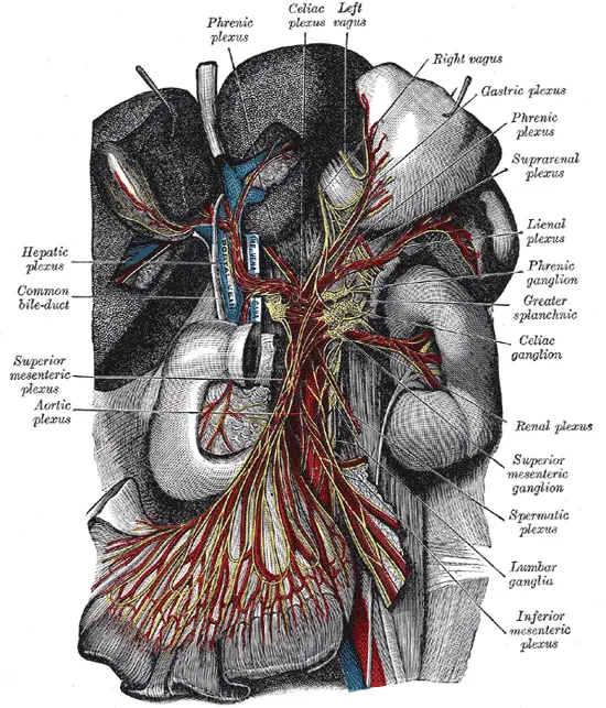
The superior mesenteric plexus is a downward continuance of the celiac prevertebral plexus and refers to a complexity of neurons that is present near the root of the superior mesenteric artery and posterior aspect of the pancreas. Various secondary plexuses get arise here, which follow the route of the arterial branches of the superior mesenteric artery. These include plexuses around jejunal and ileal arteries, and the arterial branches to the colon in the hindgut, like ileocolic, right, and middle colic arteries.
The superior mesenteric plexus accommodates parasympathetic motor neurons from the vagus nerve and sympathetic motor neurons which come through the splanchnic nerves. Additionally, visceral afferent neurons also live inside the superior mesenteric plexus, which transmits the visceral sensory information to the central nervous system through the vagus nerve and the sympathetic splanchnic nerves.
An important ganglion, called the superior mesenteric ganglion, is present within the superior mesenteric plexus, close to the root of the superior mesenteric artery. The sympathetic preganglionic neurons come from the splanchnic nerves synapse with cell bodies of post-ganglionic sympathetic neurons inside this ganglion. The post-ganglionic sympathetic neurons then supply the vessels and glands within the gastrointestinal tract, causing vasoconstriction and decreasing secretions.
FAQ
Where does the superior mesenteric plexus come from?
The superior mesenteric plexus is a continuance of the lower part of the celiac plexus, collecting a branch from the junction of the right vagus nerve with the plexus. The celiac ganglia with the sympathetic plexuses of the abdominal viscera extend from the ganglia
Is the superior mesenteric plexus sympathetic or parasympathetic?
The superior mesenteric ganglion is the synapsing point for one of the pre-and post-synaptic nerves of the sympathetic division of the autonomic nervous system (ANS).
What does the superior mesenteric plexus supply?
The superior mesenteric artery gives oxygenated blood and nutrients to the intestines. These organs are portions of the digestive system. The artery branches off of the aorta, which is the largest blood vessel in the body.
What does the superior mesenteric drain into?
The 1st jejunal branch of the superior mesenteric vein, which drains the duodenojejunal flexure and 1st jejunal loop, has been noticed to drain into either the main trunk of the superior mesenteric vein and into the left intestinal branch
Where does the superior mesenteric supply blood to?
large intestine The superior mesenteric artery supplies the midgut from the ampullary region of the second part of the duodenum to the splenic flexure of the large intestine or large bowel. The inferior pancreaticoduodenal artery arises from the superior mesenteric artery and, along with the superior pancreaticoduodenal artery, supplies the head of the pancreas.
Where does the superior mesenteric vein drain?
The superior mesenteric vein ends at the transpyloric plane (around the lower margin of the first lumbar vertebra) by combining with the splenic vein to form the hepatic portal vein.
What is the neurotransmitter in the superior mesenteric ganglia?
Parasympathetic fibers synapse close to them or within the effector organ, with acetylcholine as the neurotransmitter.
Where is the superior mesenteric plexus located?
retroperitoneum The superior mesenteric plexus is an autonomic nerve plexus and ganglia situated in the retroperitoneum region.
What spinal level is the superior mesenteric artery?
The origins of all the branches vary completely between the two vertebral levels. The celiac artery originated at T11/T12–L1/L2, subsequently the superior mesenteric artery at T12–L2, the paired renal arteries at T12/L1–L2/L3, the inferior mesenteric artery at L2–L4, and the common iliac arteries at L3–L5.
Where is the origin of the SMA?
abdominal aorta In human anatomy, the superior mesenteric artery (SMA) is an artery that originates from the anterior surface of the abdominal aorta, just inferior to the origin of the celiac trunk, and it supplies the blood to the intestine from the lower aspect of the duodenum via two-thirds of the transverse colon, as well as the pancreas.
What is the normal size of an SMV?
The superior mesenteric vein belongs to the portal venous system, presents available with a diameter of up to 1.2 cm, and is defined as aneurysmatic when it extends 1.4 cm in diameter jointly with loosing of wall parallelism
What flows into the superior mesenteric vein?
In human anatomy, the superior mesenteric vein is a blood vessel that drains blood from the small intestine (jejunum and ileum). Behind or posterior region to the neck of the pancreas, the superior mesenteric vein combines or merges with the splenic vein to form or to make the hepatic portal vein.
What happens if the superior mesenteric artery is blocked or obstructed?
In mesenteric ischemia, a blockage in an artery blocks or discontinues blood flow to a portion of the intestine. Mesenteric ischemia occurs when narrowed or blocked arteries restrict or reduce the blood flow to the small intestine. Decreased blood flow can cause permanent damage to the small intestine.
What is the Pronator Quadratus muscle? The quadrangular, short, flat, and thin muscle known as the pronator quadratus is located in the forearm’s anterior compartment. Along with the flexor digitorum profundus and the flexor pollicis longus, it is a member of the deep group of forearm flexors. The forearm flexors’ superficial group covers these three…
What is Coracobrachialis? The coracobrachialis is a long and thin muscle in the arm’s anterior compartment. It runs from the coracoid process of the scapula to the shaft of the humerus, as the term implies. The coracobrachialis muscle’s main function is to generate arm flexion and adduction at the shoulder joint. The musculocutaneous nerve innervates…
Piriformis is a muscle of the gluteal region which is located deep to the gluteus maximus. Piriformis muscle with other muscles like gemellus superior, obturator internus, gemellus inferior, quadratus femoris, obturator externus muscles are the responsible for external rotators of the hip . Piriformis attaching to the sacrum on one end and the greater trochanter…
Introduction: The orbicularis oris muscle is a complex of muscles in the lips that encircles the mouth in Human anatomy. It is a sphincter, or circular muscle, but it is actually composed of four independent quadrants that interlace and give only an appearance of circularity. It is also one of the muscles used in the…
Introduction The suboccipital nerve is also referred to as the dorsal ramus of the first cervical nerve since it arises from the posterior/dorsal ramus of the first cervical spinal nerve (C1). The suboccipital nerve runs to the posterior neck region from its origin. This nerve follows the vertebral artery along its route, which is why…
Introduction Extraocular muscles are essential for ocular movement. The four rectus muscles, superior oblique muscle, and inferior oblique work in conjunction to move the eyes in every direction. The inferior oblique is a thin, narrow eye muscle and belongs to the group of six extraocular muscles or the orbit’s extrinsic muscles. The inferior oblique muscle…