
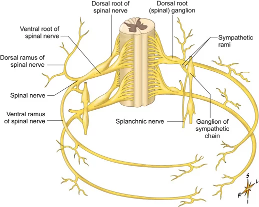
Splanchnic Nerves
The splanchnic nerves are paired visceral nerves (nerves that contribute to the supply of the internal organs), carrying fibers of the autonomic nervous system (visceral efferent fibers) as well as sensory fibers from the organs (visceral afferent fibers). All splanchnic nerves carry sympathetic nerve fibers except for the pelvic splanchnic nerves, which carry parasympathetic fibers.
Types of splanchnic nerves
The term splanchnic nerves types are:
- pelvic splanchnic nerves
- cardiopulmonary nerves
- lumbar splanchnic nerves
- thoracic splanchnic nerves(greater,and lesser)
- sacral splanchnic nerves
Pelvic splanchnic nerves
The pelvic splanchnic nerves (also called nerve originates) are the preganglionic parasympathetic nerve fibers that begin from the anterior rami of spinal nerves S2-S4 and freely spreads across the abdominal wall and pelvic cavities to supply the abdominopelvic viscera.
The main function of the pelvic splanchnic nerves is to give the parasympathetic insert to the autonomic ganglia of the pelvis. Therefore, the pelvic splanchnic nerves provide a parasympathetic supply for the majority of the pelvic organs, as well as the urinary bladder, hindgut, ureter, prostate, and urethra.
Origin and course
Pelvic splanchnic nerve
- The pelvic splanchnic nerves begin from the same nerves that help in the formation of the sacral plexus. mainly, their fibers get originate from the anterior rami of spinal nerves S2-S4. They pass through the anterior sacral foramina to undertake the presacral surface area and branch out into three main locations:
- The majority of the pelvic splanchnic fibers join the inferior hypogastric plexus, while the smaller portion of the fibers connects the hypogastric nerves and runs with them to the superior hypogastric plexus.
- The smallest portion of the pelvic splanchnic fibers runs across the pelvic brim to the retroperitoneal space to supply the mesentery of the sigmoid and descending colon.
- From the plexuses, these [still preganglionic] fibers are spread to their target organs by runs with the arteries as part of their periarterial nervous plexuses. Upon extending to the organs, the fibers acquire from pelvic splanchnic nerves supply the parasympathetic ganglions that are implanted in the walls of each organ, where they synapse with postganglionic parasympathetic neurons.
Function
The major function of the pelvic splanchnic nerves is to give the preganglionic parasympathetic nerve fibers to supply the hindgut and pelvic viscera. These nerves are in charge of the parasympathetic supply of the urinary bladder, descending colon, sigmoid colon, rectum, ureter, prostate, and urethra.
More specifically, activation of parasympathetic fibers in the pelvic splanchnic nerves helps in vasodilation of the erectile tissues, secretion in the hindgut, and motor activity in the hindgut and urinary bladder.
Cardiopulmonary nerve
Cardiopulmonary nerves are the splanchnic nerves that are postsynaptic and sympathetic. They get originate in the cervical and upper thoracic ganglia and supply the thoracic cavity.
All of the main sympathetic cardiopulmonary nerves originate from the stellate ganglia and the caudal halves of the cervical sympathetic trunks below the level of the cricoid cartilage. Parasympathetic cardiopulmonary nerves get originate from the recurrent laryngeal nerves and the thoracic vagus just distal to the laryngeal and thoracic vagus. These interlink with the sympathetic cardiopulmonary nerves to construct the ventral and dorsal cardiopulmonary plexuses
The Lumbar Splanchnic Nerve
Structure
This nerve also consists of paired nerves on each side of the human body that get originate from the sympathetic branches of the L1 and L2 nerve roots. These nerves come to the aortic plexus and synapse in the accompanying ganglia. The postganglionic fibers supply blood vessels in the same region as well as glands and smooth muscle.
Function:
The postganglionic sympathetic fibers vasoconstrict blood vessels, supply and cause secretions from sweat glands, and contract the arrector pili muscles. The nerve fibers that supply the blood vessels of skeletal muscle help in vasodilatation. Eventually, the fibers that reach the viscera maintain glandular secretions, allow for general vasoconstriction, and inhibit smooth muscle contraction of the gastrointestinal system.
The postganglionic sympathetic fibers control the secretions from the sweat glands and also give motor to the arrector pili muscle by vasoconstrictor blood vessels, while the fibers that enter the skeletal muscle give motor nerves by vasodilators.
Thoracic splanchnic nerves
The thoracic splanchnic nerves are a set of sympathetic nerves that contribute to autonomic innervates to the abdomen and pelvis. It might sound abnormal that the nerves of the abdomen and pelvis are named “thoracic”; however, this is because of the fact that these nerves all pass through the ganglia of the thoracic sympathetic trunk. The thoracic splanchnic nerves begin from the thoracic and lumbar segments of the spinal cord, T1-L2. The fibers of these nerves then run within the white rami interface and pass through the ganglia of the sympathetic trunk without synapsing. finally, they continue toward the abdominopelvic cavity as three sets of paired nerves:
- Greater splanchnic nerves
- Lesser splanchnic nerves
- Least splanchnic nerves
The thoracic splanchnic nerves predominantly accommodate the preganglionic (presynaptic) sympathetic fibers. These fibers connect with the autonomic ganglia, from which the postganglionic (postsynaptic) fibers pass the sympathetic inputs to the abdominal organs
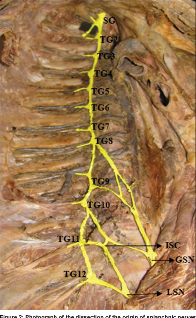
Greater splanchnic nerve
Each greater splanchnic nerve is organized by the medial branches of the 5th to 9th thoracic sympathetic ganglia. It runs along with the anterior surface of the spine and releases branches to the descending aorta. The nerve then crosses the fibers of the ipsilateral crus of the diaphragm to enter the abdomen. The majority of its fibers end by connecting with the celiac ganglia, contributing to the celiac plexus. These fibers are associated with the control of the enteric nervous system of the foregut.
A small quantity of fibers of the greater splanchnic nerve travels toward the ipsilateral suprarenal gland to supply the adrenal medulla and stimulate the release of catecholamines (epinephrine and norepinephrine).
Lesser splanchnic nerve
The lesser splanchnic nerve get arises from the medial branches of the 10th and 11th thoracic sympathetic ganglia. It runs lateral and in equal to the greater splanchnic nerves, on the anterior surface of the spine and enters the abdomen in the same manner – by crossing the ipsilateral diaphragmatic crus.
The lesser splanchnic nerves end by synapsing within the aortic renal and superior mesenteric ganglia. The corresponding postsynaptic fibers then regulate the activity of the midgut.
Least splanchnic nerve
The least splanchnic nerves get arise from the medial branches of the 11th and the 12th thoracic sympathetic ganglia.
They run lateral and parallel to the lesser splanchnic nerves, over the anterior or front surface of the spine, close to its lateral margin. It enters the abdomen by passing beneath the medial arcuate ligament and then joins the renal ganglia, provides to the sympathetic supply for the renal plexus. The fibers of the least splanchnic nerve are sometimes joined by the lesser splanchnic nerve. In this case, the lesser splanchnic nerve releases the small branches to the renal ganglia.
The Sacral Splanchnic Nerve:
Structure:
The sacral splanchnic nerves are the paired nerves that run on each side of the human body. These two parallel nerves construct the inferior hypogastric plexus to join with the sacral part of the sympathetic trunk. These branches of the splanchnic nerves run up from the inferior hypogastric plexus toward the superior hypogastric plexus and then pass to the inferior mesenteric plexus and the aortic plexus to innervate the hindgut.
Function:
The sacral splanchnic nerve gives both sensory and motor supply to the posterior aspect of the leg, foot, muscle, and skin of the pelvis region. The major branches of this splanchnic nerve join and form parts of the superior, inferior gluteal, posterior cutaneous, sciatic nerve, and finally, the pudendal nerves.
FAQ
What do splanchnic nerves carry?
The splanchnic nerves are paired with the visceral nerves (nerves that contribute to the supplies of the internal organs), carrying fibers of the autonomic nervous system (visceral efferent fibers) as well as sensory fibers from the organs
What makes a nerve splanchnic?
The splanchnic nerves are simultaneous autonomic nerves that supply abdominal and pelvic viscera. They are comprised of motor nerve fibers going to the internal organs and sensory nerve fibers coming from these organs.
Where do splanchnic nerves originate?
The thoracic splanchnic nerves get originate from the thoracic and lumbar segments of the spinal cord, T1-L2. The fibers of these nerves then run within the white rami communicates and pass through the ganglia of the sympathetic trunk without synapsing.
Where do splanchnic nerves typically terminate?
The lumbar splanchnic nerves get arise from the upper lumbar levels and end in the inferior mesenteric and hypogastric ganglia. From these prevertebral ganglia, the postganglionic fibers supply organs in the pelvis, lower abdomen, and lower limb.
What happens if the pelvic splanchnic nerve is damaged?
any injury to the pelvic parasympathetic nerves can produce a flaccid bladder with overflow incontinence and can cause erectile impotence in males. It should be noted that both parasympathetic and sympathetic autonomic nerves play important roles in sexual function.
How many pelvic splanchnic nerves are there?
The abdominopelvic splanchnic nerves, which construct the sympathetic aspect, consisting of three branches.
What does the thoracic splanchnic nerve innervate?
Thoracic Splanchnic Nerve:
The greater splanchnic nerve helps with the mobility of the foregut and gives a sympathetic supply to the adrenal medulla. Specifically, it supplies the alimentary canal, liver, gallbladder, pancreas, adrenal medulla, and spleen.
Where do the thoracic splanchnic nerves go?
Along with the thoracic course, the greater splanchnic nerves give minor branches to the aorta before reaching the diaphragm. All three paired thoracic splanchnic nerves (greater, lesser, and least splanchnic nerves) penetrate the lateral diaphragmatic crus at the vertebral level of T11 through a common foramen.
Where do splanchnic nerves typically terminate?
The lumbar splanchnic nerves get arise from the upper lumbar levels and end in the inferior mesenteric and hypogastric ganglia. From these prevertebral ganglia, the postganglionic fibers supply organs in the pelvic region, lower abdomen, and lower limb.
What do the lumbar splanchnic nerves supply?
The lumbar splanchnic nerves get arise from the upper lumbar levels and end in the inferior mesenteric and hypogastric ganglia. From these prevertebral ganglia, the postganglionic fibers supply organs in the pelvis, lower abdomen, and lower limb.
Where are the cardiopulmonary splanchnic nerves located?
The cardiopulmonary nerve is the splanchnic nerve that is postsynaptic and sympathetic. They get originate in the cervical and upper thoracic ganglia and supply the thoracic cavity.

3 Comments