Systems of the Human Body
The human body is an intricate and fascinating machine composed of numerous interconnected systems, each playing a vital role in maintaining life and enabling us to function effectively. These systems work in harmony to support our physical and mental well-being, allowing us to interact with the world around us.
Overview
The following is a list of the body’s principal organ systems.
- Heart and circulatory systems combined
- System of digestion and excretion
- The Immune system
- The Lymphatic system
- Integumentary/exocrine system
- The Endocrine system
- System of muscles
- Nervous system.
- Renal and urinary systems
- System of reproduction
- breathing apparatus
- Bone structure
Human anatomy and physiology are built on the foundation of 11 different organ systems. The respiratory system, the digestive and excretory system, the circulatory system, the urinary system, the integumentary system, the skeletal system, the muscular system, the endocrine system, the nervous system, and the reproductive system are the eleven organ systems.
The immune system, for instance, shields the body from infection, but it is not an organ system because it is not made up of organs. There are other bodily systems that are not organ systems. Some organs are involved in more than one system; for instance, the nose functions as a sensory organ in the nervous system in addition to being a part of the respiratory system; the testes and ovaries are involved in both the reproductive and endocrine systems.
The body systems, or organ groups that collaborate to create and maintain life, make up the biological machinery that is the human body. Occasionally, when learning about cells and molecules, we lose our way and fail to notice the bigger picture. Taking a step back and examining the larger anatomical picture may be beneficial.
You will receive a brief overview of the human body’s systems on this topic page so that each organ you learn in the future will build upon the foundation you have established here.
- Organ system: An assembly of organs that collaborate to carry out one or more bodily tasks.
- Musculoskeletal system: Support, posture, and movement of the musculoskeletal system
- Heart system: Circulation of nutrients, oxygen, and hormones throughout the body; removes waste products from cellular metabolism
- Respiratory system: The respiratory system controls breathing, the body’s exchange of oxygen and carbon dioxide with the surrounding air, and the acid-base balance.
- Nervous system: The nervous system is responsible for initiating and regulating essential bodily functions, sensation, and movement. Sensation, movement, and other essential bodily functions are initiated and regulated by the nervous system.
- Digestive system: Food is broken down by the digestive system, both mechanically and chemically so that the body can absorb it and use it as fuel.
- Urinary system: Urine is produced and expelled by the urinary system, which filters blood and gets rid of waste and extraneous chemicals.
- Endocrine system: The menstrual cycle, blood sugar levels, and other physiological processes are all regulated by the hormones produced by the endocrine system.
- Lymphatic system: The lymphatic system handles the flow of extra tissue fluid and the body’s immune system.
- Reproductive system: The production of reproductive cells and their contribution to the process of reproduction are the functions of the reproductive system.
- Integumentary system: Insulin synthesis, sensory perception, and physical defense of the skin are all part of the integumentary system.
Skeletal system
Bones and cartilage make up the skeletal system. The skeleton consists of two parts: axial and appendicular. The axial skeleton is made up of the bones in the head and trunk. The bones that make up the limbs as well as the structures that support the pectoral and pelvic girdles make up the appendicular skeleton.
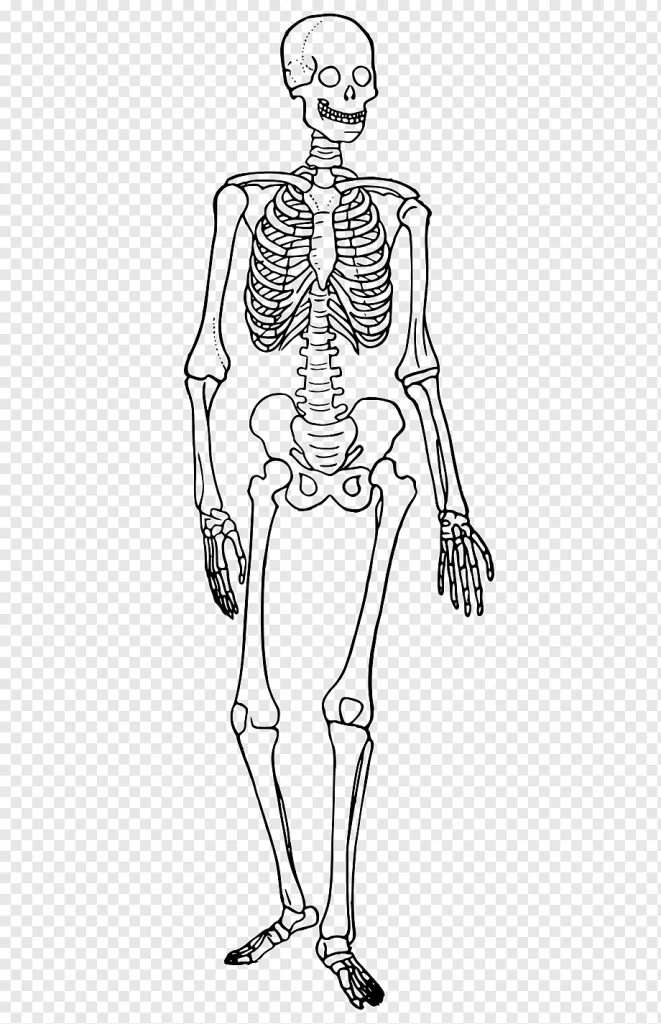
An adult human body consists of 206 bones. The term “joint” refers to the location where two bones fit together. Ligaments strengthen and cartilaginous support joints. The skeletal system is responsible for producing blood cells, moving, protecting the body, storing calcium, and regulating hormones in addition to providing mechanical support. The skeletal system’s components are adapted to the way the body part they support functions.
Thus, just as the bones of the head and neck, thorax, abdomen, and upper and lower limbs are studied topographically, so too is the anatomy of bones, joints, and ligaments. The structure that holds up the human body is called the skeleton. At birth, it has about 270 bones; by adulthood, some of those bones have fused together, bringing the total number down to about 206 bones. The skeleton’s bone mass reaches its maximum mass between the ages of 25 and 30.
Bone mass accounts for approximately 14% of total body weight, or 10–11 kg for the average person. The axial skeleton and the appendicular skeleton are the two separate parts of the human skeleton. The spine, thorax, skull, and other similar bones make up the axial skeleton. The bones of the upper and lower limbs, the pelvic girdle, and the shoulder girdle together form the appendicular skeleton, which is joined to the axial skeleton.
Six primary tasks are carried out by the human skeleton: support, mobility, defense, blood cell production, mineral storage, and endocrine regulation.
The morphology of the skull, dentition, long bones, and pelvis differ slightly between the sexes, although the human skeleton is not as sexually dimorphic as that of many other primate species. Within a particular population, female skeletal elements are typically smaller and less robust than their male counterparts. In order to aid in childbirth, the human female pelvis differs from the male pelvis. Male humans do not have penile bones, in contrast to most other primates.
Divisions
Axial
The axial skeleton, which has 80 bones, is made up of the lumbar region, which has 12 pairs of ribs and the sternum; the brain, which has 22 bones and 7 associated bones; and the vertebral column, which has 32–34 bones. Because of variations in the length of the lower two parts of the vertebral column, the sacral and coccygeal bones, there are differences in the number of bones in the vertebral column among individuals.
The axial skeleton aids in the maintenance of an upright posture in humans by transferring weight from the head, trunk, and upper extremities to the lower extremities at the hip joints. Numerous ligaments support the bones of the spine. The erector spine muscles aid in balance and provide additional support.
Appendicular
The pectoral girdles, the upper limbs, the pelvic girdle or pelvis, and the lower limbs make up the appendicular skeleton, which consists of 126 bones. They serve to facilitate movement and safeguard the primary organs involved in digestion, elimination, and reproduction.
Function
The six main purposes of the skeleton are to provide support, mobility, protection, blood cell production, mineral storage, and hormone regulation.
Support
The framework that supports and preserves the body’s shape is provided by the skeleton. The pelvic structures are supported by the muscles, ligaments, and pelvis. If the lungs were not supported by the rib cages, costal cartilage, and intercostal muscles, they would collapse.
Movement
Some of the joints that connect the bones allow for a greater range of motion than others; for example, the ball and socket joint at the neck has a wider range of motion than the pivot joint. Skeletal muscles are what propel movement; they are affixed to the skeleton at different points along the bones. The primary mechanics of movement are provided by the muscles, bones, and joints, which are all regulated by the nervous system.
It is thought that human movement dexterity and agility were compromised during the prehistoric era due to a decrease in bone density. As a result of the shift from hunting to agriculture, human bone density has significantly decreased.
Protection
The skeleton aids in preventing damage to a number of important internal organs.
- The vertebrae are what keep the spinal cord safe;
- The brain is protected by the skull.
- The main blood vessels, the heart, and the lungs are shielded by the sternum, spine, and rib cage.
Blood cell production
The process by which blood cells develop in the bone marrow is known as hemopoiesis, and it occurs in the skeleton. The marrow of the long bones, like the femur and tibia, is where hematopoiesis predominantly happens in children. The pelvis, skull, vertebrae, and sternum are the main areas where it appears in adults.
Storage
Both the bone marrow and the bone matrix have the ability to store iron in ferritin and to handle calcium, which is involved in the metabolism of both elements. It should be noted that bones are composed partially of hydroxyapatite (which makes up 70% of a bone) and chondroitin sulfate rather than just calcium. 18.5% phosphorus, 0.2% hydrogen, 39.8% calcium, and 41.4% oxygen make up hydroxyapatite by mass. Carbon and oxygen make up the majority of the sugar chondroitin sulfate.
Endocrine regulation
Osteocalcin, a hormone secreted by bone cells, plays a role in controlling glucose levels in the blood and the accumulation of fat. Osteocalcin enhances insulin sensitivity and secretion, while also boosting the number of cells that produce insulin and lowering fat stores.
Clinical significance
Skeletal disorders are categorized in a wide variety. Among the most prevalent is osteoporosis. Equally common is scoliosis, a side-to-side curvature of the back or spine that often shows up on a spine x-ray as a prominent “C” or “S” shape. Adolescence is when this condition becomes more apparent, and it affects women more often.
Arthritis
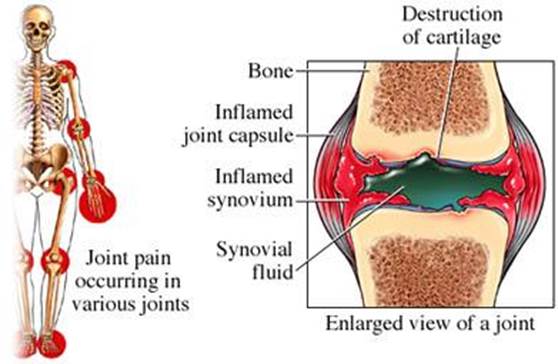
Joint disease is what is known as arthritis. It includes one or more joints that are inflamed. When a joint or joint is affected by arthritis, it may move in strange directions, hurt to move or become completely immobile. There are differences between the various types of arthritis in terms of symptoms.
Both of the skeleton’s larger and smaller joints may be impacted by osteoarthritis, the most prevalent type of arthritis. There will be a degradation, softening, and wearing away of the cartilage in the affected joints. As a result, there is less space between the bones, where cartilage normally resides, and less joint mobility.
Osteoporosis
Osteoporosis is a bone disease that lowers bone mineral density and increases the risk of fractures. The existence of a fragility fracture is included in the definition of “established osteoporosis”. The World Health Organization defines osteoporosis in women as a bone mineral density 2.5 standard deviations below peak bone mass, relative to the age and sex-matched average, as measured by dual-energy X-ray absorptiometry.

The most common form of osteoporosis in women is known as “postmenopausal osteoporosis,” which occurs after menopause. However, premenopausal women and men may also develop osteoporosis if they have certain hormonal disorders or other chronic diseases, or if they smoke or take certain medications, particularly glucocorticoids. Until a fracture happens, osteoporosis typically shows no symptoms. For this reason, individuals who have developed osteoporosis are at risk of fracture and have one or more risk factors are frequently given a DEXA scan.
Treatment for osteoporosis involves recommendations to give up smoking, cut back on alcohol, exercise frequently, and maintain a balanced diet. It may also be recommended to take calcium supplements and vitamin D. Osteoporosis may be considered when initiating hormone replacement therapy prior to beginning medication. Bisphosphonates and strontium ranelate are examples of such medications.
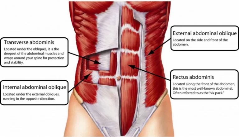
Muscular system
Every muscle in the body is a part of the muscular system. There are three different kinds of muscles: skeletal, cardiac, and smooth. Smooth muscle is present in the walls of blood vessels and hollow organs like the intestines and stomach. Heart muscle, also known as the false heart, is made up of cardiac muscle cells. The body’s bones are where skeletal muscles are attached.

Of these three, only the skeletal muscle allows us to consciously produce movement with our body; the other two muscle types function completely subconsciously and are controlled by the autonomic nervous system. Striated muscle is the term used to describe the histologically striped appearance of cardiac and skeletal muscle fibers due to their repetitive arrangement. Because it lacks repeating sarcomeres, smooth muscle is not striated.
Your body contains more than 600 muscles. It takes specific muscles to move, lift, or remain motionless. Others help with food digestion, breathing, and vision. The heart is a muscle that is in charge of pumping blood throughout your body. Muscle function can be impacted by numerous diseases and injuries. Maintain a healthy weight, eat well, and exercise frequently to keep your muscles strong.
What are muscles?
Muscles are soft tissues. Many stretch fibers make up your muscles. These comprise more than 600 muscles in your body. Different types of muscles have different functions. You need specific muscles to run, jump, and perform fine motor skills like threading a needle. You can breathe and digest food with the help of other muscles. Your heart is a very strong muscle because it beats a thousand times a day. Muscle function can be impacted by a wide range of illnesses, traumas, and injuries.
Pain, spasms, or weakness in the muscles are all a result of these conditions. More serious disorders can lead to paralysis. Cardiomyopathy and other heart diseases make it difficult for the heart to pump blood around the body. A healthy lifestyle helps muscles function as they should. You can keep your muscles strong by eating a balanced diet, exercising frequently, and maintaining a healthy weight. Be sure to visit your provider regularly to check for diseases and conditions that can cause muscle problems.
What are the types of muscles?
You control certain muscles voluntarily using your nervous system (the body’s control center). You can consider moving them prior to performing so. Other muscles work involuntarily, which means you have no control over them. They do their job automatically. To work, they take signals from other body systems, such as your digestive system or cardiovascular system.
Three types of muscle tissue compose the human body. They are:
- Skeletal: These muscles are part of the musculoskeletal system, working with bones, tendons, and ligaments. Skeletal muscles and bones are joined by tendon attachments all throughout the body. Together, they support and help you move your body weight. These are your voluntary muscles. Muscle fibers known as “twitch muscles” contract quickly and release short bursts of energy. Others move slowly, such as the back muscles that help maintain posture.
- Cardiac: The heart’s walls are lined with these muscles. They help your heart pump blood that travels through the cardiovascular system. You have no control over your heart muscles. Your heart tells them when to make a deal.
- Smooth: These muscles line organs like the bladder, stomach, and intestines. Smooth muscle is involved in numerous bodily processes, such as the respiratory, urinary, and reproductive systems of both sexes. There is no deliberate thought required for these muscles to operate. In addition to helping your lungs expand during breathing, they carry out crucial tasks like transferring waste through the intestines.
Structure
Skeletal muscle fibers are made up of a single muscle cell that is cylindrical. A single skeletal muscle may consist of hundreds or even thousands of muscle fibers joined together and wrapped in a covering of connective tissue. Each muscle is surrounded by a covering of connective tissue called the epimysium. Fascia, the connective tissue outside the epimysium, surrounds and separates muscles. Parts of the epimysium extend inward, dividing the muscle into sections.
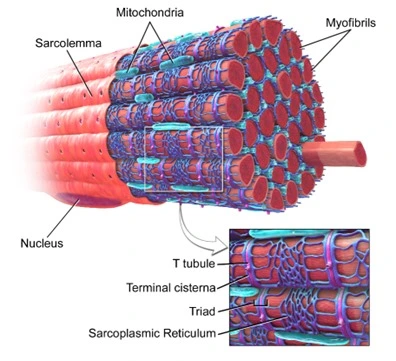
Each section contains a bundle of muscle fibers. Each bundle of muscle fibers is called connective tissue and is surrounded by a layer of connective tissue called the perimysium. In muscle tissue, each individual muscle cell, called a muscle fiber, is surrounded by a connective tissue called the endomysium. Skeletal muscle cells (fibers), like other body cells, are soft and delicate. The connective tissue covering provides support and protection to the delicate cells and allows them to withstand the forces of contraction.
The sheaths also provide pathways for blood vessels and nerves to pass through. Typically, the epimysium, perimysium, and endomysium extend beyond the fleshy part of the muscle, the belly or stomach, to form a thick rope-like tendon or a broad, flat sheet-like aponeurosis. Tendons and aponeurosis create the indirect muscle attachments to the connective tissue of adjacent to muscles or the periosteum of bones. Where the muscle often connects the bone at the joint, the tendon joins the muscle at each of the ends.
One end of the bone remains relatively fixed or stable, while the other end moves as a result of muscle contraction. Skeletal muscles have many blood vessels and nerves. It is directly related to the primary function of skeletal muscles, contraction. A nerve cell must send an impulse to a skeletal muscle fiber before it can contract. Normally, every nerve that penetrates the epimysium of a skeletal muscle is accompanied by an artery and at least one vein. Nerve and blood vessel branches follow the connective tissue components of the nerve cell muscle and one or more small blood vessels called capillaries.
Function
What do muscles do?
Muscles play a role in almost every body system and function. Different muscles help:
- Breathing, speaking, and swallowing.
- The process of food digestion and residue removal.
- Movement, sitting still, and standing straight.
- Blood circulates via the heart and blood vessels.
- Pushing the baby through the birth canal as the uterine muscles contract and relaxes.
- Sight and hearing.
Clinical Significance
What conditions and disorders affect the muscles?
Some disorders, diseases, medications, and injuries can cause problems with muscle function. They include:
- Cancer and other diseases: Muscle issues can be brought on by various cancers, including sarcoma, and other illnesses. These comprise several kinds of myopathies (muscle diseases), autoimmune illnesses including myasthenia gravis (MG), and neuromuscular conditions like amyotrophic lateral sclerosis (ALS). Muscle weakness is brought on by inflammation in the muscles, which is the result of a disease called polymyositis.
- Cardiovascular disease: Issues with the heart and blood vessels can arise from a variety of venous diseases as well as cardiovascular conditions, such as coronary artery disease. Weakened vascular smooth muscle can lead to a heart attack.
- Chronic pain disorders: All over the body, the muscles experience chronic pain due to conditions like fibromyalgia and other disorders.
- Genetic disorders: Muscular dystrophy is a hereditary condition that is handed down within families. More than thirty kinds of muscular dystrophy are present. Permanent muscle weakness is the result of the disorder.
- Infections: Muscle fibers can sustain damage from bacterial and viral infections.
- Injuries: Many different types of injuries can cause muscles to tear or become overstretched (muscle strains). Back strain is a very common injury. Muscle cramps or spasms may arise from overuse injuries, accidents, or trauma. These wounds may result in paralysis in extreme circumstances.
- Medication: Certain drugs, like those used in chemotherapy, can cause pain in the muscles. Painful muscles can also be a side effect of high blood pressure medications.
Cardiovascular system
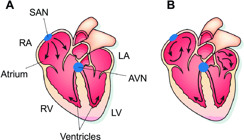
The circulatory system of blood vessels and the heart make up the cardiovascular system. The heart’s four chambers are composed of two ventricles and two atria. Blood leaves the heart by means of the left and right ventricles after passing through the upper chambers of the atria. The heart’s valves inhibit blood from circulating backward.
The heart is a bidirectional pump. Blood that has lost oxygen is pumped by the right side of the heart into the lungs’ pulmonary circulation, where it is replenished with oxygen. In addition, blood is pumped into the systemic circulation via the left side of the heart, supplying oxygen-rich blood to the peripheral tissues. The heart’s conduction system regulates the regular pumping of the heartbeat.
The vascular system, commonly referred to as the circulatory system, is made up of capillaries, veins, and arteries. All of the blood vessels in the body are linked to one another and carry the blood throughout its contents. Arteries, which eventually narrow to become smaller arterial vessels known as arterioles, are the means by which blood exits the heart. The network of even smaller vessels known as capillaries is where arterioles end. The capillary walls allow for the exchange of nutrients and gases.
On their journey from capillaries to the heart, tiny veins known as venules progressively increase in lumen before becoming veins. Blood is carried by arteries from the heart to the periphery and by veins from the periphery to the heart. While there are some histological differences between arteries and veins, their primary functional distinction is reflected in the direction in which they carry blood.
The three distinct circuits that make up the circulatory system are:-
- Blood is moved from the heart to the lungs through pulmonary circulation,
- The coronary circulation, which nourishes the heart muscle, and
- Blood is transported all over the body via systemic circulation.
The inferior vena cava and superior vena cava are the principal vein representatives in the systemic circulatory system, whereas the aorta and its branches are prominent arteries.
The most important tasks of the cardiovascular system are the transport of oxygen, nutrients, and hormones through blood throughout the body and the removal of carbon dioxide and other metabolic waste products.
The circulatory system is an organ system that includes the heart, blood vessels, and blood that circulates throughout the body of a human or other vertebrate. It includes the cardiovascular system, which consists of the heart and blood vessels (from the Greek cardia meaning heart, and the Latin vascula meaning blood vessels). The circulatory system has two sections, systemic circulation or circulation and pulmonary circulation or circulation.
In some sources, the terms cardiovascular system and vascular system are interchangeable with vascular system. Arteries are the large blood vessels of the heart, including large elastic arteries and veins. other arteries, smaller arteries, capillaries that connect to venous veins (small veins), and other veins. Since the circulatory system in vertebrates is closed, blood never exits the vascular network. Arthropods are among the invertebrates that have an open circulatory system.
Similar to sponges and comb jellies, diploblasts do not have blood circulation. Red blood cells, white blood cells, platelets, and plasma make up blood, which is a liquid. It circulates around the body, carrying oxygen and nutrients to tissues and collecting and removing waste products. Circulating nutrients include proteins and minerals, and other components include hemoglobin, hormones, and gases such as oxygen and carbon dioxide. These substances provide nutrition, help the immune system fight disease, and help maintain homeostasis by stabilizing temperature and natural pH.
In vertebrates, the lymphatic system completes the circulatory system. The lymphatic system transports excess plasma (filtered by the capillaries of the circulatory system as interstitial fluid between cells) away from body tissues through additional pathways that return the excess fluid to the bloodstream as a lymph node. The lymphatic system is a subsystem that is essential for the functioning of the circulatory system; without it, the blood would flow out.
The immune system and the lymphatic system are interdependent. Lymphs take much longer to circulate than blood, and unlike a closed (blood) circulatory system, a lymph node is an open system. Some sources describe it as the secondary circulatory system. Many cardiovascular diseases can affect the circulatory system. Cardiologists are medical professionals who specialize in the heart, and cardiothoracic surgeons specialize in operations on the heart and its surrounding areas. Vascular surgeons focus on disorders of blood vessels and lymphatic vessels.
Structure
The heart, blood vessels, and blood constitute the circulatory system. All vertebrates possess a cardiovascular system generated up of blood vessels and a heart. The pulmonary circulation and the systemic circulation are the two primary divisions of the circulatory system. The right heart’s pulmonary circulation circulates blood that is deficient in oxygen to the lungs, where it is restored and transmitted back to the left heart.
The systemic circulation is the circulatory pathway that uses the vena cava, a large vein, to return deoxygenated blood to the right heart after transporting oxygenated blood from the left heart to the remaining parts of the body. Systemic circulation can also be defined into two parts – macrocirculation and microcirculation. The average adult contains five to six liters (about 4.7 to 5.7 liters) of blood, which is about 7% of their total body weight.
Red blood cells, white blood cells, plasma, and platelets constitute the blood. The digestive system also works with the circulatory system and provides nutrients that the system needs for the heart to function. Other circulatory pathways are also involved, such as coronary blood flow to the heart, cerebral blood flow to the brain, renal blood flow to the kidneys, and bronchial blood flow to the lungs. The human circulatory system is closed, which means that the blood is in a network of blood vessels.
Nutrients travel through the small blood vessels of the microcirculation to reach the organs. Including a network of lymphatic vessels, lymph nodes, organs, tissues, and circulating lymph nodes, the lymphatic system is a vital component of the circulatory system. This subsystem is an open system. An important function is to transport the lymph node, drain it, and return the interstitial fluid to the lymphatic channels back to the heart to return to the circulatory system. Cooperating with the immune system to guard against infections is another crucial role.
Heart
The heart pumps blood to all parts of the body, which supplies each cell with nutrients and oxygen and removes waste products. The left heart pumps oxygenated blood that returns from the lungs to the rest of the body in the systemic circulation. In pulmonary circulation, blood that is deficient in oxygen is pumped from the right heart to the lungs. The human heart has one atrium and one ventricle per circulation, and both the systemic and pulmonary circulations have a total of four chambers: left atrium, left ventricle, right atrium, and right ventricle.
An upper chamber that occupies the right side of the heart is called the right atrium. Blood returned to the right atrium is deoxygenated (deoxygenated) and sent to the right ventricle to be pumped through the pulmonary artery to the lungs for reoxygenation and removal of carbon dioxide. The left atrium receives freshly oxygenated blood from the lungs and pulmonary vein, which is directed into the strong left ventricle to be pumped through the aorta to the various organs of the body.
Pulmonary circulation
The portion of the circulatory system known as pulmonary circulation serves as accountable for pumping oxygenated blood back to the heart via the pulmonary vein and deoxygenated blood to the lungs through the pulmonary artery. The right atrioventricular valve, commonly referred to as the tricuspid valve in the heart, opens the right atrium to access deoxygenated blood from the superior and inferior vena cava.
From there, it passes into the right ventricle and is pushed into the pulmonary artery by the pulmonary semilunar valve. The light Gas exchange takes place in the lungs, when carbon dioxide is released from the blood and oxygen is absorbed. Now, the left atrium receives blood that is rich in oxygen from the pulmonary vein. Blood circulates to the tissue of the lung’s larger airways by means of bronchial circulation, which varies from systemic circulation.
Systemic circulation
The systemic circulation is the circuit that transports oxygenated blood from the left heart through the aorta to the rest of the body. Two major veins, the inferior and superior vena cava, carry deoxygenated blood back to the right heart through systemic circulation. From there, it is pumped from the right atrium to the pulmonary circulation for oxygenation. Systemic circulation can also be defined into two parts – macrocirculation and microcirculation.
Function
Approximately 98.5% of the oxygen in the arterial blood sample of a healthy person breathing air at sea level is chemically bound to hemoglobin molecules. Not bonded to hemoglobin, about 1.5% is dissolved physically in other elements of blood. The hemoglobin molecule is the main oxygen transporter in vertebrates.
Clinical Significance
Many diseases affect the circulation. These include several cardiovascular diseases that affect the heart and blood vessels. Hematological diseases that affect the blood, such as anemia, and lymphatic diseases that affect the lymphatic system. Cardiologists are medical professionals who specialize in the heart, and cardiothoracic surgeons specialize in operations on the heart and its surrounding areas. Vascular surgeons focus on blood vessels.
Respiratory system
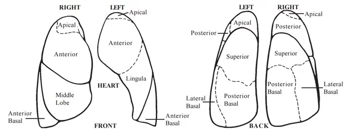
The respiratory system consists of several organs; nasal cavity, pharynx, larynx, trachea, bronchi, bronchi, and lungs (alveoli). The nasal cavity and pharynx together are called the upper respiratory tract, while the other organs form the lower respiratory system.
The respiratory system (also respiration, respiration) is a biological system consisting of certain organs and structures used for gas exchange in animals and plants. The anatomy and physiology that cause it varies greatly depending on the size of the organism, the environment in which it lives, and its evolutionary history. In terrestrial animals, the respiratory surface is inserted into the mucous membranes of the lungs.
Gas exchange in the lungs takes place in millions of small air sacs. in mammals and reptiles, they are called alveoli, and in birds atrium. These microscopic air sacs have a very rich circulation, which brings the air into close contact with the blood. These air sacs communicate with the external environment through airways or hollow tubes, the largest of which is the trachea, which branches into two main bronchi in the middle of the chest. They enter the lungs, where they gradually branch into narrower secondary and tertiary bronchi, which branch into many smaller tubes, the bronchi.
The bronchial tubes in birds are called parabronchi. Bronchioles or parabronchi usually open into microscopic alveoli in mammals and into an atrium in birds. Air must be pumped from the environment into the alveoli or atrium through the process of breathing, which involves the respiratory muscles. In most fish and many other aquatic animals (both vertebrates and invertebrates), the respiratory system consists of gills, which are external organs that are either partially or fully immersed in water. This water flows over the gills in a number of active or passive ways.
Gas exchange takes place in the gills, which consist of thin or very flat filaments and lamellae, which expose a very large area of highly vascularized tissue. Other animals, such as insects, have respiratory organs with very simple anatomical features, and even the skin plays an important role in gas exchange in amphibians. Despite plants having respiratory organs too, they may exchange gases in a reverse direction from animals. Respiration in plants involves anatomical features such as stomata found in different parts of the plant.
The respiratory organs, with the exception of the alveoli, draw air into the lungs using respiratory muscles (mainly between the diaphragm and the ribs). After stepping into the lungs, the air communicates with the blood that the pulmonary circulation conveys in the alveoli, at the point of gas exchange. Here, carbon dioxide is removed from the blood, and oxygen is returned to the blood. Thus, the main task of the respiratory system is to bring oxygen to the body and remove carbon dioxide.
Other lung functions
Local defenses
Irritation of the nasal passages or respiratory nerve endings can cause a cough reflex and sneezing. Air is pushed out of the nose or trachea just like a consequence of these reactions. In this way, irritating substances stuck to the mucus lining the airways are removed or transferred to the mouth, where they can be swallowed. During a cough, the contraction of the smooth muscles of the airway walls narrows the trachea, bringing the ends of the cartilage plates together and pushing the soft tissue into the cavity.
This increases the flow rate of exhaled air and removes any irritating particles or mucus. A number of molecules that play a role in lung assurance are frequently released by the epithelium of the airways. These consist of reactive oxygen species, reactive nitrogen species, collectins, defensins, and other peptides and proteases, in addition to secretory immunoglobulins (IgA). Through functioning as antimicrobial agents, these secretions are helpful in combating infections in the respiratory tract. Various chemokines and cytokines are also secreted that recruit normal immune cells and other cells to the site of infection.
The immune function of surfactants is largely due to two proteins: SP-A and SP-D. These proteins can bind to sugars on the surface of pathogens and thus optimize them for assimilation by phagocytes. In addition, it controls inflammatory reactions and collaborates with the adaptive immune system. Surfactant degradation or inactivation can increase susceptibility to pneumonia and infections. Most of the respiratory system is covered by mucous membranes, which contain mucosa-associated lymphoid tissue that produces white blood cells such as lymphocytes.
Prevention of alveolar collapse
Type II alveolar cells in the lungs produce surfactant, which is also referred to as surfactant lipoprotein complex (phospholipoprotein). It minimizes surface tension and water from floating on the surface of a thin layer of water that lines the inner surface of the alveoli. The surface tension of the water surface (water-air interface) causes the surface to contract. If this surface is curved, as it is in the alveoli of the lungs, the contraction of the surface reduces the diameter of the alveoli.
The sharper the curvature of the water-air interface, the greater the tendency for the alveoli to collapse. This has three effects. First, the surface tension of the alveoli prevents the alveoli from expanding (ie makes the lungs stiff or uncomfortable) during inhalation. Surfactant reduces surface tension and thus makes the lungs more flexible or less rigid than without them. Second, the diameter of the alveoli increases and decreases during the respiratory cycle.
This means that the alveoli have a greater tendency to collapse (ie, produce atelectasis) at the end of expiration than at the end of inspiration. When the alveoli shrink during exhalation, the molecules of the surfactant set closer together because they are floating on the water’s surface. Because of this, they have a greater surface tension reducer in small alveoli than in large ones (for example, at the end of inhalation, when the surfactant molecules are separated from each other).
The tendency of the alveoli to collapse at the end of exhalation is therefore almost the same as at the end of inspiration. Third, the surface tension of the curved water layer lining the alveoli tends to draw water from the lung tissues into the alveoli. The surfactant reduces this risk to negligible levels and keeps the alveoli dry. Premature babies who cannot make surfactant have lungs that tend to collapse every time they exhale.
This condition, called respiratory distress syndrome, is fatal if left untreated. Basic science experiments using chicken lung cells support the use of steroids as a means of promoting type II alveolar cells. In fact, when premature birth is at risk, every effort is made to delay delivery, and during this delay, the mother often receives multiple steroid injections to encourage lung maturation.
Affects all body functions
Pulmonary vessels have a fibrinolytic system that dissolves clots that may have entered the pulmonary circulation through an embolism, often from the deep veins of the legs. They also release various substances that enter the systemic arterial blood and remove other substances from the systemic venous blood that enter them through the pulmonary artery. Some prostaglandins are removed from the bloodstream, others are synthesized in the lungs and released into the blood when lung tissue is stretched.
The lungs activate one hormone. The physiologically inactive decapeptide angiotensin I is converted in the pulmonary circulation to the aldosterone-releasing octopeptide angiotensin II. The lungs indicate the reaction most frequently, nevertheless, it occurs in other tissues as well. Angiotensin II also directly affects the walls of the arteries, causing vasoconstriction of the arteries and thus an increase in arterial blood pressure. Large amounts of the angiotensin-converting enzyme responsible for its activation are located on the surfaces of alveolar capillary endothelial cells.
The converting enzyme also inactivates bradykinin. The blood circulation time through the alveolar capillaries is less than a second, but 70% of the angiotensin I arriving in the lungs is converted to angiotensin II in one step through the capillaries. On the surface of lung endothelial cells, four additional peptidases were recently found.
Registration
The movement of gas through the larynx, pharynx, and mouth allows people to speak or vocalize. Bird vocalization, or song, occurs through an organ located at the bottom of the trachea. In humans, sound is produced by the vibration of air flowing through the larynx (voice box), and in birds. Therefore, the movement of gas is crucial for communication.
Temperature control
Panting in dogs, cats, birds, and some other animals is a way to lower body temperature by evaporating saliva through the mouth (rather than evaporating sweat from the skin).
Clinical Significance
Respiratory disorders can be divided into several general groups:
- Diseases that narrow the airways (eg emphysema, bronchitis, asthma)
- Diseases restricting the lung (eg fibrosis, sarcoidosis, alveolar damage, pleural effusion)
- Vascular diseases (e.g. pulmonary edema, pulmonary embolism, pulmonary hypertension)
- Infectious, environmental, and other “diseases” (eg pneumonia, tuberculosis, asbestosis, particulate matter emission)
- Primary cancers (eg lung cancer, mesothelioma)
- Secondary cancers (eg cancers that originate elsewhere in the body but have spread to the lungs)
- Insufficient surfactant (eg respiratory distress syndrome in premature babies). Problems with respiration are usually handled by respiratory therapists and pulmonologists. A medical ventilator may be used for respiratory failure or inadequate breathing.
Nervous system
In biology, the nervous system is a very complex part of an animal that coordinates its activity and sensory information by sending signals to and from different parts of the body. The nervous system detects changes in the environment that affect the body and then works with the endocrine system to respond to such events. Nerves first appeared in worm-like organisms about 550-600 million years ago. It breaks down into two primary components in vertebrates: the peripheral nervous system (PNS) and the central nervous system (CNS).
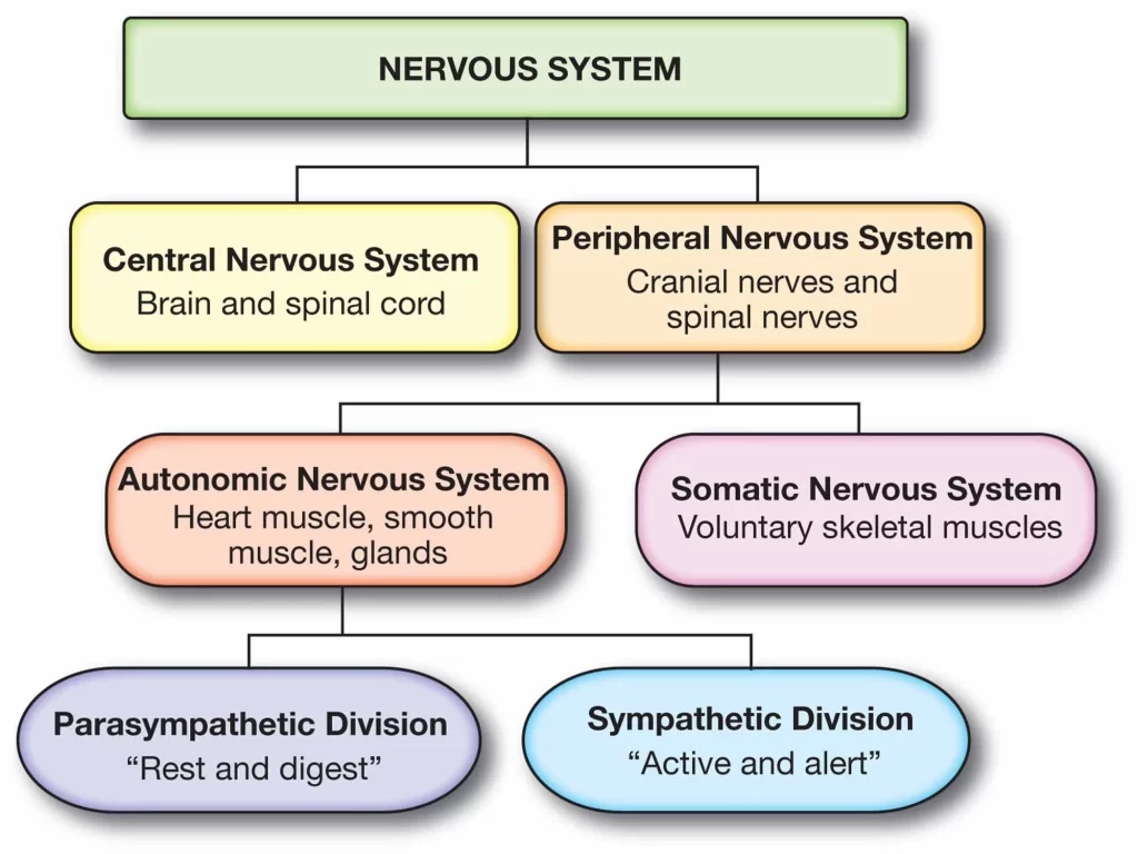
The brain and spinal cord are the two primary structures of the central nervous system. The PNS consists mainly of nerves, which are closed bundles of long fibers or axons that connect the central nervous system to all other parts of the body. Nerves that transmit signals from the brain are called motor nerves or efferent nerves, while nerves that transmit information from the body to the central nervous system are called sensory nerves or afferents. Mixed nerves that serve both purposes are spinal nerves.
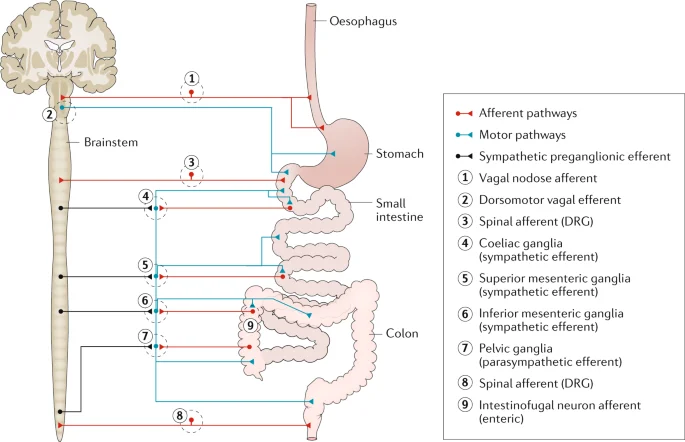
The somatic, autonomic, and enteric nervous systems are the three separate subsystems that make up the PNS. Voluntary movement is mediated by somatic nerves. Both the parasympathetic and sympathetic nervous systems are subsets of the autonomic nervous system. The sympathetic nervous system is activated in emergency situations to mobilize energy, while the parasympathetic nervous system is activated when the organism is relaxed. It is the enteric nervous system that governs the digestive tract. The nervous systems of the mouth and the autonomic nerve function reflexively.
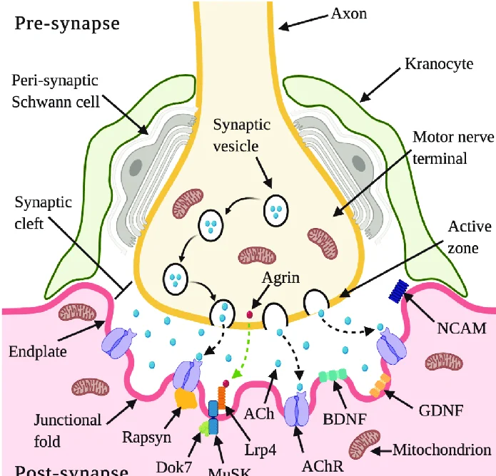
Anterior cranial nerves are those that emerge from the skull, whereas posterior cranial nerves originate from the spinal cord. Neural tissue is the unique cell type that characterizes the nervous system at the cellular level. Fast and precise signal transmission to other cells is made possible by the unique structures found in neurons. They send these signals as electrochemical impulses that travel along thin fibers called axons, which can travel through electrical synapses directly to neighboring cells or trigger chemicals called neurotransmitters at chemical synapses.
After a neuron sends a synaptic signal, a cell can be stimulated, inhibited, or subjected to other modulations. Connections between neurons can form neural pathways, neural circuits, and larger networks that create the perception of an organism of the world and determine its behavior. Along with neurons, the nervous system also contains other specialized cells called glial cells (or simply glial cells) that provide structural and metabolic support. Many nerve cells and blood vessels form a neurovascular unit that regulates cerebral blood flow to meet the high energy demands of fast-firing neurons.
Nervous systems are found in most multicellular animals, but they vary widely in complexity. The only multicellular animals that have no nervous system at all are fungi, placozoa, and mesozoans, which have very simple body plans. The nervous system of radially symmetrical organisms, ctenophores (scallops) and cnidarians (including anemones, hydras, corals, and jellyfish) consists of a plexus. All other animal species, with the exception of some types of worms, have a nervous system that includes a brain, a central cord (or two parallel cords), and nerves radiating from the brain and midbrain.
The size of the nervous system varies from a few hundred cells in the simplest worms to about 300 billion cells in African elephants. The job of the central nervous system is to send signals from one cell to another or from one part of the body to another and receive feedback. Nervous system dysfunction can be caused by genetic defects, physical damage from trauma or toxicity, infection, or simply aging. The medical department of neurology studies disorders of the nervous system and seeks means to prevent or treat them.
In the peripheral nervous system, the most common problem is impaired nerve conduction, which can be caused by a variety of causes, including diabetic neuropathy and demyelinating diseases such as multiple sclerosis and amyotrophic lateral sclerosis. The study of the nervous system is the main focus of the scientific field of neuroscience. The nervous system controls how we interact and respond to our environment by controlling the function of organs in other body systems.
The organs of the nervous system include the brain, spinal cord, and sense organs. Neural signals are transmitted throughout the body via neurons connecting them. Morphologically and topographically, the nervous system is divided into the central nervous system (CNS) and the peripheral nervous system (PNS). Functionally, the nervous system is considered divided into two parts; the somatic (SNS) or voluntary nervous system and the autonomic (ANS) or involuntary nervous system.
Central Nervous System
The definition of the central nervous system is that it receives information from the body’s environment and produces instructions, thus directing all the activities of the human body. The peripheral nervous system transmits this bidirectional flow of information to and from the central nervous system.
The central nervous system is a combination of the brain and spinal cord. The brain is placed in the neurocranium and consists of the cerebrum, cerebellum, and brainstem (pons and medulla oblongata). In the central parts of the central nervous system are spaces called ventricles, which are filled with cerebrospinal fluid (CSF). Inside the spine is the spinal cord. The spinal cord extends through the middle of the spinal cord. It connects with the brain’s ventricles and is also filled with CSF.
The central nervous system consists of nerve cells and their processes (axons). The gray matter consists of nerve cells and is found in the cerebral cortex and mid-spinal cord. White matter consists of axons that connect and build nerve pathways. Gray matter is where the instructions are generated, while white matter is the path along which the instructions travel to the organs.
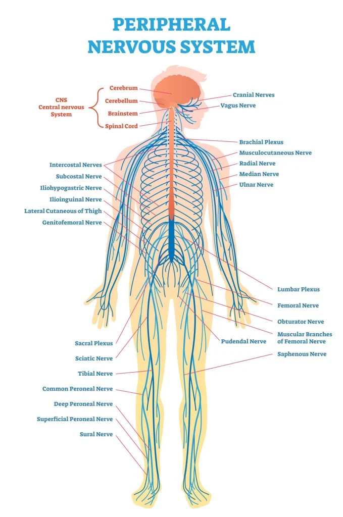
Peripheral nervous system
The definition of the peripheral nervous system is that it carries information from the central nervous system to target tissues and from target tissues to the central nervous system. It consists of nerves and their nerves. Nerves that carry information from peripheral sense organs (for example, eye, tongue, nasal mucosa, ear, and skin) to the central nervous system are called ascending, afferent, or sensory nerve fibers.
The fibers that carry information from the central nervous system to the periphery (muscles and glands) are descending, efferent, motor, or secretory nerve fibers. A ganglion is a collection of nervous tissue outside the central nervous system, consisting of the cell bodies of neurons. Ganglia can be both sensory and autonomic. Sensory nerves are connected to spinal nerves and some cranial nerves (V, VII, IX, X).
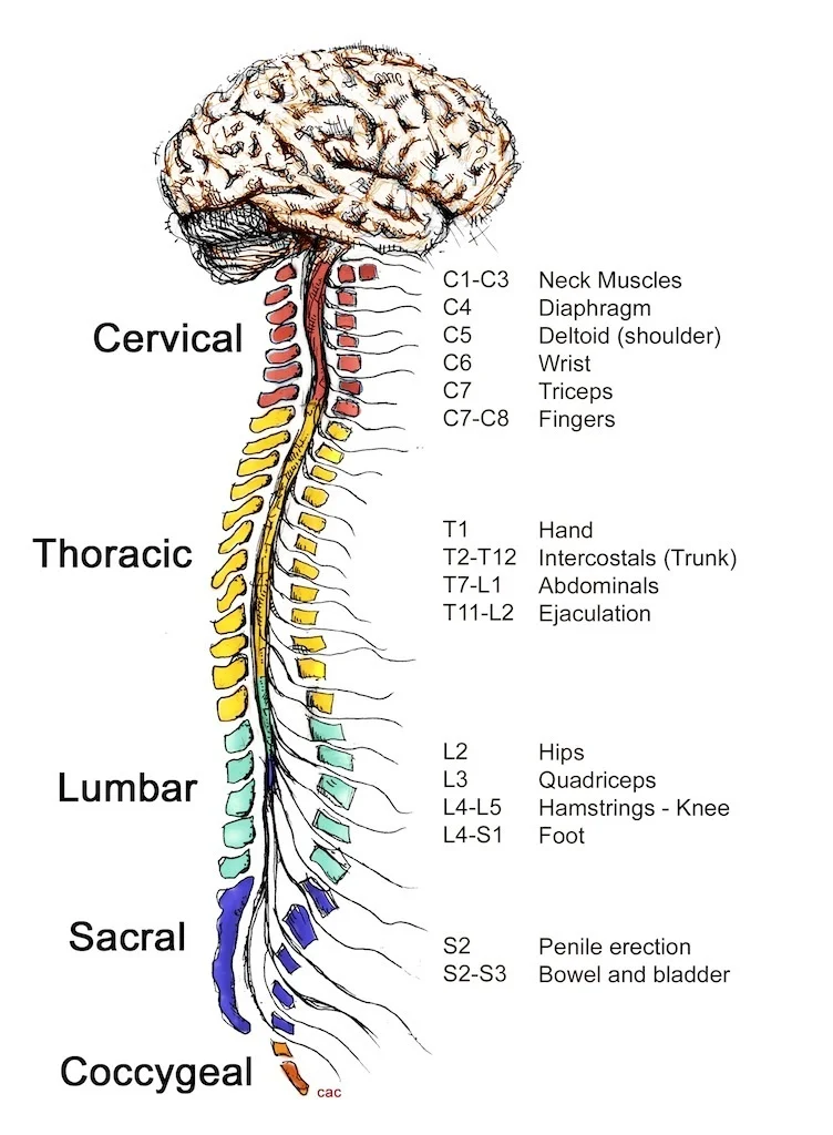
The central nervous system is the source of peripheral nerves. 12 pairs of cranial nerves leave the brain and 31 pairs of spinal nerves leave the spinal cord. Cranial nerves I-XII are named according to where they exit the skull (anterior and posterior). Spinal nerves are divided into 8 cervical, 12 thoracic, 5 lumbar, 5 sacral, and 1 posterior spinal nerves, depending on the spinal level from which they arise. In some areas of the body, peripheral nerves come together to form nerve networks called plexuses.
Notable plexuses include:
- Cervical plexus (C1-C4) – Innervates the back of the head, some neck muscles, the heart muscle, and the diaphragm via the auricle, transverse carotid nerve, lesser occipital nerve, supraclavicular, and phrenic nerves.
- Brachial Plexus (C5-T1) – Innervates the upper limbs with nerves such as the median, ulnar, radial, musculocutaneous, and axillary nerves.
- Lumbar plexus (L1-L4) – innervates the muscles and skin of the abdomen and pelvis and the thigh muscles by the iliohypogastric, lymphatic, genitofemoral, lateral femoral cutaneous, obturator, and femoral nerves.
- Sacral plexus (S1-S4, branches of L4, L5) – innervates the muscles and skin of the pelvic area, the back of the thigh, the lower leg, and the foot through the following nerves; gluteus muscle, sciatic nerve, gluteal skin, pudendal, piriformis nerve, internal obturator nerve, and quadrate femoral nerve.
Somatic and autonomic nervous system
The somatic nervous system (SNS) and the autonomic nervous system (ANS) are parts of the peripheral nervous system that transmit information through the cranial and spinal nerves.
The definition of the somatic nervous system is that it allows us to control our movements and reactions. It transmits sensory and motor information between the skin, sensory organs, skeletal muscles, and the central nervous system; connecting the human body with the environment and responding to external stimuli. The most important somatic peripheral nerves are the median nerve, the sciatic nerve, and the femoral nerve.
The definition of the autonomic nervous system is that it unconsciously controls all internal organs through their associated smooth muscles and glands. Functionally, the ANS is divided into the sympathetic (SANS) and parasympathetic (PANS) autonomic nervous systems. The definition of the sympathetic nervous system is informally known as inducing the “flight or fight” state because it is the part of the ANS that is most active during times of stress.
PANSs predominate in the resting state and are more active in “rest and digestion” or “feeding and reproduction”. “activity. SANS and PANS centers are located in the brainstem and spinal cord and are connected to SANS and PANS nerve ganglia located throughout the body. Note that there is no pure SANS or PANS nerve, but their fibers are incorporated into certain somatic nerves, causing them to mix.
Development
In vertebrates, the landmarks of the development of the embryonic nervous system are the generation and differentiation of neurons from stem cell precursors, the transition of immature neurons from their place of birth in the embryo to their final position, the growth of axons, and the growth. of vagal neurons. to their postsynaptic partners in the embryo, the formation of synapses between these axons and their postsynaptic partners, and finally lifelong changes in synapses that are thought to underlie learning and memory.
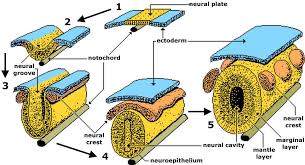
All biphasic animals form a gastrula in an early stage of development, which is polarized, with one end called the animal pole and the other the plant pole. The gastrula is disc-shaped with three cell layers, an inner layer called the endoderm, which forms the lining of most internal organs, a middle layer called the mesoderm, which forms bones and muscles, and an outer layer, called the ectoderm, which gives rise to the skin and nervous system.
In vertebrates, the first sign of the nervous system is a thin group of cells in the center of the spine called the neural plate. The inner part of the neural plate (along the midline) is for the central nervous system (CNS) and the outer part is for the peripheral nervous system (PNS). As development progresses, a fold called the neural groove appears in the midline. At the top, this fold closes when it gets more deeper. At this stage, the future CNS looks like a cylindrical structure called the neural tube, while the future PNS looks like two bands of tissue called the neural crest that run lengthwise over the neural tube.
The sequence of steps from neural plate to neural tube to neural crest is known as innervation. At the beginning of the 20th century, the famous experiments of Hans Spemann and Hilde Mangold showed that the formation of nervous tissue is “induced” through signals from a group of mesodermal cells called an organizational region. For decades, however, the nature of neural induction defeated all attempts to figure it out, until it was finally solved in the 1990s by genetic approaches.
The induction of nerve tissue requires the blocking of the so-called bone morphogenetic protein or BMP gene. In particular, the BMP4 protein appears to be involved. Two proteins secreted by the mesoderm, Noggin and Chordin, can inhibit BMP4 and thus cause the transformation of ectoderm into neural tissue. A similar molecular mechanism appears to be involved in very different types of animals, including arthropods and vertebrates.
However, in some animals, another type of molecule called fibroblast growth factor (FGF) may play an important role in induction. Induction of neural tissue causes the formation of nerve precursor cells called neuroblasts. Due to asymmetric division, neuroblasts in Drosophila produce two different products: a neuroblast and a “ganglion stem cell” (GMC). A GMC divides once to produce either a pair of neurons or a pair of glial cells. In general, a neuroblast is capable of producing an indeterminate number of neurons or glia.
As a 2008 study shows, one factor common to all bipedal organisms (including humans) is a family of secreted signaling molecules called neurotrophins that regulate the growth and survival of neurons. Zhu et al. identified DNT1, the first neurotrophin discovered in flies. DNT1 is structurally similar to all known neurotrophins and is an essential determinant of Drosophila neuronal cell fate. Because neurotrophins have now been identified in both vertebrates and invertebrates, this evidence suggests that neurotrophins were present in a common ancestor of bilaterian organisms and may represent a common mechanism of neurogenesis.
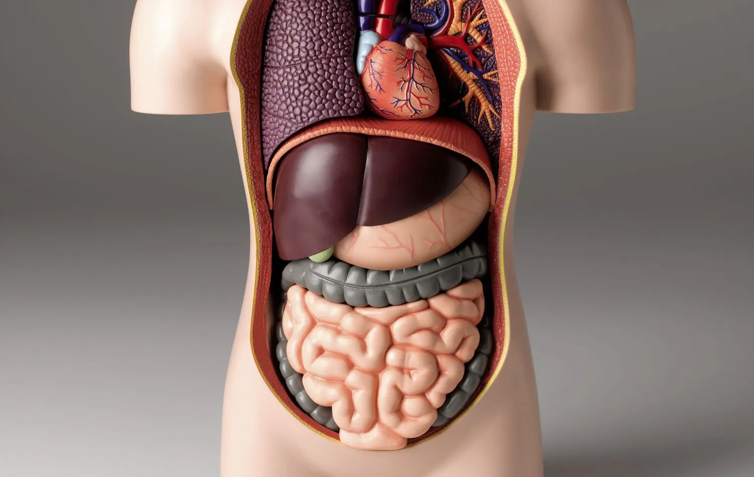
Digestive system
The digestive system and its job is to break down food into smaller and smaller compounds until they can be absorbed by the body and used as energy. It consists of a series of digestive organs and auxiliary digestive organs. The mouth to the anus is where the digestive tract’s organs lie. So it is actually a tube consisting of the mouth, pharynx, esophagus, stomach, small intestine, large intestine and anus. Additives in the digestive tract contribute to the mechanical and chemical breakdown of food.
These include the tongue, salivary glands, pancreas, liver, and gallbladder. The human digestive system consists of the digestive tract and digestive organs (tongue, salivary glands, pancreas, liver, and gall bladder). Digestion involves breaking down food into smaller components until the body can digest and absorb them. There are three stages in the digestive process: the main stage, the stomach stage, and the intestinal stage.
The first stage, the main stage of digestion, begins with the secretion of gastric glands in response to the sight and smell of food. This stage involves the mechanical breakdown of food by chewing and the chemical breakdown by digestive enzymes that occurs in the mouth. Saliva contains the digestive enzymes amylase and lingual lipase, which are secreted by the salivary glands and serum glands of the tongue. Chewing, in which food is mixed with saliva, initiates the process of mechanical digestion. This creates a bolus that is swallowed through the esophagus into the stomach.
The second stage, the gastric stage, takes place in the stomach. This is where food undergoes additional digestion by combining with stomach acid and passing through the duodenum, the first segment of the small intestine.
The third phase, the intestinal phase, begins in the duodenum. Here, partially digested food is mixed with several enzymes produced by the pancreas. Digestion is facilitated by the chewing of food by the masticatory muscles, tongue, and teeth, as well as peristalsis and segmental contractions. The production of stomach acid and mucus in the stomach is necessary for digestion to continue.
Peristalsis is a rhythmic contraction of muscles that begins in the esophagus and continues along the stomach wall and the rest of the digestive tract. This initially results in the production of chyme, which, after a complete breakdown in the small intestine, is absorbed in the lymphatic system in the form of a keel. The majority of food digestion happens in the small intestine. Water and some minerals are absorbed back into the blood in the large intestine. The waste products of digestion (feces) are excreted from the rectum through the anus.
Components
Adult digestive system
Several organs and other components are involved in the digestion of food. The accessory organs of the digestive tract include the liver, gallbladder, and pancreas. Other parts are the mouth, salivary glands, tongue, teeth, and larynx. The largest structure in the digestive tract is the GI tract. It starts from the mouth and ends at the anus and covers about nine meters. An important digestive organ is the stomach. Its mucous membranes contain millions of embedded gastric glands.
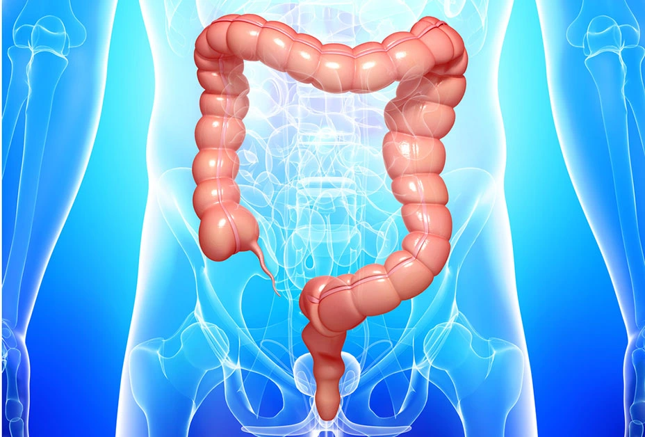
Their secretions are essential for the functioning of the body. Most of the digestion of food takes place in the small intestine, which is the longest part of the digestive tract. The large intestine makes up the majority of the digestive tract. Here the water is absorbed and the remaining waste is stored before defecation. The digestive tract contains a large number of specialized cells. These comprise taste cells, enterocytes, pancreatic duct cells, different gastric gland cells, and microvoltaic cells. One part of the digestive system that is also a part of the excretory system is the large intestine.
Development
In the early stages of embryonic development, the embryo has three embryonic layers and is confined to the yolk sac. During the second week of development, the embryo grows and begins to surround and surround parts of this sac. Coated parts form the basis of the digestive system of an adult. Parts of this foregut begin to differentiate into digestive organs such as the esophagus, stomach, and intestines. During the fourth week of development, the stomach turns.
The abdomen, originally located in the midline of the embryo, rotates so that its body remains on the left. This rotation also affects the part of the digestive tract below the stomach, which becomes the duodenum. At the end of the fourth week, the developing duodenum begins to compress a small protrusion on the right side, the hepatic diverticulum, which becomes the bile duct. Just below this is another outlet known as a cystic diverticulum, which later develops into the gallbladder.
Clinical Significance
All parts of the digestive system are susceptible to various disorders, many of which may be congenital. Oral diseases can also be caused by pathogenic bacteria, viruses, fungi, and side effects of certain medications. Oral diseases include diseases of the tongue and salivary glands. A common gum disease in the mouth is gingivitis caused by plaque bacteria. The most common viral infection of the oral cavity is gingivitis caused by herpes simplex.
A common fungal infection is candidiasis, commonly known as thrush, which affects the lining of the mouth. There are some esophageal diseases, such as Schatzki’s rings, that can restrict the passage and cause difficulty swallowing. They can also completely block the esophagus. Gastritis, peptic ulcers, and gastroparesis are examples of chronic stomach diseases. Malabsorption, ie. the abnormal absorption of nutrients in the gastrointestinal tract, can lead to a number of problems such as malnutrition and anemia.
Malabsorption can have many causes, from infection to enzyme deficiencies such as exocrine pancreatic insufficiency. It can also occur as a result of other gastrointestinal diseases, such as celiac disease. An autoimmune condition affecting the small intestine is celiac disease. This can lead to vitamin deficiency due to poor absorption of nutrients in the small intestine. Small bowel function can also be blocked by a volvulus, a loop of the bowel that twists and closes off the attached bowel.
In severe cases, this can result in mesenteric ischemia. A common intestinal disease is diverticulitis. Diverticula are small pouches that can form within the intestinal wall and can become inflamed, causing diverticulitis. This disease can cause complications when the inflamed diverticulum ruptures and becomes infected. Any infection can spread to the lining of the stomach (peritoneum) and cause potentially fatal peritonitis.
Crohn’s disease is a common chronic inflammatory bowel disease (IBD) that can affect any part of the digestive tract, but most often begins in the lower ileum. Ulcerative colitis, also known as ulcerative colitis, is another large inflammatory bowel disease that is limited to the colon and rectum. Both IBDs can increase the risk of colorectal cancer. Among IBDs, ulcerative colitis is the most prevalent. Presently, the most prevalent functional gastrointestinal disorder is irritable bowel syndrome or IBS. These are the idiopathic disorders that were defined in part by the Roman process.
The protist Giardia lamblia is the cause of the small intestine disease giardiasis. It is contained within the small intestine’s cavity and does not spread. It can frequently show no symptoms at all, but it can also frequently show several symptoms. The most prevalent harmful parasite infection in humans is giardiasis. Diagnostic tools are available to investigate gastrointestinal disorders, which most commonly involve ingestion of barium sulfate. These are called upper gastrointestinal series for imaging the pharynx, larynx, esophagus, stomach, and small intestine, and lower gastrointestinal series for imaging the large intestine.
Pregnancy
Pregnancy can predispose to certain indigestion. Gestational diabetes can develop in a mother as a result of pregnancy, and although it often causes few symptoms, it can lead to preeclampsia.
Urinary system
The urinary framework is a body waste framework containing a gathering of organs that produce and discharge urine. It consists of the bladder, urethra, ureters, and kidneys. Kidneys are matched bean-formed organs put retroperitoneally. The kidneys have a rich blood supply given by the renal course. Nephrons inside the kidneys channel the blood that goes through their snare of vessels (glomerulus).
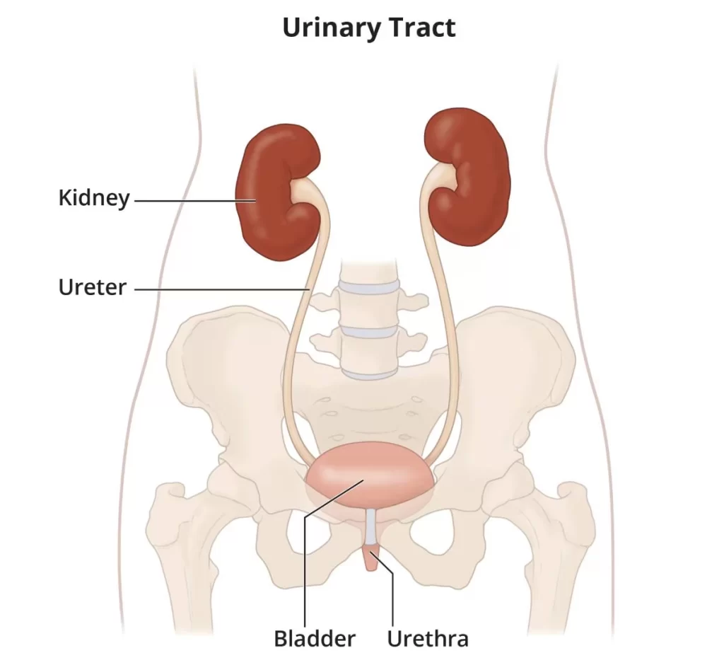
The blood filtrate then, at that point, goes through a progression of tubules and gathering channels, at last shaping the last ultrafiltrate, urine. Urine passes into the ureters, containers of smooth muscle that pass pee from the kidneys onto the urinary bladder. The bladder is an empty solid organ that gathers and stores pee before removal by pee (micturition). Elements of the urinary framework incorporate; the disposal of body squander, guideline of blood volume and pulse, guideline of electrolyte levels, and blood pH.
The excretory framework is an uninvolved natural framework that eliminates overabundance, superfluous materials from the body liquids of a living being, to assist with keeping up with inner substance homeostasis and forestall harm to the body. The double capability of excretory frameworks is the end of the side effects of digestion and depleting the group of spent and separated parts in a fluid and vaporous state.
In people and different amniotes (vertebrates, birds, and reptiles) the vast majority of these substances leave the body as pee and somewhat exhalation, Well-evolved creatures likewise oust them through perspiring. Only the organs that are specifically used for excretion are considered to fall under a part of the excretory system. The term refers to the urinary system in its restricted sense. In any case, as discharge includes a few capabilities that are just hastily related, it isn’t typically utilized in additional proper characterizations of life structures or capability.
As most sound working organs produce metabolic and different squanders, the whole life form relies upon the capability of the framework. Separating at least one of the frameworks is a serious ailment, for instance, kidney disappointment.
Urinary System
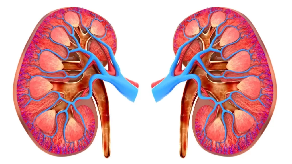
The kidneys are huge, bean-formed organs that are available on each side of the vertebral section in the stomach pit. People have two kidneys and every kidney is provided with blood from the renal supply route. The kidneys eliminate from the blood the nitrogenous squanders like urea, as well as salts and abundant water, and discharge them as pee.
This is finished with the assistance of millions of nephrons present in the kidney. The filtrated blood is out of control from the kidneys by the renal vein (or kidney vein). The pee from the kidney is gathered by the ureter (or excretory cylinders), one from every kidney, and is passed to the urinary bladder. The urinary bladder gathers and stores the urine until pees. The urine gathered in the bladder is passed into the outer climate of the body through an opening called the urethra.
Kidneys
The kidney’s essential capability is the disposal of waste from the circulation system by the creation of urine. They carry out a few homeostatic roles, for example,
1. Keep up with the volume of extracellular liquid
2. Keep up with ionic equilibrium in the extracellular liquid
3. Keep up with pH and osmotic centralization of the extracellular liquid.
4. Discharge harmful metabolic results like urea, alkali, and uric corrosive.
The manner in which the kidneys do this is with nephrons. There are more than 1 million nephrons in every kidney; these nephrons go about as channels inside the kidneys. The kidneys channel the required materials and waste. Required materials return to the circulatory system; unnecessary materials become pee and are ousted through the urethra.
Now and again, an overabundance of squanders take shape as kidney stones. They develop and can become excruciating aggravations that might require a medical procedure or ultrasound therapies. A few stones are sufficiently little to be constrained into the urethra.
Ureter
The ureters are solid pipes that push pee from the kidneys to the urinary bladder. In the human grown-up, the ureters are typically 25-30 cm (10-12 in) long. In people, the ureters emerge from the renal pelvis on the average part of every kidney prior to slipping towards the bladder on the facade of the psoas significant muscle. The ureters cross the pelvic edge close to the bifurcation of the iliac conduits (which they run over).
This “pelvic ureteric intersection” is a typical site for the impaction of kidney stones (the other being the ureterovesical valve). The ureters run posteriorly on the horizontal walls of the pelvis. They then bend anteromedially to enter the bladder through the back, at the vesicoureteric intersection, running inside the mass of the bladder for a couple of centimeters. The discharge of pee is forestalled by valves known as ureterovesical valves. In the female, the ureters go through the mesometrium en route to the bladder.
Urinary bladder
The urinary bladder is the organ that gathers squander discharged by the kidneys preceding removal by pee. It is an empty strong, and distensible (or versatile) organ, and sits on the pelvic floor. Pee enters the bladder through the ureters and ways out by means of the urethra.
Embryologically, the bladder is gotten from the urogenital sinus, and it is at first constant with the allantois. In human guys, the foundation of the bladder lies between the rectum and the pubic symphysis. It is better than the prostate and isolated from the rectum by the rectovesical exhuming. In females, the bladder sits mediocre compared to the uterus and foremost to the vagina. It is isolated from the uterus by the vesicouterine exhuming. In babies and small kids, the urinary bladder is in the midsection in any event, when void.
Urethra
In life systems, the (from Greek – urethra) is a cylinder that interfaces the urinary bladder to the beyond the body. In people, the urethra has an excretory capability in the two sexual orientations to pass.
Function
The primary elements of the urinary framework and its parts are to:
- Manage blood volume and arrangement (for example sodium, potassium, and calcium)
- Manage pulse.
- Manage pH homeostasis of the blood.
- Adds to the development of red platelets by the kidney.
- Combines calcitriol (the dynamic type of Vitamin D).
- Stores side-effects (for the most part urea and uric corrosive) before it and different items are taken out from the body.
Urine Formation
Normal urine creation in grown-up people is around 1-2 liters (L) each day, contingent upon the condition of hydration, movement level, natural variables, weight, and the singular’s well-being. Delivering excessively or too little pee requires clinical consideration. Polyuria is a state of unnecessary pee creation (> 2.5 L/day). Conditions including low results of pee are oliguria (< 400 mL/day) and anuria (< 100 mL/day).
The most vital phase in pee arrangement is the filtration of blood in the kidneys. In a sound human, the kidney gets somewhere in the range of 12 and 30% of the cardiovascular result, however it midpoints around 20% or around 1.25 L/min.
The essential underlying and useful unit of the kidney is the nephron. Its main capability is to manage the grouping of water and dissolvable substances like sodium by sifting the blood, reabsorbing what is required, and discharging the rest as pee.
In the initial segment of the nephron, Bowman’s case channels blood from the circulatory framework into the tubules. Hydrostatic and osmotic tension slopes work with filtration across a semipermeable film. The filtrate incorporates water, little atoms, and particles that effectively go through the filtration layer. Be that as it may, bigger atoms, for example, proteins and platelets are kept from going through the filtration film.
How much filtrate is delivered consistently is known as the glomerular filtration rate or GFR and sums to 180 liters each day. Around the vast majority of this filtrate is reabsorbed as it goes through the nephron and the leftover 1% becomes pee. The urinary framework is managed by the endocrine framework by chemicals like antidiuretic chemicals, aldosterone, and parathyroid chemicals. Guideline of focus and volume
Regulation of concentration and volume
The Urinary System is affected by the circulatory framework, sensory system, and endocrine framework. Aldosterone plays a focal part in managing circulatory strain through its impact on the kidney. It follows up on the distal tubules and gathering conduits of the nephron and expands the reabsorption of sodium from the glomerular filtrate. Reabsorption of sodium brings about the maintenance of water, which increments pulse and blood volume.
Antidiuretic chemical (ADH), is a neurohypophysial chemical tracked down in many well evolved creatures. Holding water in the body and vasoconstriction two essential capabilities are. Vasopressin directs the body’s maintenance of water by expanding water reabsorption in the gathering channels of the kidney nephron. Vasopressin increments water porousness of the kidney’s gathering pipe and distal tangled tubule by actuating the movement of aquaporin-compact disc water directed in the kidney nephron gathering conduit plasma layer.
Urination
Urine, likewise now and again alluded to as micturition, is the discharge of pee from the urinary bladder through the urethra to the beyond the body. In solid people (and numerous different creatures), the course of pee is under deliberate control. In newborn children, a few older people, and those with neurological injuries, pee might happen as a compulsory reflex. Physiologically, micturition includes coordination between the focal, autonomic, and substantial sensory systems. Cerebrum focuses that control pee incorporates the pontine micturition focus, periaqueductal dark, and the cerebral cortex. In placental warm-blooded creatures, the male launches pee through the penis, and the female through the vulva.
Clinical significance
Urological disease can be associated with congenital or acquired dysfunction of the urinary tract. For example, urinary tract obstruction is a urological disease that can cause urinary retention. Diseases of the kidney tissues are usually treated by nephrologists, while diseases of the urinary tract are treated by urologists. Gynecologists can also treat female incontinence. Diseases of other body systems also directly affect the work of the urogenital system.
For example, a protein released in the kidney associated with diabetes has been shown to sensitize the kidney to the harmful effects of hypertension. Diabetes can also directly affect urination due to peripheral neuropathy, which occurs in some people with poorly controlled blood sugar levels. Urinary incontinence can be caused by a weakening of the pelvic floor muscles, which is caused by, for example, pregnancy, childbirth, aging, and obesity.
Results of recent systematic reviews indicate that behavioral therapy generally improves outcomes for urinary incontinence, especially when exposed to stress and compulsions when compared to medication alone. Pelvic floor exercises, called Kegel exercises, can help with this condition by strengthening the pelvic floor. Urinary incontinence can also have medical causes, which are often treatable. In children, this condition is called enuresis. Certain cancers, such as those of the bladder, kidney, ureter, and urethral regions, also affect the urinary system. Because of the role and location of these organs, treatment is often difficult.
Endocrine system
- Endocrine systems are specialized organs (endocrine glands) scattered throughout the body that produce hormones. The main organs of the endocrine system are shown in the diagram below. Regarding the functioning of the endocrine system; hormones produced by the endocrine system regulate many different body functions, such as triiodothyronine, which regulates metabolism, or estrogen and progesterone, which regulate the menstrual cycle. Hormones secreted by endocrine glands directly into the circulatory system control the activity of distant target organs.
- The endocrine system is the body’s messaging system, consisting of feedback loops of hormones released directly through the internal glands of the circulatory system that target and regulate distant organs. In vertebrates, the hypothalamus is the nervous system control center for all hormonal systems. In humans, the most important endocrine glands are the thyroid, parathyroid, pituitary, pineal, and adrenal glands, as well as (male) testes and (female) ovaries.
- The hypothalamus, pancreas, and thymus function as endocrine glands in addition to other functions. (The hypothalamus and the pituitary gland are organs of the neuroendocrine system. One of the most important functions of the hypothalamus – it is located in the brain next to the pituitary gland – is to connect the endocrine system to the nervous system through the pituitary gland ) Also, other organs, like the kidneys, play a role in the hormonal system, because they secrete certain hormones.
- Endocrinology is known for the study of the endocrine system and its disorders. Glands that signal to each other in sequence are often called an axis, such as the hypothalamic-pituitary-adrenal axis. In addition to the specific endocrine organs mentioned above, many other organs that are part of other body systems have secondary endocrine functions, including the bones, kidneys, liver, heart, and gonads. For instance, erythropoietin is an endocrine hormone secreted from the kidneys.
- Hormones can be complexes of amino acids, steroids, eicosanoids, leukotrienes, or prostaglandins. Endocrine systems differ from exocrine glands, which secrete hormones to the outside of the body, and a system called paracrine signaling over a relatively short distance between cells. Endocrine glands do not have ducts, are vascular, and usually have intracellular vacuoles or granules that store their hormones. In contrast, exocrine glands such as salivary glands, sweat glands, and digestive glands tend to be much less vascular and have ducts or cavities. One specialization of internal medicine is endocrinology.
Function
Hormones:
A hormone is any class of signaling molecules produced by glandular cells in multicellular organisms that are transported via the circulatory system to distant organs to regulate physiology and behavior. Hormones have a variety of chemical structures that fall into three main categories: eicosanoids, steroids, and amino acid/protein derivatives (amines, peptides, and proteins). Glands that secrete hormones form the endocrine system.
The term hormone is sometimes expanded to include chemicals produced by cells that act on the same cell (autocrine or intracrine signaling) or on nearby cells (paracrine signaling). Hormones are used to communicate between organs and tissues for physiological regulation and behavioral functions such as digestion, metabolism, respiration, tissue function, sensory perception, sleep, excretion, lactation, stress, growth and development, movement, and mood. Hormones affect distant cells by binding to certain receptor proteins on the target cell, which changes the function of the cell.
This can lead to cell type-specific responses involving rapid changes in the activity of existing proteins or slower changes in the expression of target genes. Amino acid-based hormones (amines and peptide or protein hormones) are water-soluble and act on the surface of target cells through signal transduction pathways. steroid hormones, which are fat soluble, cross the plasma membranes of target cells and act in their nuclei.
Cell signaling:
A typical method of cell signaling in the endocrine system is endocrine signaling, that is, the use of the circulatory system to reach distant targets. However, there are other pathways such as paracrine, autocrine, and neuroendocrine signaling. Pure neurocrine signaling between neurons, on the other hand, belongs entirely to the nervous system.
Autocrine:
A type of signaling known as autocrine signaling occurs when a cell secretes a hormone or chemical messenger known as an autocrine substance, which binds to autocrine receptors on the same cell and causes alterations in the cell.
Paracrine:
Some endocrinologists and physicians include the paracrine system as part of the endocrine system, but there is no consensus. Paracrine works more slowly and targets cells in the same tissue or organ. Somatostatin, a substance that targets other pancreatic cells and is released by certain pancreatic cells, is an illustration of this.
Juxtacrine:
Juxtacrine signaling is a type of cell-to-cell communication mediated by oligosaccharides, lipids, or protein components of the cell membrane that can affect either the radiating cell or immediately adjacent cells. It occurs between adjacent cells with wide patches of closely spaced plasma membranes interconnected by transmembrane channels called connexons. The distance between the cells can usually be only 2-4 nm.
Clinical significance
- Endocrine disorders are common, including conditions such as diabetes, thyroid disease, and obesity. Endocrine disease is characterized by improperly regulated hormone release (productive pituitary adenoma), inappropriate response to signaling (hypothyroidism), absence of a gland (type 1 diabetes, impaired erythropoiesis in chronic renal failure), or structural enlargement in a critical location, such as. the thyroid gland (toxic multinodular goiter).
- Endocrine failure can result from loss of reserve, hyposecretion, agenesis, atrophy, or active destruction. Overactivity may be due to excessive secretion, suppression, hyperplastic or neoplastic changes, or hyperstimulation. There are three types of endocrinopathies: primary, secondary, and tertiary. Primary endocrine disease suppresses the function of the downstream glands. Secondary endocrine disease refers to a problem with the pituitary gland. Tertiary endocrine disease is associated with dysfunctions of the hypothalamus and the hormones that release it.
- Because the thyroid and hormones signal the proliferation of distant tissues, for example, the estrogen receptor has been linked to certain breast cancers. Endocrine, paracrine, and autocrine signals are all involved in proliferation, which is one of the key steps in oncogenesis. Other common diseases caused by endocrine dysfunction include Addison’s disease, Cushing’s disease, and Graves’s disease; disease Cushing’s disease and Addison’s disease are pathologies associated with adrenal gland dysfunction.
- Adrenal dysfunction can be caused by primary or secondary factors and can lead to hypercortisol or hypocortisol. Cushing’s disease is characterized by hypersecretion of adrenocorticotropic hormone (ACTH) from a pituitary adenoma, which ultimately leads to endogenous hypercortisol by stimulating the adrenal glands. Some of the clinical symptoms of Cushing’s disease include obesity, moon face, and hirsutism. Addison’s disease is a hormonal disorder caused by hypocortisolism caused by adrenal insufficiency.
- Adrenal insufficiency is important because it correlates with a reduced ability to maintain blood pressure and blood sugar, which can lead to death. Graves disease is associated with hyperactivity of the thyroid gland, which produces hormones T3 and T4. Graves disease; The effects of the disease range from excessive sweating, fatigue, heat intolerance, and high blood pressure to swelling of the eyes, which causes redness, swelling, and, in rare cases, impaired vision or double vision.
Lymphatic system
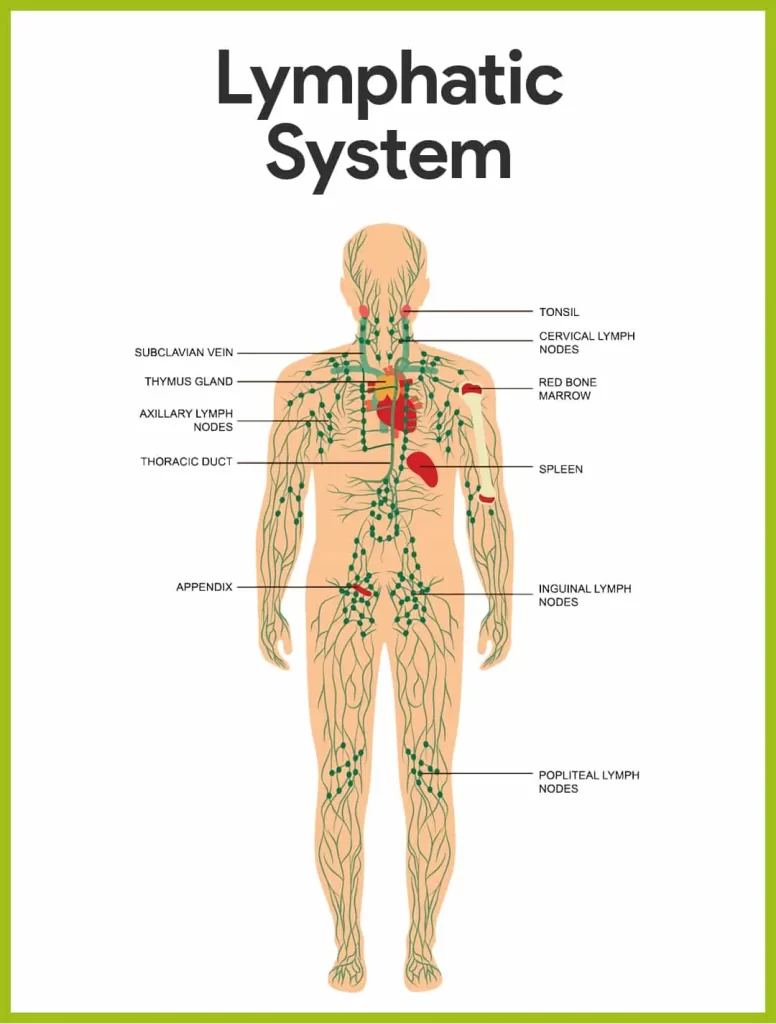
- The lymphatic system is a network of lymphatic vessels that remove excess tissue fluid (lymph) from the intercellular fluid compartment, filter it through lymph nodes, contact lymphocytes (white blood cells) of the immune system, and return the fluid to the lymphatic system. circulatory system. The lymphatic system is comprised of lymph, lymphoid organs, lymphatic plexuses, lymphatic vessels, and lymph nodes.
- The function of the lymphatic system is to transport and remove toxins and waste from the body; recycle proteins; and protect the body from microorganisms. Lymph is a watery tissue fluid with a consistency similar to blood plasma. It begins as interstitial fluid, which fills the spaces between cells. Excess fluid is absorbed and transported through the lymph nodes to the lymph vessels and passes through the lymph nodes on the way.
- Superficial lymphatic vessels are located next to veins in the subcutaneous tissue. They drain into the deep lymphatic vessels that follow the arteries. The right lymphatic duct and the chest lymphatic duct are the two main collecting ducts formed by the union of larger lymph nodes where lymph vessels empty. The thoracic duct begins at the cisterna keel and collects lymph from the left side of the head, neck, and chest, left upper limb, abdomen, and both lower limbs and empties it into the left vena cava (the junction of the left internal jugular). vein and the left inferior clavicular vein).
- The right lymphatic duct drains the rest of the body and merges into the right vena cava. The lymph node cleaned from the venous corners is returned to the circulatory system in the blood fluid. Note that the central nervous system was not previously thought to have lymphatic vessels. However, recent studies have shown that its lymph node drains through lymphatic vessel-like structures in the meninges. One can distinguish between primary and secondary organs within the lymphatic system.
- Through the primary lymphatic vessels, generated lymphocytes circulate into the vessels. The red bone marrow and lymph nodes are the two primary lymphoid organs. There are several lymph nodes, tonsils, appendix, and spleen that are secondary lymphatic organs. A lymphoid mass with lymphocytes affixed to lymphatic vessels is called a lymph node. The things that lymph nodes filter are extra tissue fluid, foreign pathogens, leaking plasma proteins, and cell debris.
- The aortic root, joint, femoral, cervical, axillary, tracheal, and deep nodes are among the central points of the body where there are clusters of lymph nodes. In vertebrates, the lymphatic system is an organ system that concludes the circulatory system and is an important part of the immune system. It consists of a large network of lymph nodes, lymph nodes, lymph nodes, lymph nodes, lymph nodes, and lymph nodes.
- Lymphatic vessels carry the transparent fluid known as the lymphatic system back to the heart where it is recycled. (The lympha, the Latin word for lymph, is related to the goddess “Lympha” of freshwater). The lymphatic system is open, in contrast to the circulatory system, which is closed. The human circulatory system uses capillary filtration to remove plasma from the blood, processing 20 liters of blood on average per day. Approximately 17 liters of filtered blood are reabsorbed directly into the blood vessels, while the remaining three liters remain in the interstitial fluid.
- One of the main functions of the lymphatic system is to provide an additional three liters of blood circulation. Another key function is immune protection. With respect to the waste products and cell debris, proteins, and bacteria that are present, lymph is remarkably similar to blood plasma. Lymphocyte cells are mostly lymphocytes. Associated lymphoid organs consist of lymphoid tissue and are sites of either lymphocyte production or lymphocyte activation.
- These include the lymph nodes (which have the highest concentration of lymphocytes), the spleen, the thymus, and the tonsils. Lymphocytes are originally produced in the bone marrow. Lymphatic organs also contain other types of cells, such as stromal cells. Lymphoid tissue is also associated with mucous membranes, such as mucosa-associated lymph nodes (MALT). Fluid from the circulating blood leaks into the tissues of the body through capillaries, carrying nutrients to the cells.
- The fluid bathes the tissues as interstitial fluid, collecting waste products, bacteria, and damaged cells, and then flows as lymph into the capillaries and lymphatic vessels of the lymph nodes. These blood vessels carry lymph throughout the body, passing through many lymph nodes that filter out unwanted materials such as bacteria and damaged cells. The lymphatic vessels then branch into much larger lymphatic vessels called lymphatic ducts.
- The right lymphatic duct drains the right side of the area, and the much larger left lymphatic duct, known as the thoracic duct, drains the left side of the body. To get back into the bloodstream, the ducts empty into the subclavian veins. When muscles contract, lymph flows through the system. Some vertebrates have a lymphatic heart that pumps lymph into a vein. Olaus Rudbeck and Thomas Bartholin first described the lymphatic system independently in the 17th century.
Development
Lymphatic tissues begin to form at the end of the fifth week of embryonic development. Lymphatic vessels form from lymph sacs that arise from developing blood vessels originating in the mesoderm. The first lymph nodes to arise are the paired jugular lymph vessels at the junction of the internal jugular vein and subclavian vein.
From the lymph nodes in the neck, lymph nodes spread to the chest, upper limbs, neck, and head. Some plexuses enlarge and form lymphatic vessels in their areas. Each jugular lymph node has at least one connection with its jugular vein, the upper thoracic duct develops from the left. The spleen is formed from mesenchymal cells between the layers of the dorsal mesentery of the stomach. The thymus gland arises as an outgrowth of the third pharyngeal sac.
Function
The lymphatic system performs multiple interconnected tasks.
- Its responsibility is to get rid of interstitial fluid from tissues.
- It absorbs and transports abundant fatty acids and fats from the digestive system
- It transports white blood cells to the lymph nodes and from there to the bones
- The lymph node transports antigen-presenting cells, such as dendritic cells, to the lymph nodes where the immune response is stimulated.
Absorption of fat
Lymphatic vessels called lacteals are located at the beginning of the digestive tract, mainly in the small intestine. The bulk of other nutrients absorbed in the small intestine enter the portal venous system and pass through the portal vein to the liver for processing, whereas fats, or lipids, travel to the lymph nodes, then through the chest and into the bloodstream.
Pathway (There are some exceptions, such as fatty acid esters of glycerol called medium-chain triglycerides, which passively diffuse into the portal system from the gastrointestinal tract.). Lymph that gets noticeably enlarged by small intestine lymph nodes is referred to as a “keel”. The liver breaks down nutrients that have made their way into the bloodstream and throughout the body.
Immune system functioning
Because it is the primary location for T and B cells and other cells that are part of the adaptive immune system, the lymphatic system is vital to both the body and the immune system. Lymphatic system cells respond to antigens presented or found by cells directly or by other dendritic cells.
Once an antigen is recognized, an immunological cascade begins, involving the activation and recruitment of more and more cells, the production of antibodies and cytokines, and the recruitment of other immunological cells such as macrophages.
Reproductive system
The reproductive system, frequently referred to by the term the reproductive organs, is comprised of the external and internal reproductive organs that work collectively to support reproduction. Unlike other organ systems, the reproductive system has significant gender differences.
The human reproductive system includes the male reproductive system, which produces and stores sperm. and the female reproductive system, which produces eggs and protects and nourishes the fetus until birth. Humans have a high level of gender differentiation. Typical secondary sex characteristics vary widely, in addition to variations in nearly every reproductive organ. Sexual contact and internal fertilization are two common steps in the human reproductive process.
In this process, the male inserts his penis into the female and into her vagina and ejaculates, which contains sperm. A small number of sperm travel through the cervix into the uterus and then into the fallopian tubes to fertilize an egg. One sperm is all that requires action to fertilize an egg. After successful fertilization, the fertilized egg, or zygote, moves from the fallopian tube into the uterus, where it implants in the uterine wall. It marks the beginning of pregnancy, better known as pregnancy, which lasts about nine months until the fetus develops.
When the fetus has developed to a certain point, the pregnancy ends with parturition, which is accompanied by childbirth. During childbirth, the muscles of the uterus contract and the cervix dilates for hours, and the baby emerges from the vagina. Babies are completely dependent on their caregivers and require a high level of parental care. Babies depend on their caregivers for comfort, cleanliness, and food. Food can be given through breastfeeding or feeding.
The female external genitalia, also known as the genitalia, are the labia, the clitoris, and the vaginal opening. The uterus, vagina, fallopian tubes, and ovaries are the internal reproductive organs. IUDs provide access and protection of the vagina and uterus, as well as sufficient warmth and moisture to aid its sexual and reproductive functions. In addition, it is important for sexual excitement and orgasm in women.
The uterus (cervix) and the exterior of the body are connected by a tube known as the vagina. The ovaries secrete hormones and produce eggs, which are transported to the fallopian tubes of the uterus. The uterus provides protection, nutrition, and waste disposal for the developing embryo and fetus. In addition, contractions of the muscular wall of the uterus promote the expulsion of the fetus at the time of birth.
The male external genitalia are the testicles and penis, while the internal ones are the epididymis, ductus deferens, and adrenal glands. In terms of functionality, they can be divided into three categories. The first category involves sperm production (testes) and storage (epididymis). Organs of the second class produce ejaculation; ductus deferens and accessory glands (seminal vesicles and prostate gland). The last category are those used for mating and depositing sperm, including the penis, urethra, and ductus deferens.
Development
The development of the reproductive system and the development of the urinary tract are closely related to the development of the human fetus. Despite the differences between the adult female and male, derived from the middle mesoderm. [clarification needed] [no main verb] The three most important fetal precursors of the reproductive organs are the Wolffian duct, the Müllerian duct, and the gonads.
One well-known and essential regulatory element in the regular distinguishing procedure of the reproductive system is endocrine hormones. The Wolffian duct virtually vanishes in the female reproductive system, but it forms the epididymis, vas deferens, ductus deferens, ejaculatory duct, and seminal vesicle in the male reproductive system. The opposite is true for the Müllerian duct, as it essentially disappears from the male reproductive system and forms the fallopian tubes, uterus, and vagina in the female body.
The gonads continue in developing the ovaries and testicles in both sexes; these organs are frequently referred to as jointly homologous because they originate in the same immature structure. There are several other homologous structures between the male and female reproductive systems. The female and male fallopian tubes share similar blood vessels and functions, but they are not homologous; rather, they are analogs of one another because they develop from distinct fetal structures.
Disease
There are four main categories of human reproductive disorders. They are:
- Genetic or congenital abnormalities.
- Cancers.
- Infections, which are often sexually transmitted diseases.
- Varicose veins
Functional problems are caused by environmental factors, physical damage, psychological problems, autoimmune disorders, or other causes. The two most well-known functional issues are infertility and sexual dysfunction, which are general terms that cover a wide range of disorders with various underlying causes.
Certain reproductive disorders are often symptoms of other diseases and disorders or have multiple or unknown causes, making them difficult to classify. Examples of unclassified diseases are Peyronie’s disease in men and endometriosis in women. A few of these are Bloom syndrome, cystic fibrosis, Klinefelter’s syndrome, and Turner syndrome.
Integumentary system
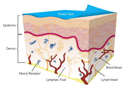
- The integumentary system is a collection of organs that make up the outer shell of the body. This includes the skin, appendages, sweat glands, and sensory receptors.
- The assortment of organs that comprise the animal’s outermost covering is known as the integumentary system. It consists of the skin and its appendages, which act as a physical barrier between the external environment and the internal environment, which it protects and preserves in the organism of the animal. It is primarily the skin on the body, including the outer skin.
- The integrated system includes skin, hair, scales, feathers, hooves, and claws. It has several additional functions: it can maintain water balance, protect deeper tissues, excrete waste, regulate body temperature, and is the site of attachment for sensory receptors that detect pain, sensation, pressure, and temperature.
- The human body’s largest organ is the skin. The epidermis, dermis, and hypodermis are its three main layers. The epidermis is a thick keratinized epithelium made of multiple cell layers. The blood vessels and nerves that supply the skin are located in the dermis, a layer of connective tissue beneath the epidermis. The hypodermis, or underlying fascia, is made up of skin appendages such as hair, nails, sebaceous and sweat glands, as well as fat and connective tissue.
- The inner membrane performs distinct functions. It creates a continuous layer of defense against the body’s many ills, including cuts from the outside world, loss of heat and moisture, and UV radiation that can cause cancer. It also facilitates the synthesis of vitamin D, eliminates waste, and has sensory receptors for pressure, temperature, pain, and sensation.
Functions
The integrated system is accountable for a number of bodily and balance-related roles. All body systems cooperate with each other to maintain the internal conditions necessary for the body to function. The skin has the important function of protecting the body and acts as the body’s first line of defense against infections, temperature changes, and other problems affecting homeostasis. Its main features include:
- Protects the body and internal living tissues and organs
- Protects infectious organisms from invading
- Prevents the body from becoming parched
- Protects the body against sudden temperature changes, maintains homeostasis
- Help eliminate waste through sweat
- Serves as a receptor for pressure, heat, cold, touch, and pain (see somatosensory system)
- Prevents sunburn by releasing melanin into the body.
- Make vitamin D when you receive ultraviolet radiation.
- Reserve water, fat, glucose, vitamin D
- Maintaining body shape
- Formation of new cells from the stratum germinativum to repair small wounds
- Protects against UV rays.
- Regulates body temperature
- It isolates, separates, and protects the organism from the environment.
In aquatic or continuously moist environments, small-bodied invertebrates use their integument as an outer layer to help them breathe. This gas exchange system, where gases simply diffuse into and out of the interstitial fluid, is called an integumentary exchange.
Clinical significance
Possible diseases and injuries of human internal organs are as follows:
- Eruption
- Yeast
- Athlete and leg
- Infection
- Sunburn
- Skin cancer
- Albinism
- Winnie the Pooh
- Herpes
- Herpes labialis, often called cold sore
- Impetigo
- Rubella
- Cancer
- Psoriasis
- Rabies
- Rosacea
- Atopic dermatitis
- Eczema
FAQs
What are the human body’s eleven systems?
The human body’s 11 systems are as follows:
1. The skeletal system;
2. The muscular system;
3. The nervous system;
4. The endocrine system;
5. The circulatory system;
6. The digestive system;
7. The respiratory system;
8. The excretory system; and
9. The immune system
10. System of reproduction
11. System of integument
How can one keep their body and system healthy?
Here are a few tips:
Eat a healthy diet that is low in processed foods, sugar, and unhealthy fats, Get regular exercise, Get enough sleep, Manage stress, Don’t smoke, and Limit alcohol consumption. See your doctor for regular checkups.
Which of the body’s systems is the most essential?
In order to maintain our health and existence, every system in the human body plays a crucial role. That being said, some systems are valued higher than others. To govern our thoughts, movements, and emotions, for instance, the nervous system is required. In addition to delivering nutrients and oxygen to the cells, the circulatory system is also required to eliminate waste materials and carbon dioxide. Our cells would not survive in the absence of a circulatory system. For food to be broken down into nutrients the body can absorb, the digestive system is needed. Humans cannot survive without the nutrients that our digestive systems provide.
How do the different systems work together?
The human body’s many systems interact intricately and synchronically to maintain our health and survival. For example, the skeletal system provides support for the muscular system, which allows us to move. The nervous system controls the muscles, and the endocrine system regulates many body functions, including muscle growth and development. The circulatory system delivers oxygen and nutrients to the muscles, and the digestive system breaks down food into nutrients that the muscles can use. The respiratory system exhales carbon dioxide, which is a waste product of cellular respiration. The excretory system removes waste products from the blood, and the immune system protects the body from infection. The reproductive system allows humans to reproduce, ensuring the continuation of the human species. Body temperature regulation and environmental protection are provided by the integument system.
References
List of systems of the human body. (2014, March 2). Wikipedia. https://en.wikipedia.org/wiki/List_of_systems_of_the_human_body
Vasković, J. (2023, November 3). Human body systems. Kenhub. https://www.kenhub.com/en/library/anatomy/human-body-systems
Muscle Types | SEER Training. (n.d.). https://training.seer.cancer.gov/anatomy/muscular/types.html
Professional, C. C. M. (n.d.). Muscle. Cleveland Clinic. https://my.clevelandclinic.org/health/body/21887-muscle