Zygomaticotemporal nerve
Introduction
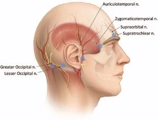
The zygomatic nerve is the branch of the maxillary nerve, which by itself is a branch of the trigeminal nerve (CN V). It runs from the orbit & splits into the zygomaticotemporal & the zygomaticofacial nerve. It gives sensory supply to the skin over the zygomatic bone & the temporal bone. either It also carries postganglionic parasympathetic axons to the lacrimal gland. It might be blocked by anesthetizing the maxillary nerve.
It ascends between the bone, & substance of the Temporalis muscle, pierces the temporal fascia just 2.5 cm. above the zygomatic arch, & is supplied to the skin of the side of the forehead, & communicates with the facial nerve & with the aurićulotemporal branch of the mandibular nerve.
As it penetrates the temporal fascia, it gives off a slender spring, which runs in the middle of the two layers of the fascia to the lateral angle of the orbit.
Structure
The zygomatic nerve is a branch of the maxillary nerve (CN V2), by itself a branch of the trigeminal nerve (CN V). Its branches are at the pterygopalatine ganglion. It runs from the pterygopalatine fossa through the inferior orbital fissure to penetrate the orbit. In the orbit, it runs anteriorly along with the lateral wall.
Branches of zygomaticotemporal nerve
Soon after the zygomatic nerve enters the orbit it split into its branches. These include:
the zygomaticotemporal nerve.Then passes through the zygomaticotemporal foramen in the zygomatic bone.
the zygomaticofacial nerve. This proceeds through the zygomaticofacial foramen in the zygomatic bone.
where It gives the communicating branch to the lacrimal nerve.
Variation
occasionally, the zygomatic nerve does not give branches within the orbit. alternatively, it enters a single foramen in the zygomatic bone known as the zygomatic-orbital foramen. In this case, it splits within the bone into the zygomaticotemporal nerve & the zygomaticofacial nerve.
II. Physical Examination
A. Inspect the face & scalp for soft tissue injuries.
B. Assess soft touch sensation to the face & neck prior to administration of local anesthesia.
- Trigeminal nerve distributions
a.Ophthalmic (V1): supraorbital, supratrochlear, infratrochlear, external nasal, & lacrimal nerves
b.Maxillary (V2): infraorbital, zygomaticofacial, zygomaticotemporal nerves
c.Mandibular (V3) divisions: mental, buccal, auriculotemporal nerves - Terminal cutaneous branches of C2-3
a.Greater auricular and transverse cervical nerves - Locally inspect wounds if a sensory deficit is appreciated.
a.Repair a severed nerve trunk with microsurgical technique if proximal & distal ends can be found.
b.New nerve endings will grow & renew sensation typically within 6 months if the nerve trunk is intact.
C. Assess the function of the facial nerve distributions previous to the administration of local anesthesia.
- Ask the patient to raise the eyebrows, close both eyes, smile, pucker the lips, & show the lower teeth.
- Any Injuries to the facial nerve anterior to the level of the lateral canthus will probably resuscitate.
- Any Injuries to the facial nerve posterior to the level of the lateral canthus need exploration & microsurgical repair within
2 to 3 days after injury. - Obtain an electromyogram if the physical examination is undetermined.
a.Findings of the muscle amplitude in the weak muscles show nerve to stretch or onychomycosis.
b.The lack of electrical signal suggests a transaction of a branch of the facial nerve.
Function of zygomaticotemporal nerve
The end branches of the zygomatic nerve contain sensory axons. These give sensation to the skin over the temporal bone & the zygomatic bone.
The zygomatic nerve also carries postganglionic parasympathetic axons. These axons possess their cell bodies in the pterygopalatine ganglion. They travel from the ganglion to the zygomatic nerve, & then to the lacrimal nerve through a communicating branch. From the lacrimal nerve, they enter the lacrimal gland & give a secretomotor supply.
Clinical significance
The zygomatic nerve can be blocked indirectly by anesthetizing the maxillary nerve (CN V2). The zygomatic nerve & its branches might be damaged by a fracture to the zygomatic bone.
FAQ
What is the function of the zygomaticotemporal nerve?
The zygomaticotemporal nerve runs along with the lateral wall of the orbit & gives sensation to the lateral side of the forehead. The zygomaticofacial nerve exits the skull at the zygomaticofacial foramen inferolateral to the orbit, giving sensation to the malar prominence of the cheek.
What passes through the Zygomaticotemporal foramen?
The zygomaticotemporal branch of the zygomatic nerve, which is a branch of the maxillary division of the trigeminal nerve, runs along with the lateral wall of the orbit. It passes through the zygomaticotemporal foramen & then travels in the temporal fossa
What does the zygomatic nerve affect?
The pterygopalatine group includes the zygomatic nerve, which gives sensory supply to the temporal & lateral zygomatic region, & the sphenopalatine branches, which help to supply the mucosa of the maxillary sinus, upper gums, upper molars, & mucous membranes of the cheek.
What is the zygomatic nerve used for?
The zygomatic nerve is a branch or part of the maxillary nerve, by itself a branch of the trigeminal nerve (CN V). It runs through the orbit & splits into the zygomaticotemporal & the zygomaticofacial nerve. It gives sensory supply to the skin over the zygomatic bone & the temporal bone.