Patellar Tracking Disorder
What is Patellar Tracking Disorder?
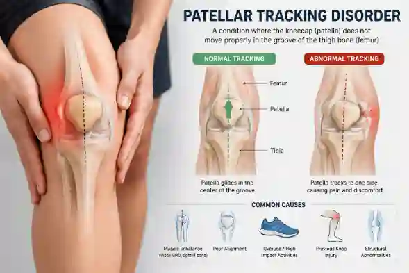
Patellar tracking disease, also known as patellar maltracking, is a condition in which the kneecap (patella) slides laterally out of its groove when the leg is bent or straightened.
If you’ve ever experienced grinding, popping, or severe pain around your kneecap, you could have Patellar Tracking Disorder (PTD). Consider your kneecap (the patella) to be a train that should travel smoothly down a track—a groove at the end of your thigh bone (the femur). When you bend and straighten your leg, the kneecap should fit precisely in this groove. However, with PTD, the train derails. Instead of gliding straight, your kneecap shifts, often to the outside of your knee.
This imbalance generates several issues. It places unequal pressure on the cartilage beneath the kneecap, causing ache, inflammation, and pain. This condition, sometimes referred to as “runner’s knee,” is especially common in sportsmen but may affect anybody. The sensation can range from a subtle aching that worsens with exercise to a searing pain that makes it difficult to go up stairs, squat, or sit for extended periods of time.
Recognizing that the issue is about alignment rather than a fundamental tension is the first step toward finding the appropriate remedy. A good orthopedic diagnosis is essential for determining the specific reason and setting you on the route to rehabilitation.
Anatomy of the Knee
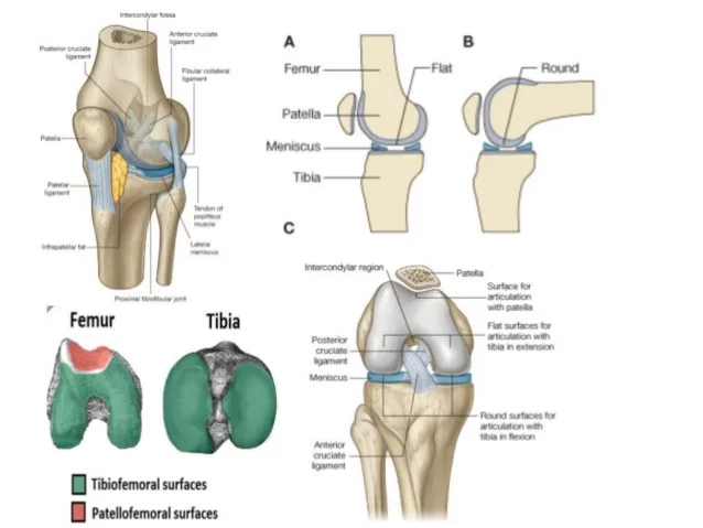
The knee is composed of four bones. The bone that connects the hip to the knee is called the femur, or thighbone. The tibia, or shinbone, connects the ankle and knee. The patella (kneecap) is a tiny bone in front of the knee that rests on the knee joint as it bends. The fibula is a shorter, thinner bone that runs parallel to the tibia on the exterior. The joint functions like a hinge, but with considerable rotation.
The knee is a synovial joint, which means it’s lined with synovium. The synovium generates fluid that lubricates and nourishes the interior of the joint. The smooth surfaces at the extremities of the tibia and femur are referred to as articular cartilage.
- Femur
- It is the weight-bearing bone in the thigh. It serves as an attachment point for the majority of knee muscles.
- Condyle
- The two femoral condyles account for the rounded end of the femur. The tibial (shinbone) meniscus may be effortlessly crossed by the femur due to the smooth articular surface.
- Tibia
- The tibia (shinbone), the body’s second biggest bone, is the weight-bearing bone in the leg. The menisci only partially cover the tibia’s superior surface where it meets the femur. The menisci operate as shock absorbers, preserving the tibia’s articular surface while also helping in knee rotation.
- Fibula
- The fibula, while not a weight-bearing bone, serves as attachment points for the Lateral collateral ligaments (LCL) and the biceps femoris tendon.
- The articulation of the tibia and fibula also allows for a small amount of mobility, allowing some flexibility in response to the activities of muscles attached to the fibula.
- Patella
- The patella (kneecap), which is connected to the quadriceps tendon above and the patellar ligament below, sits on the anterior articular surface of the lower end of the femur to protect the knee joint. The patella serves as a fulcrum for the quadriceps, keeping the tendon off the lower end of the femur.
- Menisci
- The medial and lateral menisci are thin C-shaped fibrocartilage layers that partly cover the surface of the tibia where it connects to the femur. Because the majority of the meniscus lacks a blood supply, it cannot heal from injuries in the same manner as the rest of the body. The menisci operate as shock absorbers, preserving the tibia’s articular surface while also helping in knee rotation. The intact menisci operate as secondary stabilizers, interacting with the ligaments’ stabilizing role and being most effective when the surrounding ligaments are intact.
- Anterior Cruciate Ligament (ACL)
- The ACL is the knee’s principal stabilizing ligament. The ACL connects the tibia (shin bone) and femur (thigh bone) by passing through the center of the knee joint. The ACL stops the femur from slipping rearward onto the tibia. The ACL, together with the PCL, rotates the knee to stabilize it. Thus, if one of these ligaments is severely damaged, the knee will become unstable when planting the foot of the injured limb and rotating, causing the knee to collapse and give way.
- Posterior cruciate ligament (PCL)
- The posterior cruciate ligament (PCL) has received significantly less attention than the ACL due to its rarity of injury.
- The PCL keeps the femur from shifting too far forward over the tibia. The PCL is the knee’s primary stabilizer and is nearly twice as powerful as the ACL. It offers a central axis around which the knee may spin.
- Collateral Ligaments
- Collateral ligaments prevent hyperextension, adduction, and abduction.
- Superficial MCL (Medial Collateral Ligament) links the medial epicondyle of the femur to the medial condyle of the tibia, resisting valgus strain.
- The Deep MCL (Medial Collateral Ligament) connects the medial epicondyle of the femur to the medial meniscus. The LCL (Lateral Collateral Ligament) is independent from the articular capsule and connects the lateral epicondyle of the femur to the head of the fibula, resisting varus force.
Causes of patellar tracking disorders
- Weakness of the thigh muscles
- Increased stress on the kneecap due to twisting knee motions
- Traumatic injury to the knee
- Damage to the cartilage, tendons, ligaments
- Improper exercise technique
- Obesity
- Flat feet
- Structural deformity of the knee
Risk factors for patellar tracking disorder
- Weight – Overweight people use additional pressure on the knee joint and are more likely to develop a patellar tracking problem.
- Patients who run or play sports that require kneeling, knee bending, or jumping are more likely to develop patellar tracking disorder.
- Conditions described in causes — If you have any of the conditions listed in the causes of patellar tracking disorder, you are more likely to develop the problem.
Symptoms of patellar tracking disorder
Symptoms differ according to the severity of the medical condition and include:
- Pain in the front of the knee when walking up or down stairs, bending, or jumping
- Catching or locking of the knee in specific postures
- Walking might produce a popping or grinding feeling.
- Sensation that the knee might collapse at any time
- Swelling around the joint
Diagnosis of Patellar Tracking Disorder
The symptoms of patellar tracking disorder might be confused with those of numerous other medical conditions, making diagnosis challenging. The most typical diagnostic procedure includes:
- Physical examination: Your doctor will ask you how the pain started and what makes it worse, as well as observe and feel the knee when you perform particular activities.
- X-ray: Radiographs of the knee may be obtained to assess any injury to the bone surfaces.
- MRI: This test creates pictures of the body’s soft tissues and can assist in diagnosing knee ligament and tendon damage.
Treatment for Patellar Tracking Disorder
Conservative methods:
- RICE Method: Your physician will advise you to elevate, compress, relax, and apply ice to your knee. This is a frequent way to relieve inflammation and pain.
- Knee taping: This technique includes strapping a specific tape across your knee to prevent improper patella movement.
- Physical therapy: Your doctor will recommend stretching and flexibility exercises to help strengthen the thigh and other muscles surrounding the knee.
Surgery:
- Surgery is typically unnecessary. If your doctor advises surgery to ease your pain, be sure you completely understand why. Be aware that there is contradictory research about the effectiveness of surgery for patellar tracking dysfunction.
- One widely advised operation is to sever the lateral retinaculum, a ligament that supports the patella’s outer border.
- Another operation is the repair of the medial patellofemoral ligament (MPFL), which connects to the inner side of the kneecap and prevents it from slipping outward.
- Both of these may be performed with arthroscopic surgery with a tiny incision. You may need to stay at home for a week or longer following surgery. A full recovery usually takes four to six months to a year.
Exercises for Patellar Tracking Disorder
Stretching Exercises
IT band stretch
How to perform:
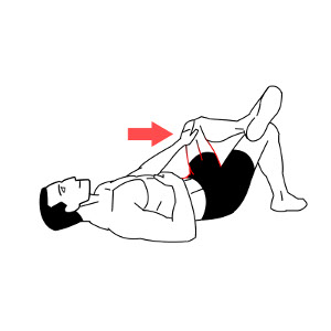
- Lie on your Back (Supine Position).
- Raise your upper leg a few inches over your lower leg while keeping your knee flexed.
- Allowing your upper leg to sweep back a few inches should result in a small stretch on the side of your thigh.
- Hold for thirty seconds.
- On the other side, repeat.
- Five times over, repeat.
Hip flexor stretch
How to perform:
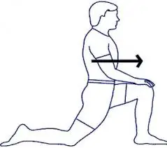
- Kneel on the affected leg and extend your other leg out in front of you while keeping your foot flat on the ground. If you have pain in the front of your knee, place a towel beneath it.
- Keeping your back straight, softly push your hips forward. Your rear leg, hip, and upper thigh should all feel stretched.
- For a minimum of 20 to 30 seconds, hold the stretch.
- Do this two to four times.
Hamstring stretch
How to perform :
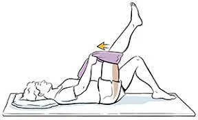
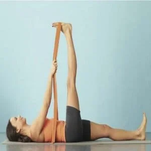
- With your legs outstretched, lie down on the floor or a mat.
- Bend the right leg and place the strap over the thigh of the right foot to stretch it.
- Hold the strap with both hands.
- Maintain the left foot flexed and the leg stretched on the ground. As a result, the thigh and calf should go toward the floor.
- Stretch the right leg slowly while flexing the foot. The right leg should be straight with a little bend at the knee. Your foot should be toward the ceiling at the bottom.
- Gently pull the strap until your hamstrings begin to slightly extend.
- Hold the stretch for 10 to 30 seconds.
- Switch legs. Repeat 2-4 repetitions per leg.
Quadriceps stretch
How to perform:
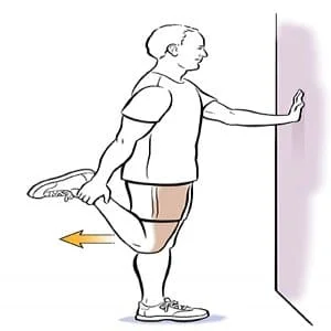
- Stand with your side against the wall, one hand on the wall for balance.
- Keeping your knees and thighs together, elevate your outer foot toward your back while holding it with your outside hand.
- The front of your thighs should feel slightly to moderately stretched.
- After holding for a relaxation breathing cycle, go to the opposite foot.
Calf stretch
How to perform:
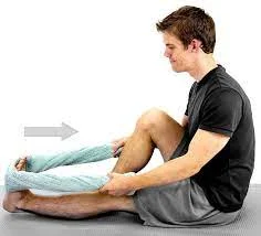
- While seated with your legs straight, wrap a towel around the ball of your foot.
- Keeping your knee straight, draw the towel’s ends towards you.
- Hold for 30 seconds. Repeat three times per side. Three times a day.
Strengthening Exercises
Static quadriceps exercise
How to perform:
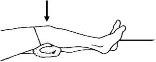
- Sit on the ground or on the plinth.
- Just below your left knee, place a thin, rolled towel.
- Keeping your right foot level on the ground, gently bend your right knee. Now flex your left foot and tense your upper thigh muscles.
- On your right knee, press the cloth on the floor. Push the towel towards you with your toes.
- Avoid arching your back or bending your shoulder joint.
- Hold the knee press for 7-10 seconds. Then relax your leg.
- Repeat this 10-15 times.
Seated knee extension
How to perform:
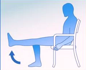
- Sit tall in a strong chair, feet flat on the floor, hands lightly resting on the chair’s sides for support.
- To extend your leg in front of you, raise your foot and slowly straighten one knee.
- Grip the chair slightly at the top as it supports your thigh.
- Lower your foot down with control.
- Repeat 5 to 10 times, then swap legs.
Straight leg raises
How to perform:
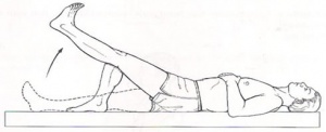
- Lie on your back, hips square, legs comfortable on the floor.
- Put your unaffected leg’s foot flat on the ground and bend its knee to a 90-degree angle.
- Contract your quadriceps to stabilize your straight leg muscles.
- Inhale gently and elevate your straight leg five to six inches off the ground.
- Hold for 3 seconds.
- Breathe out slowly and drop the leg to the floor with control.
- Relax and repeat ten more times.
Mini squats
How to perform:
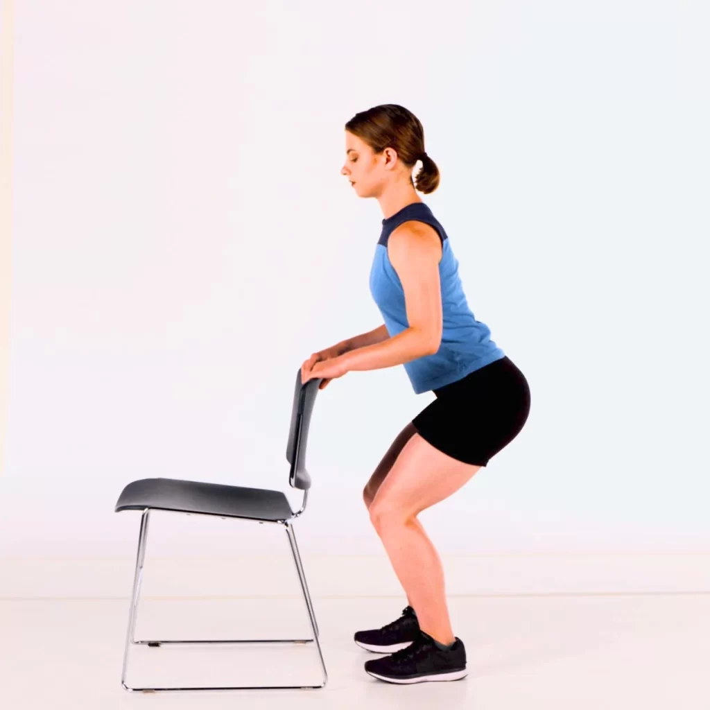
- Start by placing your feet comfortably apart. (For support, you can grasp a chair or a wall.)
- Move your hips back and bend your knees softly, as if you were just getting into a chair.
- Squeeze your thigh muscles while holding this position.
- To return to standing, push through your feet.
Wall sits
How to perform:
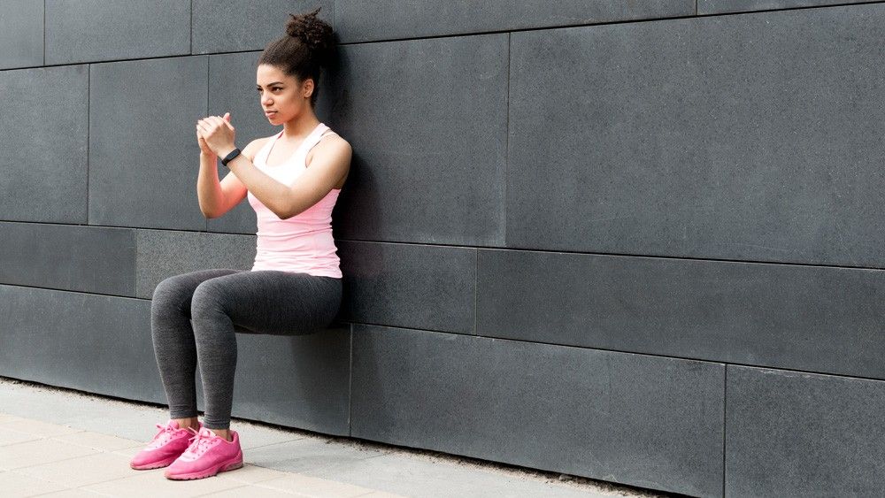
- Stand with your back flat against a wall.
- Your knees and hips should be at a 90-degree.
- Hold as long as possible while keeping appropriate form.
- To go back to the beginning position, push up through your heels.
Clamshells
How to perform:
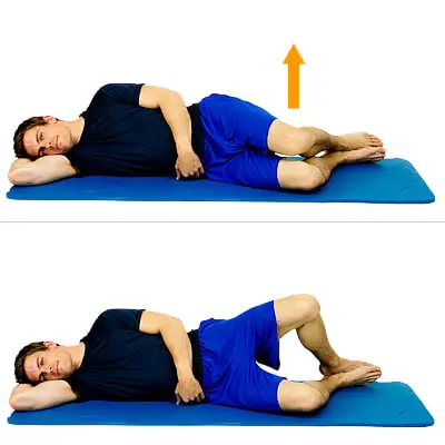
- With your legs elevated and your knees bent at a 45-degree angle, lie on your side.
- Use your upper arm to support your body while resting your head on your lower arm. Because the upper hip tends to bounce backward, make sure your hipbones are stacked on top of one another.
- Pulling in your belly button may assist in stabilizing your spine and pelvis.
- Keep both feet together and raise your upper knee as high as you can raise it without moving your hips or pelvis. Avoid raising your lower leg off the ground.
- After pausing, place your higher leg back on the ground. Perform 20 repetitions on each side.
Side-lying leg raises
How to perform:
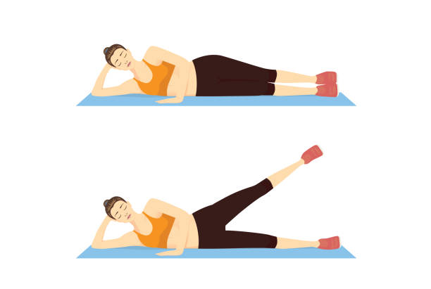
- To do a side-lying leg lift, lie on your side with your torso straight. For added comfort, bend the lower knee slightly.
- Gently brace your abdominal muscles. Without bending backward or twisting your pelvis, slowly raise the higher leg to the side. The movements should be smooth and controlled. Lower your leg with the same control. Breathe normally throughout.
Glute bridges
How to perform:
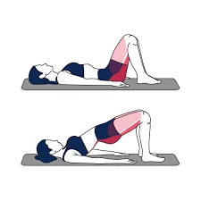
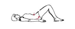
- Lie on the floor with both legs flexed and feet flat on the floor. Your feet should be hip-width apart, toes pointed straight ahead, and heels roughly 6-8 inches from your glutes. With your palms toward the ceiling, put your arms by your sides.
- Squeeze your glutes and abs as you elevate your hips to the ceiling.
- Raise your hips as high as you can without bending your back. Elevate your hips until your shoulder, hip, and knee are in one straight line.
- At the top position, tighten your glutes as much as you can, then hold the posture for two seconds.
- Slowly descend your hips to the floor, maintaining tension in your glutes and core.
Balance and Proprioception Training
Single-leg balance
How to perform:
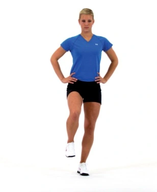
- Stand tall, feet together.
- Keeping the knee slightly bent, slowly raise one foot off the ground.
- Maintain a straight line of sight.
- Hold the posture for 30-60 seconds and then swap legs.
Balance board exercises
Front-to-back
How to perform:
- Slowly tilt the board forward until it reaches the floor.
- Slowly tilt the board back until it reaches the floor behind you. Your arms might be placed at your sides or in front of you.
- Tilt forward and back slowly for 30 seconds.
Side-to-side
How to perform :
- Stand in the initial balance posture.
- Tilt the board slowly to the right, then left.
- Tilt from side to side for thirty seconds.
What Exercises Should You Avoid When You Have Patellar Tracking Disorder?
- Basketball, volleyball, and jumping rope
- High-impact aerobics
- Lunges and deep squats (90 degrees or more)
- Running, especially inclines and trails
- Stair stepper machines
Benefits of Exercises in Patellar Tracking Disorder
- Enhance your kneecap’s alignment so that it remains centered in its groove when you bend and straighten your leg. By doing this, the grinding, popping, and pain that result from pulling the patella off course are avoided.
- Strengthening the particular muscles that control your patella. This includes your glutes and other hip muscles, as well as your quadriceps, particularly the inner portion known as the vastus medialis obliquus (VMO).
- Preventing future injuries: The goal of workouts is to train your body to move intelligently and intentionally. Precision is more important than power or speed. You may educate your muscles to activate in the proper order and stabilize the entire knee joint by moving slowly and methodically.
- Improve knee movement
Prevention of Patellar Tracking Disorder
Maintaining strength, flexibility, and activity can help avoid patellar tracking problems and improve joint health. Here is what Hinge Health’s physical therapists recommend.
- Maintain hip and gluteal strength. These muscles serve to maintain leg posture and knee function.
- Make gradual progress in your activities. Sudden increases in time or intensity may be beyond your knee’s current capabilities. Starting gently and gradually helps your body to adjust and increase capability.
- Pay attention to your movement patterns. Work with a physical therapist to identify and correct movement behaviors that may be contributing to knee pain.
- Address muscular tension. Regularly stretching your hip flexors, IT band, quadriceps, hamstrings, and calves improves flexibility and kneecap mobility.
- Take movement breaks. Breaking up extended sitting keeps your knees lubricated and minimizes muscular tension. Move every 30-45 minutes, even if it’s only fidgeting or performing knee extensions in your seat.
- Wear proper footwear. Supportive shoes can help you maintain better alignment and improve knee joint health, especially while conducting repeated activities like jogging.
When to see a doctor
Patellar tracking issues may typically be improved with focused workouts and at-home therapy. However, if your knee pain is significant, worsening, or interfering with everyday activities, consult a healthcare professional or physical therapist.
It’s also advisable to get treatment if you have:
- Sudden acute pain or popping feeling
- Knee lock or buckling
- Significant edema that does not improve.
- Trouble bending or straightening your knee.
- Bearing weight might be difficult.
- Pain that persists after 4-6 weeks of focused workout
Conclusion
Patellar tracking dysfunction might cause pain and prevent you from participating in your favorite sports or recreational activities. Home cures and physical treatment may typically bring you back to them in a few weeks or months.
If physical therapy isn’t working for you, think about what else may be going on. Other causes of knee pain may exist, including something as basic as your footwear.
Surgery is the final resort. Make sure to consult your doctor and an expert about what is best for you. Before undergoing surgery, get at least a second opinion.
FAQs
Are “runner’s knee” and “patellar tracking disorder” similar?
Yes, both names are frequently used to describe the same problem. Officially known as patellofemoral pain syndrome, “runner’s knee” is a common phrase for the pain that surrounds your kneecap. Patellar tracking disorder is a more specific diagnosis that explains why you’re experiencing pain—your kneecap isn’t sliding properly in its groove. Therefore, even if both show the same pain, one is a symptom, and the other is the mechanical cause.
Is patellar tracking a lifetime issue?
Patellar tracking disorder might be challenging, but be patient. Most patients report feeling better after a few months of therapy. In general, the longer you’ve had this condition, the longer it will take to heal.
Can patellar tracking be corrected without surgery?
Most patellar tracking disorders can be successfully addressed without surgery. Rest, frequent stretching and strengthening exercises, taping or bracing the knee, ice application, and short-term use of nonsteroidal anti-inflammatory medicines (NSAIDs) are all possible non-surgical treatments.
How long does it take to correct patellar tracking?
Patellar tracking can be fixed in a matter of weeks or months. Conservative therapy may reduce initial pain in 1 to 2 months, but it usually takes 1 to 6 months to properly cure the underlying muscle imbalances and return to high-impact activities.
Is running harmful for patellar tracking disorder?
Weak thigh muscles (quadriceps). Imbalanced/tight/loose muscles, ligaments, or tendons in the hip, leg, or foot can induce patellar tracking disorder by producing biomechanical issues. Overuse of the knee, particularly while carrying a significant load through it, such as sprinting or jumping.
How can exercise improve patellar tracking?
Quadriceps strengthening is the most typical therapy for patellar tracking problems. Exercises that promote flexibility and strengthen the muscles surrounding the hip can also assist.
References:
- Clam Shells. (n.d.). [Video]. Hingehealth. https://www.hingehealth.com/resources/articles/patellar-tracking-disorder/
- Hecht, M. (2019a, February 14). About Patellar Tracking Disorder. Healthline. https://www.healthline.com/health/patellar-tracking-disorder#takeaway
- Patellar Tracking Disorder/Patellar Maltracking | Dr Amit Momaya | Orthopedic Surgeon Birmingham | Sports Medicine. (n.d.). https://www.momayamd.com/patellar-tracking-disorder-patellar-maltracking-orthopedic-sports-medicine-specialist-birmingham-huntsville-montgomery/
- McCormick, F. (2025, November 21). 5 key exercises to fix patellar Tracking Disorder. Dr Frank McCormick. https://drfrankmccormickmd.com/5-exercises-fix-patellar-tracking/
- Physio Inq. (n.d.). The Ultimate Guide to Patellar Tracking Disorder. https://www.physioinq.com.au/blog/patellar-tracking-disorder-guide
- https://www.mercy.com/health-care-services/orthopedics-sports-medicine-spine/specialties/knee-leg/conditions/patellar-tracking-disorder
- Knee Surgery Las Vegas | Knee Specialist | Knee Pain Henderson. (n.d.). John Baldauf MD. https://www.baldaufortho.com/normal-anatomy-of-the-knee-joint/