Common Peroneal Nerve
Common Peroneal Nerve Anatomy
The common fibular nerve (common peroneal nerve; external popliteal nerve; lateral popliteal nerve) is a nerve in the lower leg that provides sensation over the posterolateral part of the leg and the knee joint.
It divides at the knee into two terminal branches:
The superficial fibular nerve and deep fibular nerve, which innervate the muscles of the lateral and anterior compartments of the leg respectively. When the common fibular nerve is damaged or compressed, foot drop can ensue.
Description Of Common Peroneal Nerve:
The common peroneal nerve is the smaller and terminal branch of the sciatic nerve which is composed of the posterior divisions of L4, 5, S1, S2.
It courses along the upper lateral side of the popliteal fossa, deep to biceps femoris and its tendon until it gets to the posterior part of the head of the fibula.
It passes forwards around the neck of the fibula within the substance of fibularis (peroneus) longus, where it terminates by dividing into the superficial and deep fibular (peroneal) nerves.
The nerve can be palpated behind the head of the fibula and as it winds around the neck of the fibula. The common fibular (peroneal) nerve gives articular branches to the knee and superior tibiofibular joint.
The lateral cutaneous nerve of the calf supplies the posterolateral side of the proximal two-thirds of the leg. It usually arises in common with the fibular (peroneal) communicating branch which joins the sural nerve in the middle third of the leg.
Branches of Common Peroneal Nerve:
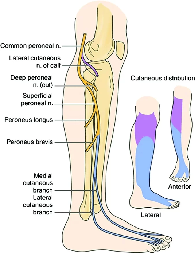
While it still lies in the popliteal fossa, the common peroneal nerve gives off:
- The Genicular branches to the knee joint
- The Lateral cutaneous nerve of the calf
- A sural communicating branch
The two terminal branches includes :
- Superficial peroneal nerve (L5,S1,2)
- Deep peroneal nerve (L4,5,S1,2)
Superficial branch: It runs in and supplies the muscles of the lateral (peroneal) compartment of the leg.
In addition it supplies the skin over the lateral lower two-thirds of the leg and the whole of the dorsum of the foot except for the area between the 1st and 2nd toes, which is supplied by the deep peroneal nerve.
Deep branch: It runs with the anterior tibial vessels over the interosseous membrane into the anterior compartment of the leg and then over the ankle to the dorsum of the foot.
It supplies all of the muscles of the anterior compartment as well as providing a cutaneous supply to the area between the 1st and 2nd toes.
Function of Common Peroneal Nerve:
Nerve roots: L4 – S2
Motor:
- Innervates the short head of the biceps femoris directly. Also supplies (via branches) the muscles in the lateral and anterior compartments of the leg.
Sensory:
- Innervates the skin over the upper lateral and lower posterolateral leg. Also supplies (via branches) cutaneous innervation to the skin of the anterolateral leg, and the dorsum of the foot.
Motor Functions:
- The common fibular nerve innervates the short head of the biceps femoris muscle (part of the hamstring muscles, which flex at the knee).
- In addition, its terminal branches also provide innervation to muscles:
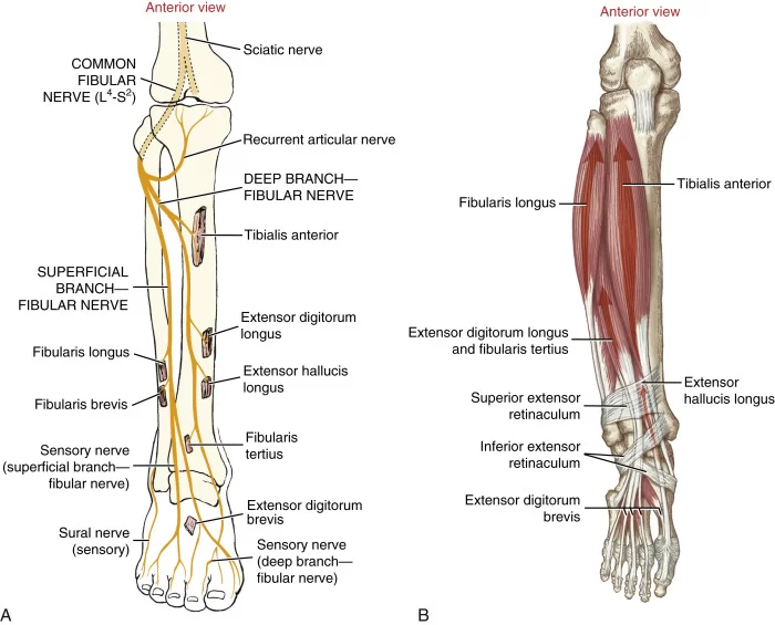
- Superficial fibular nerve: Innervates the muscles of the lateral compartment of the leg; fibularis longus and brevis. These muscles act to evert the foot.
- Deep fibular nerve: Innervates the muscles of the anterior compartment of the leg; tibialis anterior, extensor digitorum longus and extensor hallucis longus.
- These muscles act to dorsiflex the foot, and extend the digits. It also innervates some intrinsic muscles of the foot.
- If the common fibular nerve is damaged, the patient may lose the ability to dorsiflex, evert the foot, and extend the digits.
Sensory Functions:
- There are two cutaneous branches that arise directly from the common fibular nerve as it moves over the lateral head of the gastrocnemius.
- Sural communicating nerve: This nerve combines with a branch of the tibial nerve to form the sural nerve.
- The sural nerve innervates the skin over the lower posterolateral leg.
- Lateral sural cutaneous nerve: Innervates the skin over the upper lateral leg. In addition to these nerves, the terminal branches of the common fibular nerve also have a cutaneous function.
- Superficial fibular nerve: Innervates the skin of the anterolateral leg, and dorsum of the foot (except the skin between the first and second toes).
- Deep fibular nerve: Innervates the skin between the first and second toes.
Anatomical Variations
The common fibular nerve has multiple variations in its route and surrounding architecture, as is the case with most areas of the human anatomy. These variations are important to understand, especially for surgeons who may have to decompress the nerve. A few significant variations were discovered in a study comparing the anatomy of surgically decompressed nerves and cadavers. These variances may also enhance or decrease the likelihood of compressed nerves. Underneath the superficial head of the peroneus longus is a band-like structure made of fibrous tissue in the first one. The following variation also has fibrous tissue that resembles a band, but it is situated on the deep head of the peroneus muscle’s superficial surface.
The following variation likewise has fibrous tissue that resembles a band, however, it is situated on the deep head of the peroneus longus muscle’s superficial surface. The final variation mentioned comprises two muscles together with their unusual origin and junction. Though some people begin together at the fibular head and separate as they move distally, the soleus muscle and the fibularis longus muscle typically originate separately on the fibular head.
Clinical Relevance:
The common peroneal nerve is in a particularly vulnerable position as it winds around the neck of the fibula. It may be damaged at this site by
- Trauma or injury to the knee
- TKA
- Compression of the fibula head during surgery e.g. tourniquet
- Fracture of the fibula
- Fracture to tibial plateau
- Use of a tight plaster cast of the lower leg
- Crossing the legs regularly
- Pressure to the knee from positions during deep sleep or coma
- Patellar dislocations ( 33% chance of nerve damage)
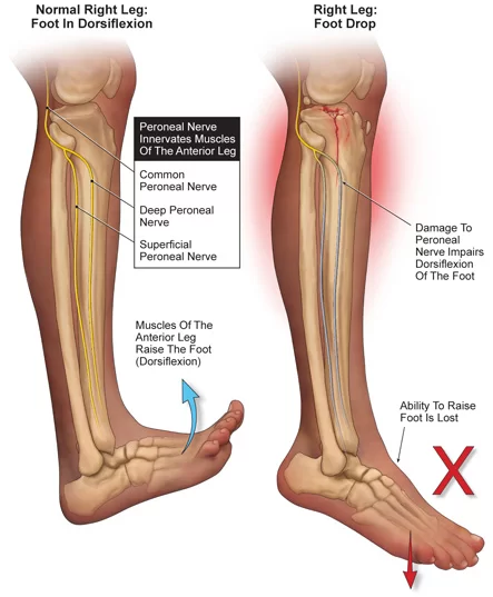
- Common peroneal nerve injury is often seen in people who are very thin, and predisposed to autoimmune conditions such as rheumatoid arthritis.
- Nerve damage secondary to chronic health issues eg Diabetes, Alcoholism, Charcot -Marie Tooth disease, or a significant knee valgus or varus.
- Damage to this nerve is followed by foot drop (due to paralysis of the ankle and foot extensors) and inversion of the foot due to paralysis of the peroneal muscles with unopposed action of the foot flexors and invertors).
- There is also anaesthesia over the anterior and lateral aspects of the leg and foot, although the medial side escapes, since this is innervated by the saphenous branch of the femoral nerve.
- Neuropathic pain can also be a common symptom of peroneal nerve neuropathy which can be managed with analgesia.



8 Comments