
Extensor digitorum longus muscle
What is the Extensor digitorum longus muscle?
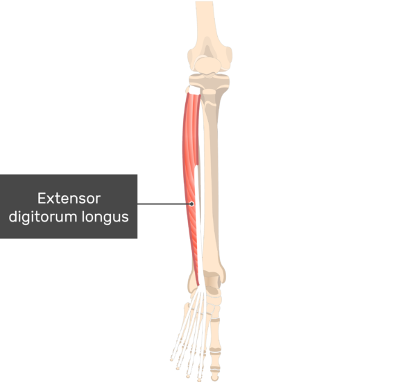
The extensor digitorum longus muscle is a skeletal muscle located in the anterior (front) compartment of the lower leg. It is one of the muscles that make up the deep layer of muscles in the front of the leg.
The muscle originates from the lateral condyle of the tibia (a bone in the lower leg), the anterior surface of the fibula (another bone in the lower leg), and the interosseous membrane (a fibrous tissue that connects the tibia and the fibula). Then it descends and inserts into the toe tendons, more specifically those of the second through fifth toes.
The feather-like extensor digitorum longus (EDL) muscle is located in the anterior (extensor) compartment of the leg. This compartment also contains the tibialis anterior, extensor hallucis longus, and fibularis (peroneus) tertius muscles, in addition to the EDL muscle.
Since this large number of muscles cross the dorsal part of the lower leg joint, their normal capability is the dorsiflexion of the foot. As the extensor digitorum longus moreover crosses the subtalar, metatarsophalangeal, and interphalangeal joints of the foot, it furthermore everses the foot and expands the toes.
Origin of Extensor digitorum longus muscle
The lateral tibial condyle, the upper portion of the anterior surface of the interosseus membrane, and the anterior surface of the fibula are the primary sites of EDL.
Insertion
The ligament of EDL passes under the inferior and superior extensor retinaculum at the anterior surface of the lower leg joint before arriving at its distal insertion.
It divides into four slips that join the proximal and distal phalanges of the foot’s lateral four digits, digits 2 through 5, respectively.
Relations
This muscle lies laterally to the tibialis anterior and extensor hallucis longus, making it the most lateral of all the muscles in the extensor compartment. The extensor digitorum longus and the tibialis anterior are connected by the anterior tibial artery and vein.
The muscle’s distal end crosses the lower leg joint’s anterior side within the inferior extensor retinaculum. While crossing the retinaculum, the ligament of the extensor digitorum longus sits medially to the ligament of fibularis tertius and horizontally to the extensor hallucis longus ligament. The extensor digitorum brevis muscle is connected to the tendons of the muscle by a superficial course in the dorsum of the foot.
Innervation
The deep fibular nerve (L5, S1), a branch of the common fibular nerve, innervates the extensor digitorum longus.
Blood supply
The leg part of the muscle is provided by two veins of the leg; The anterior tibial artery supplies the proximal portion, while the fibular artery supplies the distal portion.
The anterior lateral malleolar, lateral tarsal, metatarsal, plantar, and digital arteries vascularize the tendons of the muscle.
Function of Extensor digitorum longus muscle
Extending the lateral four toes at the metatarsophalangeal joint is the essential function of the extensor digitorum longus. This implies that while acting freely, it can’t expand the whole length of the toes, extending just at the metatarsophalangeal, while at the interphalangeal joints, the toes remain flexed. In any case, contracting along with lumbricals which are the principal extensors of the interphalangeal joints, this muscle adds to expansion at each joint between the bones of the horizontal four toes.
Acting in collaboration with the tibialis anterior, extensor hallucis longus, and fibularis tertius, this muscle takes part in the dorsiflexion of the foot when its proximal connections are fixed. At the point when the distal connections are fixed and the body is in the physical position, every one of the four muscles carries the storage compartment and lower limb to the front. The body weight-bearing point is moved from the proximal to the distal part of the foot by this action.
The gait cycle depends on each of these actions; The toe extension keeps the toes extended until the heel touches the ground, while the dorsiflexion angulates and lifts the foot above the walking surface.
Clinical relevance
To make up for a tibialis anterior that is inhibited, the extensor digitorum longus tends to be overactive and tight. Extending and myofascial release of the muscle along with activation tibialis anterior is shown to recover muscle balance and further develop functional lower leg dorsiflexion.
Extensor digitorum longus muscle stretching
Kneeling Shin Stretch
The shins can be gradually stretched by kneeling.
You should have great knee flexion to play out this stretch as you will be sitting on the heels. Do not perform it if it causes knee pain.
Bow on a mat with the highest points of the feet level on the ground and the buttocks over the impact points.
You can feel the shin stretching.
Maintain for 10-20 seconds.

Seated Shin Stretch
Relax in a chair.
As in the standing stretch, lower your knee so that the toe of your foot is extended into the ground.
Similar to the standing stretch done seated, slowly pull forward with the toe on the floor.
Maintain for 10-20 seconds.
Continue with both feet.

Extensor digitorum longus muscle strengthening
Seated Elastic Band Exercise
An elastic resistance band is required for the seated Elastic Band Exercise.
You must sit on the floor with one leg extended in front of you to perform the seated elastic band exercise. You can also sit in a chair with your foot propped up on another chair right now.
An ankle is covered with a resistance band. Join one finish to a steady item like the leg of a table, and secure the other around the foot close to the toes. So that the heel of the foot doesn’t rub against the floor, it might be helpful to rest the lower leg on a small pillow.
Pull the toes and foot up while keeping the knee extended for this exercise. As you flex the foot up, the ankle may move. Pull the foot up as much as you can and hold the end position for two seconds.
Back to the starting position, slowly relax.
Perform this exercise for 10 to 20 repetitions or until you are unable to flex your ankle up and the anterior tibialis muscle has worn out.
Cuff Weight Exercise
For the Cuff Weight Exercise, you want a cuff weight.
A cuff weight is a cushioned weight that you can fold over the lower leg. You must sit in a chair and wrap a cuff weight around your toes for the Cuff Weight Exercise. Verify that it is safe. Sitting with the cuff weight on the foot and the foot resting on the floor, begin the exercise by flexing the ankle so that the foot and toes move up toward the knee.
Hold the position for two seconds when the foot is flexed up. Gradually lower your toes back to the starting position.
Recount the activity for 10-20 counts.
Isometric Exercise
A form of exercise known as isometric exercise involves pushing against an immovable object. It can help strengthen the muscle in particular ankle ROMs and is simple to perform.
To do isometric anterior tibialis boosting you need to Sit in a seat and rest.
With the affected leg on the bottom, cross one leg over the other.
Put the foot that you want to exercise on top of the ankle.
Place your weak foot’s top into the other foot’s sole. To resist it, press down with the powerful foot. Keep in mind that the foot, not the ankle, controls movement.
After remaining in this position for five seconds, slowly release. Two to three times per day, complete the exercise for approximately 10 to 15 times. Muscles can be strengthened with isometric exercise, but strength only comes from exercising in a particular range of motion (ROM). As a result, you can perform the exercise with a variety of ankle positions.
FAQ
Where is the extensor digitorum longus ache?
The most typical symptom of foot extensor tendonitis is pain in the top of the foot. Normally, it is right close to your shoestrings. This ache may be sensed while running or walking. On an injured or inflamed extensor tendon, there may be visible swelling or a bump.
What causes pain in the longus extensor digitorum?
Overuse is thought to be the cause, as it can result in minor tendons injuries that can be painful. The normal healing process slows down as a result of the micro-trauma and repetitive stress, resulting in a painful forefoot/extensor tendon.
How can the extensor digitorum longus be relaxed?
Plantarflexion and inversion of the foot stretch the extensor digitorum longus. Balance can be achieved by using a table or wall. Plantarflexion and inversion of the foot stretch the extensor digitorum longus. Balance can be achieved by using a table or wall.
How should you massage the longus extensor digitorum?
Place the ball underneath the muscle while sitting cross-legged. Use your hands to apply pressure to the foot, but only if you need more. You can sit bending only the leg you are massaging if you are unable to sit cross-legged.


3 Comments