Laryngeal Vein
Introduction
The laryngeal veins are important additives of the venous drainage device of the larynx, which is a key discernment within the respiration and vocal tools of the human structure. These veins are trustworthy for amassing deoxygenated blood from the larynx and ferrying it back to the heart through massive venous channels.
The laryngeal veins play a critical function inside the circulatory system by way of draining blood from the larynx, an organ situated within the anterior fragment of the neck, which houses the vocal cords and is worried in respiration, sound presentation, and shielding the trachea against feasts aspiration.
What is the anatomical location of the nasal cavity?
The lymph nodes are positioned within the throat and represent a full-size factor of the human lymphatic drainage machine. This is a radical map in their real location.
Anatomic place of the laryngeal vein
Every location: The larynx, which is located in the front of the throat, is close to the vocal cords, which are associated with voice and breathing in anatomical structure.
Elevated pulmonary arteries:
- Location: The human bronchial roof is where the superior bronchial tubes end.
- Route: It passes through the thyroid gland and the superior pulmonary artery. It also controls blood flow from the apex vocal twine veins.
Lower thoracic nerve:
- Location: The decreased portion of the lungs is dehydrated by the inferior pulmonary arteries in human
- Route: It passes through the inferior pulmonary artery, lies beneath the cricoid cartilage, and also collects the blood from the pulmonary arteries and veins
Water Supply Systems:
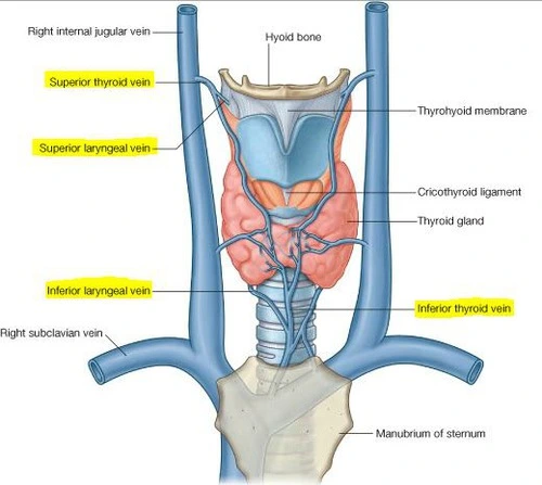
- The superior thyroid vein and the superior laryngeal vein typically connect in the human anatomy.
- The inferior pulmonary artery usually communicates with the brachiocephalic artery and drains into the inferior thyroid artery.
Relationships with other systems:
- Proximity: The lymph nodes are nearly affiliated with the thyroid gland and the spinal cord of the human body. They are usually found in the vicinity of the recurrent lung.
- Comparable muscular condition: usually connected by their associated muscles (caudal and brachial) and located next to or near these muscles
Understanding the anatomical location of the nasopharynx is critical for medical procedures, including surgery involving the thyroid gland and larynx, in the diagnosis and treatment of conditions affecting the laryngeal region.
How does the laryngeal vein unite with further veins in the neck?
The laryngeal veins hook up with other veins in the neck via a network that enables the return of deoxygenated blood from the larynx to systemic circulation. Here’s how the laryngeal veins combine with the venous machine of the neck:
Connections of the Laryngeal Veins
Superior Laryngeal Vein:
- Drainage: The superior laryngeal vein distributes blood from the upper larynx in the human body.
- Combination of features: Superior thyroid vein- The anterior laryngeal vein commonly drains into the advanced thyroid vein in the human frame.
which is an integral part of the internal jugular vein?
The thyroid gland: The superior thyroid gland also innervates various types of glands in humans. The internal jugular vein is a giant vessel that produces blood from the top and neck to the heart.
Inferior Laryngeal Vein:
- Drainage: The inferior laryngeal vein collects the blood from the decreased portion of the larynx.
Connections:
- Inferior Thyroid Vein: Usually, the inferior laryngeal vein becomes dehydrated the inferior thyroid vein. The inferior thyroid glands are the main streams of venous goback from the thyroid gland and adjacent structures of the human body.
- Brachiocephalic Vein: The brachiocephalic vein, moreover called the innominate vein, receives drainage from the inferior thyroid vein. The heart returns blood to the normal atrium of the heart The superior vena cava has long been established as a useful junction of the left and right brachiocephalic vessels.
Overall Venous Return Pathway:
Laryngeal Veins → Superior/Inferior Thyroid Veins → Internal Jugular/Brachiocephalic Veins → Superior Vena Cava → Right Atrium of the Heart
Clinical Relevance:
- Surgical Considerations: Understanding these connections is critical during surgical procedures regarding the thyroid gland or larynx to avoid inadvertently disrupting venous drainage, which may cause headaches.
- Diagnostic Procedures: Assessment of venous return through imaging studies can help diagnose many conditions, such as ulcers, blood clots, irritation affecting pulmonary arteries or related systems
- These vessels allow blood from near the lungs and surrounding areas to go green, adding to the high flow of blood in the system.
Are there multiple laryngeal veins, and if so, how are they distributed?
Yes, there are a couple of laryngeal veins, and they may be generally distributed into superior and inferior laryngeal veins. Here’s how they’re organized and disbursed:
Distribution of Laryngeal Veins
Superior Laryngeal Veins:
- Number and Distribution: There are commonly one or more advanced laryngeal veins on each facet of the larynx.
- Drainage Area: These veins more often than not drain blood from the higher part of the larynx, inclusive of the supraglottic region (the place above the vocal cords).
- Pathway: The superior laryngeal vein is situated inside the thyroid cartilage and runs parallel to the advanced laryngeal arteries. The advanced thyroid gland, which empties into the inner jugular vein, is wiped out via the way of the superior pulmonary artery in human beings.
Lower thoracic nerve:
- Number and distribution: There are commonly one or inferior veins on the person aspect of the trachea
- Drainage Site: This gland drains the narrowed part of the larynx, consisting of the infraglottic space (the region under the vocal cords).
- Procedure: The inferior pulmonary arteries are removed and placed above and below each cricoid artery. In humans, the inferior thyroid vein drains into the inferior vena cava before reaching the brachiocephalic vein.
All the nodes in the anxiety machine:
The superior pulmonary artery supplies the inner pulmonary artery and the upper pulmonary artery. The inferior vein interacts with the brachiocephalic vein via draining the inferior thyroid vein.
Variation and Clinical Relevance:
- Anatomic Variation: While the overall shape is relatively constant, the number and precise pattern of nerve fibers can vary. For instance, new veins or changes may also occur in the drainage areas.
- Clinical issues: Surgeons and physicians have to be privy to those adjustments to avoid complications in the course of surgical operation regarding the thorax or thyroid gland. Correct identification of those vessels is vital to save you excessive bleeding or inadvertent harm for the duration of surgery or examination.
- Understanding the distribution of lymph nodes and their role in revascularization contributes to understanding and making plans for medical techniques associated with the lymphatic system.
What is the laryngeal vein’s main purpose?
The number one function of the laryngeal veins is to facilitate the venous drainage of deoxygenated blood from the larynx lower back to the heart. This manner is important for retaining the right blood movement and ensuring that the tissues of the larynx acquire adequate oxygenation and vitamins. Here’s a greater outlook on the function:
Primary Functions of the Laryngeal Veins
The number one feature of the pulmonary artery is to return blood with low oxygen content material from the lungs to the heart. This manner is crucial for correct stream and to make sure that the lung tissue gets good enough oxygen and vitamins Here is an in-depth evaluation of this system.
Basic features of pulmonary arteries
Urinary tract:
- Blood Collection: The pulmonary arteries accumulate oxygenated blood from lung structures which include arteries, veins, and other tissues
- Supply to massive vessels: This blood is pumped to huge vessels including the advanced and inferior thyroid vessels, and then to the inner jugular and brachiocephalic veins
Recurrent root device:
- Continuous blood waft: By efficaciously pumping blood from the pulmonary location, the pulmonary artery helps maintain balance and average integrity in the head and neck nervous machine
- Improve circulation: Good venous movement helps to return blood to the heart, ensuring that blood from the lungs does now not weaken the tumor or motivate complications.
Supportive lung function:
- Supply of oxygen and nutrients: Good circulation helps maintain the optimal condition of the jaw muscles, which are essential for their normal function, including voice production and breathing
- Preventing edema: Good drainage prevents blood clots in the lungs and prevents the formation of edema, which can otherwise compromise lung function and overall respiratory health.
Clinical Relevance:
- Surgical Considerations: During surgeries entangling the larynx or thyroid gland, consisting of thyroidectomy or laryngectomy, cautious managing of the laryngeal veins is fundamental to avoid headaches related to venous drainage.
- Diagnosis and Treatment: Issues with the laryngeal veins, together with thrombosis or obstruction, also can affect laryngeal function and can want to be addressed to prevent headaches like vocal wire dysfunction or respiration difficulties.
In terms of analysis, the main purpose of the pulmonary arteries is to guarantee the effective return of blood depleted of oxygen from the lungs to the heart. This maintains arterial blood flow and pulmonary circulation, enabling human muscles to work as intended.
What feature do capillaries play within the circulatory gadget?
In laryngeal venous drainage, the laryngeal veins accumulate oxygenated blood and convey it to the top veins, wherein it’s far then reciprocated to the heart. This device’s form describes its contributions in terms of intensity.
Muscles from the lungs help
- Collecting blood lost in oxygen:
- From the pulmonary arteries: The pulmonary arteries are used to pump blood from the lungs to the mucosa lining the larynx and the muscles that feed the vocal cords. They then travel into the surrounding tissue. In the laryngeal venous drainage, the laryngeal vessels keep and deliver oxygenated blood to the high-quality vessels, which ultimately pump the blood back to the heart. This contribution is fully explained by the architecture of the system.
The vascular secretions from the lungs’ contribution
- Collection of deoxygenated blood:
- From the pulmonary arteries: The pulmonary arteries move into the surrounding tissue after collecting blood from various areas of the lungs, such as the lymph nodes that supply the vocal cords and the diaphragm, which covers the lungs.
Areas Drained:
- Superior Laryngeal Veins: Drain the upper parts of the larynx, such as the supraglottic vicinity (above the vocal cords).
- Inferior Laryngeal Veins: Drain the lower parts of the larynx, which include the infraglottic area (underneath the vocal cords).
Connection with Larger Venous Structures:
Superior Laryngeal Veins:
- Pathway: These veins commonly drain into the superior thyroid vein in the human body.
- Extra Drainage: The superior thyroid vein empties into the inner jugular vein, a large vein that supplies blood to the heart from the head and neck.
- Pathway: Inferior Laryngeal Veins The inferior thyroid vein is regularly in which these veins empty.
- Additional Drainage: The inferior thyroid vein is joined with the resource of the brachiocephalic vein, moreover referred to as the innominate vein, before it merges with the superior vena cava.
- The superior vena cava supplies blood to the coronary coronary heart’s appropriate atrium.
Facilitation of Venous Return:
- Maintaining Circulatory Balance: By successfully draining blood from the laryngeal area, the laryngeal veins assist regulate venous stress and prevent congestion in the larynx and surrounding systems.
- Supporting Functions: Proper drainage supports the health and characteristics of the oral mucosa, making sure of oxygen and nutrient intake and preventing edema (infection) and other situations that could impair lung function.
- Connecting the arteries:
- Venous network: The laryngeal vein is an in-depth venous community that includes the superior and inferior thyroid veins, the internal jugular veins, and the brachiocephalic veins This community guarantees that blood from the lungs will touch veins if it comes returned properly in the head and neck.
Clinical Relevance:
- Surgical and Diagnostic Considerations: Knowledge of oral mucosa and function is important in surgical procedures involving the spleen or thyroid to avoid complications such as insufficient blood flow or obstruction Furthermore, conditions affecting these muscles following thrombosis or infection, may affect lung function and normal manipulation Important
To put it simply, the laryngeal veins aid in venous drainage by collecting blood from the larynx and directing it into bigger vein systems that transport blood from the lower back to the heart. This strategy is vital for preserving the right blood flow and making sure of the healthiness and features of the laryngeal tissues.
What connection exists between the laryngeal vein and the larynx’s blood supply?
The interchange of the laryngeal veins with the larynx’s arterial supply is necessary for maintaining adequate blood flow and making sure that the laryngeal tissues acquire the essential vitamins and oxygen. Here’s a detailed rationalization of the manner these veins and arteries interact:
Interaction Between Laryngeal Veins and Arterial Supply
Arterial Supply:
- The superior laryngeal artery feeds blood to the upper portion of the larynx as well as the mucosa tissue and muscular tissue. It is a branch of the advanced thyroid artery, which is a branch of the outer carotid artery.
- Inferior Laryngeal Artery: The inferior laryngeal artery in people is a part of the inferior thyroid artery. (a branch of the human subclavian artery of the thyrocervical trunk). In people, it elements blood to the lower part of the larynx.
Venous Drainage:
- Superior Laryngeal Vein: Drains blood from the better larynx, just like the place provided with the useful resource of the advanced laryngeal artery.
- Inferior Laryngeal Vein: Drains blood from the decreased larynx, similar to the area provided by using the use of the inferior laryngeal artery
Blood Flow Dynamics:
- Oxygen and Nutrient Delivery: The arterial delivery offers oxygenated blood and vitamins to the laryngeal tissues. The laryngeal veins then accumulate the deoxygenated blood from the only tissues and ship it all over again to the coronary heart.
- Efficient Drainage: Each laryngeal vein commonly accompanies its corresponding artery, making sure that the deoxygenated blood is effectively eliminated from the region in which received arterial blood is introduced.
Coordination of Arterial and Venous Systems:
- Concurrent Supply and Drainage: The superior laryngeal artery and vein paintings in tandem, as do the inferior laryngeal artery and vein. This coordination guarantees that blood is efficiently brought to and eliminated from the laryngeal tissues.
- Regulation of Blood Flow: The proper feature of every arterial and venous system allows for holding suitable blood with the float and strain inside the laryngeal place. Any imbalance or obstruction on this gadget can cause headaches, together with edema or impaired laryngeal functions.
Clinical Considerations:
- Surgical Procedures: During surgeries regarding the larynx or thyroid, information on the relationship between those arteries and veins is critical. Care has to be taken to keep away from damaging these vessels to save you from immoderate bleeding and ensure proper recuperation.
- Pathological Conditions: Conditions collectively with tumors or vascular abnormalities will have an impact on the stability of arterial supply and venous drainage, probably critical to symptoms and symptoms and signs and symptoms and signs like swelling or voice adjustments.
Summary: The laryngeal veins and arteries interact carefully to make sure that the laryngeal tissues achieve a regular supply of oxygenated blood and that deoxygenated blood is correctly decreased lower back to the coronary heart.
The superior veins and inferior veins drain the blood from properly-organized locations via the connective tissue, making coordinated and efficient blood paintings within the lungs This communication is necessary for lung component regularity and regular circulatory health of the human body.
What are not unusual conditions or diseases that can affect the laryngeal vein?
Many conditions and diseases can affect the pulmonary glands, affecting their properties, and are certainly the main cause of headaches. Here are a few not uncommon things that can be related to pulmonary arteries:
Thrombosis of bleeding
- Justification: A thrombosis is a blood clot that can obstruct an artery and cause blood flow problems.
- Effects on pulmonary arteries: Damage to the arteries supplying the lungs due to thrombosis in the pulmonary arteries may result in edema, discomfort, and elevated lung pressure.
- Associated Conditions: These may include conditions where the thyroid is the primary site of malignancy, disorders involving excessive bleeding, or serious infections.
- Symptoms:
- Swelling: Localized swelling in the neck or laryngeal location because of impaired venous drainage.
- Pain: Discomfort or ache in the laryngeal province.
- Voice Changes: Hoarseness or changes in voice are extraordinary due to pressure on the vocal cords or surrounding structures.
- Difficulty breathing: Breathing can be affected if thrombosis causes swelling or severe obstruction.
Different types of products
- Explanation: Veins that exhibit abnormal dilation are known as varicose veins.
- Effects on pulmonary arteries: Increased pressure in the pulmonary arteries can cause edema, which in turn can cause bleeding, voice changes, or difficulty breathing
Associated Conditions: Often visible inside the context of portal hypertension or systemic venous congestion.
Symptoms:
- Bleeding: Varices can lead to spontaneous bleeding or bleeding with minimum trauma, especially if they rupture.
- Swelling: Observable swelling inside the laryngeal place because of elevated venous strain.
- Voice Changes: a trade-in voice or hoarseness brought on by laryngeal system compression.
Inflammation (Phlebitis)
- Phlebitis (Inflammation): Phlebitis, a common term for muscle infections, is a result of infectious or inflammatory illnesses.
- Effects upon pulmonary tissue: Inflammation can cause aches, impaired muscle homes, and infection within the proximal lung.
- Related occasions: These will be linked to infections that affect tissues or autoimmune sicknesses.
Signs:
- Burning and redness: Localised burning and redness inside the throat might also develop if the infection is intense.
- Inflammation: Prolonged dehydration and irritation motivate swelling in the lungs.
- Pain: The affected vicinity is smooth and hurts.
- Systemic signs and symptoms: fever, lightheadedness, and dizziness if the illness is the source of the contamination
Tumors or Masses
- Explanation: Lung tissue may also be invaded or compressed by tumours or growths in or across the lungs.
- Effects on pulmonary arteries: The tumor can block or attack the lung arteries, interfering with daily blood flow and causing symptoms such as wheezing, stiffness, or difficulty breathing
- Related conditions: Lung cancer or thyroid cancer May involve lung glands.
Symptoms:
- Hoarseness: A persistent or progressively developing condition brought on by invasion or compression of the laryngeal muscles.
- Respiratory Problems: Airway obstruction, especially shortness of breath (respiratory issue).
- Lump: A lump or mass located everywhere inside the throat or larynx.
- Pain: Soreness or pain when the tumor is causing pressure or sharp changes in the proximal end of the diaphragm.
Trauma
- Description: Damage to the veins can also result from bodily trauma to the larynx or neck.
- Impact on the Larynx: Trauma can modify the residences of the laryngeal veins further generating thrombosis, hemorrhage, or infection.
- Associated Conditions: Unintentional harm, surgical techniques, or precise trauma circumstances
Signs:
- Discomfort and swelling: Following an injury, excruciating neck or chest discomfort and swelling
- Bleeding: Pulmonary embolism or sporadic bleeding.
- Vocal Changes: Vocal twine or surrounding nerve injury ensuing in hoarseness or distortion of the voice.
- Complications associated with respiratory: A clogged or injured airway is viable.
Surgical Complications
- Description: A Strategy concerning the thyroid or larynx can also inadvertently affect the laryngeal veins.
- Impact on Laryngeal Veins: Surgery may additionally result in bleeding, thrombosis, or damage to the veins, probably generating difficulties like swelling or impaired venous go back.
- Associated Conditions: Complications from thyroidectomy, laryngectomy, or unique neck surgical approaches.
Symptoms:
- Bleeding: Postoperative bleeding within the neck or laryngeal location.
- Swelling: Edema in the laryngeal area or neck.
- Voice Changes: adjustments to voice or vocal cord qualities after surgical operation.
- Infection: Signs of contamination, consisting of fever and redness, if the surgical internet web page online becomes infected.
Infections
- Description: Infections of the lung region or adjacent structures might also affect the nerves.
- Effects on pulmonary arteries: Infection can cause infection, irritation, and thrombosis of the pulmonary arteries.
- Associated conditions: pneumonia, cellulitis, or sore throat.
Symptoms:
- Pain and Tenderness: Localized pain or tenderness in the laryngeal place.
- Swelling: Oedema caused by contamination inside the neck or larynx.
- Fever: Fever-like symptoms occur when an infection is mild or systemic.
- Voice changes: If the vocal cords or larynx are compromised, it may be difficult to talk.
Hospital management:
- Diagnosis: Diagnostic imaging (together with ultrasonography and CT scans) and clinical examination are used to diagnose issues affecting the pulmonary vasculature
- Treatment: Depending on the underlying sickness, treatment options might also include medications (together with anticoagulants), surgical procedure, or monitoring for related issues to be had
Methods of analysis:
- Imaging: Techniques such as ultrasound, CT, and MRI scans can be used to visualize lung tissue and be aware of abnormalities
- Endoscopy: Direct imaging of the lung and related systems may be used for prognosis.
- Blood tests: In case of the presence of lymphocytic leukemia or infection, appropriate blood exams can be completed to evaluate the honor of the clot or symptoms associated with infection
- In the end, thrombotic episodes, tumors, infections, and different issues can all affect the laryngeal muscle organizations. Each condition can affect muscle characteristics and health, especially signs and symptoms and ability challenges that require appropriate prognosis and remedy.
What diagnostic techniques are used to evaluate the health of the laryngeal vein?
Diagnostic techniques can also be used to evaluate the lung tissue and identify manageable troubles, these strategies are popular in monitoring coronary heart price, and blood drift, and detecting any abnormalities. A comprehensive explanation of the diagnostic procedures utilized is furnished below.
Imaging strategies
- Ultrasound has been operated directly to identify pulmonary veins and monitor blood drift.
- How it really works: It pieces together pictures of the encompassing regions and tissues through using high-frequency sound waves
- It gives noninvasive benefits, affords actual-time imaging, and might degree blood glide.
Computed Tomography (CT) Scan:
- Purpose: Give a simple cross-sectional illustration of the throat and lungs.
- Benefits: Offers high-resolution imaging and makes it simpler to pick out tumors, structural changes, or severe neurologic abnormalities.
MRI stands for magnetic resonance imaging.
- Goal: Offers pulmonary arteries and tender tissues with high selection visualization.
- In fact, it gathers sure pictures employing strong magnetic fields and radio waves. An MRI can examine the tendon’s structure and ability pathologies.
- Benefits: Outstanding for figuring out the dimensions of tumors or other lesions and for looking the touchy tissue community.
Endoscopic Examination
Laryngoscopy:
- Purpose: The reason for laryngoscopy is to reveal the lung and its surroundings without delay.
- How it works: The lungs are evaluated through the nose or mouth with a blunt or blunt endoscope within the human body
- It allows us to estimate the fame of the pulmonary techniques, along with the vocal cords and any edema
- Advantages: Provides clean specimens and can be used for biopsy if necessary.
Doppler studies
- Ambition: Follow Doppler and confirm with it to check his nostril space blood seepage.
- How it works: A Doppler ultrasound that measures circulation velocity and contraction, allowing you to see obstructions that arise on examination or issues that may be associated with bleeding.
- Benefits: Provides circulatory information and can stimulate vascular abnormalities in the skin.
Venography
- Venography serves the purpose of visualising and identifying venous abnormalities.
- How It Works: After injecting an assessment dye right into a vein, the veins are seen on X-ray snapshots. This technique is an awful lot less generally used for the laryngeal veins however may be beneficial in the best instances.
- Advantages: provides accurate images of the vein system and may be useful in understanding problems in the structure or clotting.
Blood Tests
- Purpose: To study the general fitness of the patient and discover conditions that might affect the veins.
- How It Works: It includes tests that include complete blood rely on number (CBC), coagulation profiles (e.g., PT, aPTT), and particular markers for infection or contamination.
- Edges: Provides supportive statistics on concerns like thrombosis or infections that might also affect the laryngeal veins in humans.
Biopsy
- Ambition: the cause of a biopsy is to research tumors or illnesses concerning the lung tissue of the human.
- How it works: The Tissue instances are accumulated employing endoscopic or surgical procedures and analyzed for cancerous or normal cells.
- Advantages: providing a thorough diagnostic for ailments like lung cancers.
Summary- These diagnostic treatments offer a thorough approach encompassing imaging, visualization, blood waft assessment, and tissue evaluation in assessing the fitness of the laryngeal veins. The desire for technique relies upon the scientific presentation, the suspected condition, and the need for particular information. Proper analysis is important for powerful remedy and control of conditions affecting the laryngeal veins.
What surgical procedures might involve the laryngeal vein?
The laryngeal veins can be concerned at once or in a roundabout way in a variety of surgical operations. The anatomy of the laryngeal veins is important to prevent troubles throughout these operations, which normally target the thyroid gland, the larynx, or other organs. This is a thorough overview of novel surgical techniques that may potentially impact the veins within the larynx.
Thyroidectomy
Thyroidectomy Description: During a thyroidectomy, the thyroid gland is surgically removed, both totally or in element. Engagement of the larynx veins: The laryngeal veins are closely associated with the superior and inferior thyroid veins, which similarly drain into the brachiocephalic and internal jugular veins. Those veins must stay open at some stage in thyroidectomy to prevent excessive bleeding and ensure adequate venous drainage.
Laryngectomy
- Laryngectomy Description: Most advanced laryngeal malignancies are surgically eliminated from the larynx.
- Participation of Laryngeal Veins: As a factor of the circulatory delivery to the larynx, the laryngeal veins are currently impacted.
- Surgeons have to carefully control those veins to govern bleeding and ensure surgical consequences.
Thyroid biopsy or exceptional-needle aspiration (FNA)
- Description: Methods of acquiring samples from the thyroid gland for prognosis.
- Involvement of thyroid glands: Although much less invasive, those methods can connect or not directly have an effect on the thyroid gland and oral mucosa and need appropriate control to lessen bleeding and save you tissue damage
Parathyroidectomy
- Description: Remove a parathyroid gland or glands, which may be located near the thyroid.
- Involvement with Laryngeal Veins: Similar to thyroidectomy, this technique calls for careful management of the superior and inferior thyroid veins, that are connected to the laryngeal veins.
Endoscopic Procedures
- This endoscopic treatment involves the endoscopic excision of oral tumors or foreign substances.
- Lung Muscle Involvement: To avoid unintentional harm, precision is crucial during endoscopic procedures as devices may come into contact with or even be near the pulmonary muscles.
Laser Surgery for Vocal Cord Lesions
- Description: Laser technology is used to remove lesions from the vocal cords or vocal cords.
- Involvement of pulmonary arteries: Instructions regarding pulmonary arteries should be given with caution to avoid blood clots or loss
Surgical intervention for nasopharyngeal stenosis or stenosis
- Definition: Accurate suctioning or suctioning techniques that prevent respiratory or vocal symptoms.
- Involvement of the pulmonary artery: Surgical advances may require running close to the pulmonary artery, which should be well tolerated to prevent complications
Dissection of the throat
- Description: Usually finished for head and neck cancer, this procedure involves removing tissue surrounding blood arteries from the neck.
- Pulmonary artery involvement: This procedure may require a proximal incision and needs care to avoid excessive bleeding or damage to the pulmonary artery
Tracheostomy
- Description: The surgical creation of an opening inside the trachea to offer an airway.
- Involvement with Laryngeal Veins: While often focusing on the trachea, the process may additionally contain the laryngeal area and its veins, especially if complications arise or if there is substantial anatomical disruption.
Surgical Considerations:
- Anatomical Knowledge: Surgeons have to have an intensive understanding of the laryngeal vein anatomy to keep away from unfavorable these vessels at some stage in surgical operation.
- Intraoperative Management: Techniques like careful hemostasis, use of cautery, and meticulous dissection are employed to manipulate and preserve the laryngeal veins in the course of surgical treatment.
- Postoperative Care: Management of headaches together with bleeding, swelling, or voice adjustments is vital within the postoperative length to restore some ordinary function
In summary, many surgical approaches, mainly the ones related to the thyroid, lung, or throat vicinity, can also involve pulmonary arteries. Understanding and coping well with their frame all through surgical operation is critical to save you headaches and ensure outcomes.
Are there any specific complications related to the laryngeal vein during neck surgery?
Yes, several particular complications related to the laryngeal veins can arise at some point in neck surgical operation. These complications can affect the very last consequences of the surgical procedure and the affected person’s average recuperation. Here are a few key complications associated with the laryngeal veins at some point during neck surgery:
Hemorrhage (Bleeding): Inadvertent breakage of the laryngeal veins or their tributaries during surgical operations might result in excessive bleeding.
- Possible Causes: Accidental injury to the veins during retraction or dissection.
- Insufficient control of bleeding vessels.
- Implications: This can result in huge blood loss, hematoma formation, or extended surgical danger. Immediate interest is needed to control bleeding and manipulate blood loss.
Thrombosis
- Explanation: Blood clots in the pulmonary artery or in the surrounding attached tissue.
- Potential Causes: A Stasis of blood drift because of compression or surgical trauma.
- Postoperative immobility or one-of-a-kind threat elements.
- Implications: This can also result in headaches consisting of pain, swelling, and impaired venous drainage. Moreover, it may raise the risk of pulmonary embolism (PE) or deep vein thrombosis (DVT).
Venous Obstruction
- Laryngeal vein constriction or blockage may additionally obstruct everyday venous drainage.
- Possible Reasons: Edema or scarring following a surgical procedure.
- Compression due to surgical substances or hematomas.
- Consequences: This can also result in localized edema, accelerated venous strain, and adjustments in voice patterns or breathing issues.
Hematoma Formation
- Description: Accumulation of blood inside the tissue areas due to bleeding.
- Potential Causes: Uncontrolled bleeding throughout the surgical treatment. Inadequate hemostasis.
- Implications: Hematomas can compress nearby systems, inclusive of the laryngeal veins, main to additional headaches like airway obstruction, discomfort, or voice changes.
Infection
- Postoperative infections might also impact the surrounding areas or the laryngeal veins.
- Possible Reasons: Contamination that persists after surgical approach. A negative recovery from wounds.
- Implications: It Can also irritate, and expand the threat of thrombosis, and poor recuperation. There can be fever, redness, swelling, pains, etc among the signs and symptoms.
Injury to Adjacent Structures
- Description: Proximal involvement of the larynx including vocal cords, thyroid gland, or damage to the lungs.
- Possible causes: Unintentional damage during a period of interruption or withdrawal.
- Surgical complications.
- Indications: It can affect lung function, especially voice changes, difficulty swallowing, or respiratory issues.
Postoperative Edema
- Description: An accumulation of fluid in the mouth causes swelling.
- Possible causes: Inflammatory reaction after surgery. Disruption of normal circulation.
- Indications: It May enhance symptoms such as hoarseness, respiratory distress, and pain. Pharmaceuticals or interventions might also be needed to diminish inflammation.
Management and Prevention:
- Surgical treatment: Precautionary Procedure- Careful incision and hemostasis are performed to minimize tissue loss.
- Hemostatic drug use: Employ strategies and suppliers to manage to bleed and regulate blood vessels.
- Avoid excessive contraction: To minimize trauma to surrounding muscles and tendons.
Postoperative assessment:
- Routine checkups: Look for signs and symptoms of bleeding, vomiting, or signs and symptoms of infection.
- Immediate Intervention: Addressing headaches right away to prevent the worsening of signs and symptoms.
Patient management:
- Rapid mobilization: To encourage rapid movement to reduce the risk of thrombosis and other complications.
- Medications: Administer anticoagulants or specific pharmaceutical formulations as desired, depending primarily on the circumstances and threat factors in the affected individual.
See Headache related to pulmonary arteries at some point at some stage in neck surgical treatment can cause bleeding, hemorrhage, arterial occlusion, bleeding, contamination, and damage to adjacent proper Surgical methods, precautions to comply with, and containment control are key to reducing the dangers of these merchandise and making sure ideal recovery.
How is the laryngeal vein managed at some point of thyroid or laryngeal surgeries?
Monitoring of lung tissue for the duration of thyroid or thoracic surgical operation is crucial to minimize complications and ensure a high-quality outcome. Surgeons need to be vigilant in dealing with these vessels to save you problems along with bleeding, hemorrhage, or damage. These techniques illustrate the same old control of pulmonary embolism.
Preoperative making plans
Anatomy evaluate:
- Imaging: Preoperative imaging (eg, ultrasound, CT check) can help visualize tissues and plan the surgical approach.
- Map: Identifying the critical arteries of the pulmonary artery allows us to avoid accidental injury.
Patient Assessment:
- Coagulation Profile: Assessing the patient’s coagulation status to identify any preexisting conditions that may affect the risk of bleeding.
- Risk list: Assessment of all surgical risks and planning accordingly.
- Surgical techniques
Gentle cuts:
- Reduce discomfort: Use precautionary jaw-trimming techniques to avoid damage to lung tissue and surrounding structures.
- Accurate Instrumentation: The use of good, precise surgical instruments to reduce the risk of accidental injury.
Blood clots:
- Use of cautery: cautery (laser or electric-powered) is used to maintain nerves and decrease bleeding.
- Blood thinners: Blood clotting agents (e.g., surgical gel, dressings) are used to prevent rapid bleeding and increase surgical complexity
- Suture surgery: Tighten the sutures to prevent excessive blood loss.
- Avoiding overextension: Reduce stress: Use proper stretching techniques to avoid putting too much pressure on the muscles, which can cause stiffness or obstruction.
Intraoperative Monitoring
Communication tracking and real-time evaluation:
- Visual assessment: The surgical website is continuously monitored for bleeding or symptoms of nerve harm.
- Doppler ultrasound: In a few cases, actual-time Doppler ultrasound can be used to display blood flow and ensure positive proper arterial traits.
Implementation challenges:
- Immediate Response: Address any signs of bleeding, bleeding, or obstruction straight away to prevent comparable complications.
- Process optimization: The working system is optimized as wanted based totally on real-time analysis.
Challenges to manage:
- Routine examination: The pulmonary arteries are monitored closely for bleeding, inflammation, or complications.
- Imaging: Postoperative imaging (such as ultrasound) might be employed to catch problems such as hematoma or thrombosis and others.
Controlling nausea and vomiting:
- Medications: Allocate antibiotics or general anesthesia to prevent inflammation after surgery.
- Increase: Elevates the head to diminish inflammation and enhance vascular perfusion in humans.
Voice and breathing assessment:
- Voice monitoring: Assessment of the patient’s voice and vocal function, such as surgery near the nasal cavity might also affect these functions.
- Breathing test: Checks for difficulty in breathing problems due to postoperative slit in the diaphragm.
Special Considerations
Prudent management of the thyroid gland: The superior and inferior thyroid glands, which are intimately connected to the pulmonary artery, should be monitored to prevent excessive bleeding and to ensure proper tissue regeneration
Organizing and preserving: Identifying key musculoskeletal systems to ensure appropriate treatment and avoid injury.
- Laryngeal Surgery:
- Direct software: It is injected at once into the pulmonary artery within the presence of or around the pulmonary artery to avoid headaches such as excessive bleeding or obstruction
- Postoperative care: Specific care to manage capacity troubles with pulmonary characteristics and remedy.
In summary, meticulous preoperative planning, precise technique, intraoperative evaluation, and postoperative measures are important in the management of lung tissue during thyroid or lung transplantation surgery to help prevent complications and ensure a good recovery for the patient.
How does the nasal mucosa compare to similar muscles in other parts of the body, such as the larynx?
Although they are a part of the head and neck drainage system, the larynx and larynx have unique anatomical positions, roles, and connections with the larynx.
Anatomy, location, and drainage
Principal diaphragm head:
Where: The lymph nodes that drain blood from the spleen and its related organs are typically found close to the spleen.
- Urinary system The thyroid gland is responsible for controlling the function of the superior and inferior thyroid glands. It also circulates within the internal jugular glands, brachiocephalic glands, and Shirakawa in the upper nerves.
Pharyngeal Veins:
- Location: The oral hollow space of the pharyngeal mucosa stretches from the nose to the esophagus. These muscle groups are part of the muscular tissues that connect to the wall of the nasal hollow space.
- Drainage: The nasopharynx particularly drains the pterygoid plexus, which communicates with the inner jugular vein and contributes to the go-back of fluid within the mouth and nose
Functions
- Laryngeal Veins:
- Function: The main symptoms are deoxygenated blood from the lungs and surrounding tissues. They play an important role in retaining ordinary blood flow and stress within the laryngeal area.
- Clinical Importance: Proper control is important at some stage in surgeries concerning the larynx to prevent bleeding and complications associated with voice and respiratory.
- Pharyngeal Veins:
- Function: The nasal wall and related structures produce fluid that is secreted by these glands. They belong to a sizable gland that forms a portion of the larynx and drains the nose. Significance for clinical practice: When it comes to diseases and infections of the oral cavity, the mucous membranes play a crucial role in the drainage of the oral cavity.
Venous Connections and Relationships
- Laryngeal Veins:
- Communication: The internal jugular glands are joined by way of the advanced and inferior thyroid glands, which can be in communication with the thyroid gland. This relationship is important for the return of muscle from the pulmonary region to the heart.
- Relationship: Closely associated with the thyroid gland, vocal cords, and other lung structures. Care should be taken during thoracic surgery and thyroid surgery of humans.
- Pharyngeal Veins:
- Communication: The pulmonary nerve enters the pterygoid plexus after which the inner jugular nerve. These fibers link to nerves located in the nasal cavity, larynx, and mouth.
- Communication: Attached to the lungs, larynx, and nasal mucosa. It is also inspired by the aid of nerve endings in the one’s regions and might have been impacted by way of gum abscesses or infections.
Clinical Considerations
- Surgical risks for laryngeal heads: The whole lymphatic gadget is harmed in the course of thyroid or thoracic surgical operation, which may result in adverse effects like hemorrhage, edema, stiffness, or malfunction.
- The conditions which influence the pulmonary vasculature include anemia, infections, and muscle spasms.
Pharyngeal Veins:
- Surgical Risks: Periodontal tissue is vulnerable to oral surgery or other treatments involving the surrounding structures, and damage may result in bleeding or additional consequences.
- Infections: Disorders that impact the nasal mucosa encompass infections that can unfold to the pterygoid plexus or motivate the mucosa’s internal muscular tissues to weaken.
summary- Although the pulmonary and nasal vessels are concerned with blood go with the flow from the top and neck, they range in their anatomic places, capabilities, and clinical importance The pulmonary veins cope with the venous going back from the thoracic area specifically, whilst the nasopharynx is a part of the broader communication. It’s crucial to identify the related conditions.
Are there notable differences in the laryngeal vein among different species?
Yes, there are notable differences in lung tissue between different species, reflecting anatomical and physiological differences. These differences can be important when comparing species such as mammals, birds, reptiles, and amphibians. There are a few simple mechanisms by way of which pulmonary vasculature can vary among species.
Physical adjustments inside the frame
Mammals:
- Humans: In human beings, the adrenal glands are typically composed of the superior and inferior glands, which cause the superior and inferior thyroid glands These glands are the structures of the thyroid gland, and spleen species are cautiously related.
- Other mammals: While the basic anatomical structure is similar, the size and specific branching of the diaphragm may differ in mammals e.g., in some larger mammals such as the horse and cow, the muscles the structure around the lung is more complex and there may be another branch
Birds:
- Common differences: Compared to mammals, birds have less well-defined lung tissue. Birds have different jaw structures and generally much more complex muscle structures. Nerves in this region are usually associated with the trachea and syrinx (vocal organs in birds).
- Species-specific differences: Species may exhibit neurophysiological differences in terms of vocal requirements and size.
Reptiles:
- Venous System: Reptiles normally have a much less complex laryngeal vascular gadget than mammals and birds. The muscles surrounding the lungs in reptiles are commonly weaker and may be much less specialized or much less specialized than the ones in mammals.
- Functional flexibility: The nervous system in reptiles adapts to their respiratory and vocal demands, which can vary greatly among species.
Aquatic and terrestrial animals:
- Basic Structure: Amphibians such as frogs have relatively simple lung tissue. The musculature surrounding the pelvis is generally primitive and relatively undifferentiated compared with other vertebrae.
- Differences The structure and complexity of the amphibians may vary depending on the species and their precise adaptations for respiratory and vocalization
Functional Differences
- Pulmonary function: In vocal species along with human beings and some birds, the pulmonary artery develops considerably to support the blood expansion required for the renovation of lung function and structure.
- Respiratory variations: In reptiles and amphibians, which might also have extraordinary breathing and vocal needs, the muscle groups across the larynx adapt as a result, often inflicting muscle rest
Clinical and Surgical Implications
- Human Medicine: Detailed knowledge of pulmonary vascular anatomy is critical for surgical methods related to the lung, inclusive of thyroidectomy or laryngectomy
- Comparative anatomy: An expertise in the laryngovenous anatomy of various species is critical for veterinary treatment and comparative research, especially while animal surgery or diagnosing situations
Overview- The worried tool indicates an extremely good species range, reflecting differences in anatomy, characteristics, and evolutionary variants. These variations are stimulated by the precise breathing and vocal necessities of every species, resulting in differences in lung tissue complexity and form Understanding those versions is important for scientific and veterinary exercise.
How does the laryngeal vein develop during embryogenesis?
The growth of lung tissue during embryonic improvement necessitates an integrated pattern of tissue improvement, which takes place when the lung and its environment get better and are prepared for the lung to grow from an embryonic stage to maturity.
Premature root development
Formation of the preliminary root system:
- Week 3-4: Muscle structure starts to broaden from mesodermal precursor cells in the course of early embryogenesis. The progenitor develops as part of the complete frightened gadget to subsequently supply blood to the growing tissues, which includes the lung place.
- Basic Structure: The cervical lymph nodes consist of important vessels that, to begin with, form the principal lymphatic drainage machine. These vessels are liable for pumping oxygen-bad blood from the growing fetus’s lower back to the heart.
Development of the Laryngeal Region
- How to make the foam: Weeks 4-6: The branchial valves, particularly the fourth and sixth arches, are where the lungs start to form. As the lung grows, it becomes more dense, and the surrounding tissues begin to adapt to support the growing lung tissue.
- Tissue modifications: The muscle tissue and different nearby tissues start to differentiate and alter to the brand new frame because the lung takes form.
Differentiation of Laryngeal Veins
- 7–12 weeks: This is whilst the tissue that surrounds the growing lung begins to show more. Within the nerve sooner or later the first large vascular apparatus eventually develops into the lower ascending pulmonary artery.
- Superior laryngeal vein: originates from changes in the embryonic venous plexus in the laryngeal space and ultimately to the superior thyroid vein
- The inferior pulmonary artery originates from the lower part of the cervical nervous system and eventually joins the inferior thyroid gland.
Remodeling and Maturation
- 13-20 weeks: As the developing child grows, the lung tissue adapts to allow the lungs to grow and function normally. Regarding the thyroid and pancreatic tumors, the glands are accurately located and characterized.
- Drainage: The superior and inferior pulmonary arteries establish their distinct drainage pathways to the superior-inferior thyroid gland, which then drains into the internal jugular vein during the ninety-nineties
- Vascular growth: The vasculature is functional and a well-installed system for casting off oxygen-terrible blood from the lungs.
Postnatal Development
- Postnatal Changes: After the start, the vascular system continues to mature. The laryngeal veins adapt to the useful wishes of the postnatal larynx as the breathing and vocal functions grow to be operational.
- Functional Adaptation: The veins continue to grow and adapt to adjustments in blood glide and pressure as the infant’s frame and larynx mature.
Summary- The improvement of the laryngeal veins at some point of embryogenesis includes numerous key ranges:
- Early Formation: Initial improvement from the primitive venous machine.
- Laryngeal Development: Formation and edition of veins to the growing larynx.
- Differentiation: Organization of the superior laryngeal vein and inferior laryngeal veins and their connections to the thyroid veins.
- Remodeling and maturation: final changes in the morphology of the developing soft larynx.
- Postnatal changes: After initiation, growth, and translation continue.
- Understanding the intracellular discipline of pulmonary fibrosis is essential for understanding the normal and pathological physiology of the fibers and may provide insight into congenital abnormalities affecting the lung field.
Are there any prevalent anatomical variations in the laryngeal vein?
Yes, there are several common anatomical variations of the laryngeal vein, that can vary among individuals and throughout distinctive populations. These variations can influence surgical planning and clinical results. Here’s an outline of a number of the not-unusual anatomical variations:
Number of Laryngeal Veins
Single vs. Multiple Veins:
- Variation: Some individuals may have a single laryngeal vein draining the laryngeal area, even as others may also have more than one vein. Typically, there are superior and inferior laryngeal veins, but versions inside the quantity and branching patterns can arise.
- Clinical Implication: The presence of more than one vein can also affect surgical processes and the control of bleeding.
Connection to Thyroid Veins
Superior Laryngeal Vein:
- Variation: The advanced laryngeal vein can hook up with the superior thyroid vein without delay or via an intermediate vein.
- Clinical Implication: Variations within the drainage sample can have an effect on the venous return from the larynx and might influence surgical technique.
Inferior Laryngeal Vein:
- Variation: The inferior laryngeal vein may also drain into the inferior thyroid vein immediately or through a network of smaller veins.
- Clinical Implication: Variability within the drainage pattern can impact the management of venous bleeding at some point in the thyroid or laryngeal surgical operation.
Anatomical Course and Position
Change of course:
- Differences: The course of pulmonary nodules can vary, and there are differences in their specific course compared to systems such as recurrent pulmonary nodules, thyroid nodules, and intrathoracic vessels
- Clinical Implications: It is important to understand those variables to avoid unintended complications during a phase of operations.
Position Relative to Other Structures:
- Variation: The laryngeal veins can also be located differently relative to enveloping anatomical structures, together with the trachea and esophagus.
- Clinical Implication: These positional versions can also affect the surgical technique and the threat of complications.
Drainage Patterns
Differences in drainage:
- Differentiation: The pulmonary arteries may also exhibit changes in the flow rate directly or indirectly from other arteries to the internal pulmonary arteries
- Clinical implications: Differences in drainage patterns might also affect overall resorption and intraoperative venous pressure monitoring.
Presence of Accessory Veins
Helpful or supplemental chapters:
- Differences: Some individuals may have accent glands that drain extra fluid from the lungs. These accent vessels may be related to the primary pulmonary arteries or different adjacent vessels.
- Clinical Implications: Nerve involvement can complicate surgical operations if no longer diagnosed and controlled correctly.
Genetic abnormalities
Separate drains:
- Differentiation: Congenital abnormalities may additionally bring about abnormalities in neural drainage, along with direct drainage into the brachiocephalic nerve or odd communication with different nerve structures have
- Clinical Implications: Recognition of congenital abnormalities is important to avoid headaches and appropriate surgical intervention.
Summary– Laryngeal glands can show a variety of anatomical variations, such as differences in the number of glands, relationships with thyroid glands, form and position, drainage, presence of accessory glands, and congenital anomalies, and these variations are important considerations in surgical planning and execution Understanding these differences Expected capacity challenges help ensure safe and effective surgery.
Which important veins empty into the laryngeal vein?
Most of the number one innervation of the thoracic nerves includes the advanced and inferior veins. Here’s a more distinct study of those muscles and their connection to the laryngeal muscle mass.
Superior thyroid gland
Connection of the nasal mucosa:
- Superior laryngeal vein: The advanced laryngeal vein normally drains into the advanced thyroid vein.
- These glands are responsible for drainage from the higher larynx, along with the vocal cords and surrounding systems.
- Anatomy: The ascending thyroid gland aids in the removal of blood from the upper spleen and thyroid gland. The blood is subsequently returned to the heart through the internal jugular vein.
Function and Importance:
- Urinary tract: Effective removal of deoxygenated blood from the contents of the upper lung and thyroid gland. Therapeutic Implications: It is important to closely monitor the elevated thyroid gland during laryngeal or thyroid gland surgery to avoid an increased risk of bleeding
Inferior Thyroid Vein
Connections between the nasal mucosa:
- Inferior laryngeal vein: The inferior laryngeal vein drains into the hypothyroid vein and the Fluid from the base of the diaphragm and enveloping structures is handled by these inferior glands.
- Anatomy: As the inferior thyroid gland attracts blood from the thyroid gland and the greater subordinate part of the larynx, it drains inward to the jugular vein or in a few cases without delay to the brachiocephalic vein.
Purpose and Significance:
- Drainage: Helps in the removal of oxygen-depleted blood from the thyroid gland and the bottom of the diaphragm.
- Clinical Context: To save you from hemorrhage and ensure rapid venous conduction within the course of surgical operation in the thoracic and thyroid regions, cautious monitoring of the inferior thyroid vein is critical
Different Veins Making a Contribution to the Venous Larynx
- Venous Plexus: The parathyroid plexus, which encircles the thyroid gland, may aid in the larynx’s venous drainage. In humans, there is involvement of both the superior and inferior thyroid glands.
- The inferior and superior thyroid veins may be supplied by the venous network known as the Laryngeal Plexus, which runs through the larynx.
- In precis, the number one exit factors into the pulmonary artery are the advanced and inferior thyroid glands.
- Superior laryngeal vein: The larynx drains into the superior larynx, which then connects to the internal larynx.
- The inferior thyroid vein empties into the inferior laryngeal vein, which usually connects to the inner jugular vein.
These veins are important to the venous drainage of the thyroid gland and larynx, and preserving their management for the duration of surgical tactics regarding these regions is important to stopping bleeding complications. Planning and wearing out secure surgical operations are made feasible by an understanding of their relationships.
How does the laryngeal vein contribute to the overall venous return from the head and neck?
The pulmonary arteries play an important role in the revascularization of the head and neck by acting as a conduit to drain deoxygenated blood from the pulmonary region Here they contribute to overall revascularization here are the details.
Role in venous regurgitation
Drainage of the laryngeal machine:
- Superior laryngeal vein: Superior laryngeal vein: The advanced laryngeal vein drains the top of the larynx, in addition to the encompassing lymph nodes. It collects oxygenated blood from the lungs and substances to the superior thyroid gland.
- The larynx and its surrounding tissues are tired when the inferior laryngeal vein is absent. From this point, oxygen-poor blood is pumped to the inferior thyroid gland.
Connections among thyroid glands:
- Superior thyroid gland: It takes blood from the pulmonary artery which is placed above and pumps it to the artery located within the lungs.
- Inferior thyroid vein: the inferior thyroid vein receives blood from the inferior pulmonary artery and additionally to the inner jugular vein of the human body.
Combinations in systemic venous return:
- The pulmonary artery serves as the final point of contact for the inferior thyroid glands and superior thyroid vein, which draw blood from it in the body structure.
- Brachiocephalic vein: The jugular veins on both aspects of the neck are part of the brachiocephalic veins, which then be a part of to form the superior vena cava in humans.
Importance in Venous Circulation
Maintaining muscle anxiety:
- Pressure manipulation: Through the drainage system and the thyroid vicinity, the diaphragmatic muscle mass assists adjust venous strain in the head and neck. Adequate drainage is important to maintain ordinary tissue strain and glide.
Supporting cardiovascular paintings:
- Blood return to the heart: The pulmonary arteries assist in efficaciously returning oxygen-negative blood from the head and neck to the coronary heart through connecting with the thyroid gland and the inner pulmonary arteries This is essential for blood flow movement and usual cardiovascular health.
Effects of obstruction or contamination:
- Clinical Implications: Conditions affecting the pulmonary artery or its tributaries (e.g., thyroid ailment, bronchitis) can also affect the regenerative process, and can motivate headaches which include localized pneumonia or extended arterial strain in head and neck fruit
Surgical and Clinical Relevance
Surgical considerations:
- Thyroid and laryngeal surgery: During thyroid or laryngeal surgery, careful manipulation of the laryngeal muscle mass is crucial to save you immoderate bleeding and to make sure the right tissue regeneration
- Venous get right of entry to: In a few clinical techniques, getting entry to the inner pulmonary artery or its tributaries (receiving blood from the pulmonary arteries) is used for crucial venous catheterization or control.
Diagnostic assessments:
- Imaging: Techniques including ultrasound or CT scans may be used to assess the patency and features of the pulmonary artery, especially if there are symptoms of blockage or other neurological problems
Summary- The laryngeal vein contributes extensively to the return of all veins to the head and neck via are as follows:
- Removal of deoxygenated blood: from the diaphragm and periphery to the superior and inferior thyroid glands.
- Connections between major neural pathways: These eventually lead to the peripheral nerves, then to the brachiocephalic nerves and superficial nerves
- Supporting cardiovascular function: by maintaining an appropriate continuous arterial pressure from the head and neck to the heart.
- Understanding the role of the pulmonary artery is important to manage conditions that affect revascularization, plan surgery, and ensure adequate cardiac function.
Disclaimer
The facts given approximately for nasal congestion are most refined for standard prescription drugs and should not be considered medical recommendations. The physical features, abilities, and clinical manifestations described are largely based on modern knowledge but may not be all individual variations or specific medical conditions.
Important Information: Consult a Healthcare Professional: Continue to seek advice from an authorized healthcare professional for any clinical problems, conditions, or treatments related to the gums or specific body systems.
Changes: Human hair and body systems become more inflamed. The data furnished may not be located in each case. Updates and Accuracy: Although every attempt is taken to guarantee the accuracy of the figures, clinical knowledge is ever-changing. For the most up-to-date and comprehensive advice, please consult with cutting-edge research or a healthcare professional.
Notice: This content’s writers and distributors disclaim all liability for any mistakes, omissions, or effects stemming from the use or interpretation of this information. Always consult a healthcare practitioner before sharing any medical information. You acknowledge that this material is not a replacement for a qualified medical examination and care by using it.
FAQs
What is the laryngeal vein?
The laryngeal vein is the vein that drains blood from the voice box, or larynx, and related systems. The inferior laryngeal vein drains the lower part of the larynx, while the advanced laryngeal vein drains the top section. The larynx is made up of these two veins, the superior and inferior laryngeal veins.
What veins do the laryngeal veins connect to?
The superior laryngeal vein ends in the internal jugular vein after typically connecting to the superior thyroid vein. Inferior laryngeal vein: It generally empties into the inferior thyroid gland, which is related to the internal jugular vein by the inferior pulmonary artery
Are there multiple laryngeal veins?
Yes, there are commonly essential glands within the lungs: Superior laryngeal vein: drains the higher larynx. Lower portion of the larynx is drained by the inferior laryngeal vein. Some people may additionally increase additional small vessels or adjust the variety and branching of these vessels.
What role does the laryngeal vein play in vocal cord function?
The laryngeal vein drains blood from the laryngeal tissues, which is essential for maintaining the health and characteristics of the vocal cords, although it has no direct effect on vocal wire characteristics. Voice production may be impacted through swelling and congestion, which may be prevented with effective venous drainage.
Are there any evolutionary variations in the laryngeal vein throughout the species?
Yes, the laryngeal vein varies amongst species. In people and other mammals, the laryngeal vein is properly evolved to assist complex vocalization. In birds, which even have a vocal organ (the syrinx), the veins are less described and adapted to their unique vocalization desires. Due to their unique vocalization and breathing needs, reptiles and amphibians have less complicated vein structures.
How is the laryngeal vein worried in the management of head and neck cancers?
In head and neck cancer treatment, in particular with tumors in or across the larynx, facts the laryngeal vein’s anatomy is crucial. Tumors might also additionally invade or compress the veins, affecting venous go back and probably inflicting complications. Accurate mapping of the laryngeal veins allows in surgical making plans and minimizes the danger of hemorrhage.