
Pisiform Impigment Syndrome
Definition
Pisiform impingement syndrome refers to a condition where the pisiform bone, a small bone located in the wrist, becomes irritated or compressed, often causing pain and discomfort in the surrounding area. This can result from repetitive motions or pressure on the wrist, leading to inflammation and potentially affecting the adjacent tendons and structures.
In pisiform impingement syndrome, there is compression or irritation of the structures surrounding the pisiform bone, known as nerves, ligaments, and tendons. This could be a result of various factors, including repetitive wrist motions, overuse of the wrist, trauma to the wrist, or certain anatomical variations.
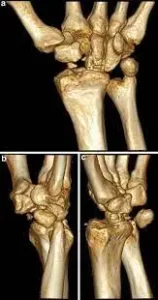
Pisiform impingement syndrome, also known as pisotriquetral syndrome, is a condition in which there is compression or irritation of the pisotriquetral bone and its surrounding structures, particularly the pisotriquetral joint.
Anatomy
Pisiform impingement syndrome involves irritation or compression of the pisotriquetral bone and surrounding the structures, particularly the pisotriquetral joint in the wrist joint. To better understand this condition, let’s look once at the relevant anatomy.
Small bone:
The pisiform is a tiny sesamoid bone located on the ulnar (pinky) side of the wrist joint. It is situated in the tendon of the ulnar Carpi ulnaris muscle and articulates with the palmar surface of the three-dimensional bone.
Pisotriquetral joint:
The Pisotriquetral joint is formed by the joint between the tricuspid bone in the wrist and longitudinal bone in the wrist joint. This joint allows smooth movement and stability between the two bones, which contributes to the function of the wrist joint.
Flexor Carpi Ulnaris Tendon:
The flexor carpi ulnaris (FCU) is a wrist flexor muscle situated on the elbow side of the forearm. Its tendon passes through the wrist joint and attaches to a long bone in the wrist.
When impingement syndrome includes the pisiform bone in the wrist joint, it is normally caused by a repetitive range of motion or direct trauma to the wrist joint area, causing irritation and inflammation in the area. Many possible anatomical factors that might contribute to pisiform impingement syndrome include:
A. Repetitive motion: Activities that include repeated flexion and extension of the wrist joint could stress the shape of the wrist joint and surrounding structures, causing inflammation and attack.
B. Anatomical variations: Mant people might have different bone shapes, sizes, or locations, or the surrounding bones and ligaments, which could increase the risk of impingement.
C. Tendon changes: Degenerative changes or inflammation in the knee flexor tendon could also contribute to compression of the petiole.
Causes of Pisiform Impingement Syndrome
Overuse and Repetitive Motion: Engaging in activities that involve repetitive wrist movements, such as typing, using a computer mouse, playing musical instruments, or certain sports, can lead to irritation and inflammation around the pisiform bone.
Gymnastics and Weightlifting: Activities that involve weight-bearing on the hands, like gymnastics or weightlifting, can put excessive pressure on the wrist, leading to pisiform impingement.
Fractures or Injuries: A previous wrist fracture or injury can alter the alignment of the wrist bones, potentially causing the pisiform to impinge on surrounding structures.
Anatomic Variations: Individuals with certain wrist anatomy or bone structures may be more prone to pisiform impingement. For example, a smaller-than-average pisiform bone or variations in the shape of adjacent bones can contribute to impingement.
Inflammatory Conditions: Conditions like rheumatoid arthritis or gout can lead to inflammation and swelling in the wrist, increasing the likelihood of pisiform impingement.
Improper Technique: Poor technique during activities that involve the wrist, such as improper form during weightlifting, can lead to imbalanced pressure on the wrist bones, contributing to impingement.
Carpal Tunnel Syndrome: In some cases, carpal tunnel syndrome (compression of the median nerve) can lead to altered wrist mechanics and contribute to pisiform impingement.
Occupational Factors: Certain professions that require repetitive wrist movements or prolonged periods of wrist use, such as assembly line work or manual labor, can increase the risk of pisiform impingement.
It’s important to note that while these factors can contribute to the development of pisiform impingement syndrome, each individual’s situation may be unique. Proper diagnosis and treatment by a medical professional are essential for effective management of the condition.
Symptoms of Pisiforme Impingement Syndrome
Wrist Pain: Persistent or intermittent pain on the ulnar (pinky finger) side of the wrist, particularly around the pisiform bone area.
Swelling and Tenderness: Swelling, inflammation, and tenderness around the pisiform bone and the surrounding soft tissues.
Painful Gripping and Movements: Pain when gripping objects, making a fist, or performing wrist movements, especially those that involve bending the wrist towards the little finger side.
Weakness and Limited Range of Motion: Weakness in the hand and wrist, as well as a decreased range of motion due to discomfort or pain.
Numbness or Tingling: Some individuals may experience numbness or tingling sensations in the ulnar nerve distribution, which can extend to the pinky finger and ring finger.
Painful Weight-Bearing Activities: Pain during weight-bearing activities that involve the hands, such as push-ups or weightlifting.
Pain with Pressure: Pain when pressure is applied directly over the pisiform bone area.
Aggravation with Repetition: Symptoms may worsen with repetitive wrist movements or activities that strain the wrist.
It’s crucial to consult a Doctor if you experience any of these symptoms in your hand. Proper diagnosis and treatment can assess and alleviate discomfort and prevent further complications
Epidemiology of Pisiform Impingement Syndrome
This condition is generally uncommon, and research on its prevalence and incidence is limited compared to more well-known wrist joint and hand joint conditions.
Pisiform impingement syndrome is considered a rare or rare condition, which means that it is not often seen in clinical practice compared to some common wrist disorders called carpal tunnel syndrome or wrist fractures in the hand.
It is often underdiagnosed or misdiagnosed due to its rarity and familiarity with symptoms with other wrist conditions.
The scarcity of epidemiological data on pisiform impingement syndrome(PIS) makes it challenging to provide precise pictures of its prevalence and incidence.
However, it is generally considered to be an infrequent condition that might affect a small percentage of individuals with wrist pain or related symptoms.
Risk factors for developing pisiform impingement syndrome might include participation in certain sports or activities involving repetitive wrist movements, occupations with repetitive wrist trauma, wrist strain, and anatomical variations in the wrist structures.
We recommend consulting medical professionals, and research publications, or seeking guidance from healthcare professionals who stay up-to-date with the latest medical literature.
Diagnosis
pisiform impingement syndrome typically involves a combination of clinical evaluation and diagnostic imaging. Here’s how it is usually diagnosed:
Medical History: Your doctor will discuss your symptoms, medical history, and any relevant activities or injuries that might be contributing to your wrist pain.
Physical Examination: A physical examination of your wrist will be conducted to assess pain, tenderness, swelling, range of motion, and strength. Your doctor may also perform specific tests to reproduce your symptoms.
Imaging: X-rays or other imaging studies, such as MRI or ultrasound, may be ordered to visualize the pisiform bone, surrounding structures, and any potential anatomical abnormalities or signs of impingement.
Diagnostic Injections: In some cases, your doctor may recommend a diagnostic injection of anesthetic or anti-inflammatory medication directly into the pisiform area. If the injection provides temporary relief, it can help confirm the diagnosis.
Electromyography (EMG): In instances where nerve involvement is suspected, an EMG test might be conducted to assess nerve function and rule out other possible causes of symptoms
Differential Diagnosis of Pisiforme Impingement Syndrome
Several conditions can have similar symptoms to pisiform impingement syndrome. When evaluating your wrist pain, a healthcare professional will consider these potential differential diagnoses. Some of these conditions include:
Ulnar Wrist Pain: Ulnar-sided wrist pain could be caused by conditions like ulnar impaction syndrome, triangular fibrocartilage complex (TFCC) injuries, or ulnar collateral ligament injuries.
Carpal Tunnel Syndrome: This condition involves compression of the median nerve at the wrist, leading to symptoms such as pain, numbness, and tingling in the hand.
De Quervain’s Tenosynovitis: Inflammation of the tendons at the base of the thumb can cause pain and discomfort on the radial (thumb) side of the wrist.
Wrist Tendonitis: Inflammation of tendons in the wrist, such as extensor or flexor tendons, can result in pain and limited mobility.
Ganglion Cyst: A fluid-filled lump that can develop near a joint or tendon, causing pain and discomfort.
Rheumatoid Arthritis: An autoimmune condition that can cause joint pain, swelling, and stiffness, including in the wrist.
Osteoarthritis: Degeneration of joint cartilage, leading to pain, stiffness, and limited movement.
Fractures or Sprains: Previous wrist fractures, sprains, or ligament injuries can cause ongoing pain and discomfort.
Referred Pain: Pain originating from another area, such as the neck or elbow, can sometimes be perceived as wrist pain.
Infections or Abscesses: Infections in the wrist joint or surrounding tissues can lead to pain, swelling, and localized symptoms.
Complex Regional Pain Syndrome (CRPS): A condition characterized by severe and chronic pain, often following an injury or trauma.
Medical Treatment of Pisiform Impingement Syndrome
Medical treatment for pisiform impingement syndrome aims to alleviate pain, reduce inflammation, and improve wrist function. Here are some potential medical treatment options:
Rest and Activity Modification: Giving the wrist time to rest and avoiding activities that aggravate the symptoms can help reduce irritation and promote healing.
Wrist Splinting: Wearing a wrist splint or brace can provide support and immobilization, allowing the inflamed area to heal. A splint may be worn during activities that worsen symptoms or at night to prevent inadvertent movements.
Anti-Inflammatory Medications: Nonsteroidal anti-inflammatory drugs (NSAIDs), such as ibuprofen or naproxen, may be recommended to reduce pain and inflammation.
Corticosteroid Injections: A corticosteroid injection directly into the pisiform area can help decrease inflammation and provide temporary relief from symptoms.
Physical Therapy: A physical therapist can design exercises and stretches to improve wrist strength, flexibility, and overall function. They may also use techniques like ultrasound or manual therapy to address soft tissue tightness.
Modalities: Heat or cold therapy, ultrasound, or other modalities may be used to reduce pain and promote healing.
Customized Ergonomics: Making adjustments to workstations, tools, or equipment to reduce wrist strain and improve ergonomics may be beneficial.
Activity and Technique Modification: Learning proper techniques for activities that involve the wrist and modifying movements can help prevent exacerbation of symptoms.
Medication Review: If certain medications are contributing to wrist symptoms, your doctor may consider adjusting or changing those medications.
Electromyography (EMG) or Nerve Conduction Studies: These tests may be performed to evaluate nerve function and rule out nerve-related causes of symptoms.
Lifestyle Changes: Making lifestyle adjustments, such as managing weight and staying hydrated, can contribute to overall joint health.
Referral to a Specialist: In cases where conservative measures are not effective, a referral to an orthopedic surgeon or hand specialist may be considered. Surgical options might be explored to alleviate impingement if warranted.
Physiotherapy Treatment
Physiotherapy can play a crucial role in the treatment of pisiform impingement syndrome by helping to relieve pain, improve wrist function, and prevent further complications. Here’s how physiotherapy treatment may be approached:
- Initial Assessment: A physiotherapist will conduct a thorough assessment of your wrist, taking into account your medical history, symptoms, and any relevant activities. This assessment assesses tailor a specialized treatment plan.
- Pain Management: Modalities such as ice or heat therapy, ultrasound, or electrical stimulation may be used to help reduce pain and inflammation in the wrist.
- Manual Therapy: Hands-on techniques like joint mobilizations, soft tissue massage, and stretching can help improve wrist mobility, decrease muscle tension, and promote healing.
- Range of Motion Exercises: Gentle and controlled exercises are introduced to improve wrist flexibility and range of motion, gradually progressing as the wrist heals.
- Strengthening Exercises: Specific exercises are prescribed to strengthen the muscles around the wrist and forearm, helping to provide better support and stability.
- Stretching Exercises: Targeted stretches help alleviate muscle tightness and improve overall wrist flexibility.
- Neuromuscular Re-education: Exercises that focus on enhancing proprioception (awareness of joint position) and coordination can improve wrist control and movement patterns.
- Posture and Ergonomics Education: A physiotherapist can provide guidance on maintaining proper posture and ergonomic principles to prevent excessive strain on the wrist during daily activities.
- Activity Modification: Learning how to modify activities that exacerbate symptoms can help protect the wrist and prevent further irritation.
- Taping and Bracing: Taping techniques or custom-made braces may be recommended to provide additional support and stability to the wrist.
- Home Exercise Program: A physiotherapist will design a customized home exercise program to continue your rehabilitation and maintain progress between sessions.
- Gradual Return to Activities: As symptoms improve, the physiotherapist will guide you through a gradual return to functional activities and sports, focusing on proper technique and minimizing strain on the wrist.
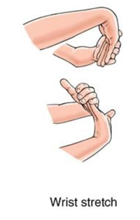
Exercise For Pisiforme Impigment Syndrome
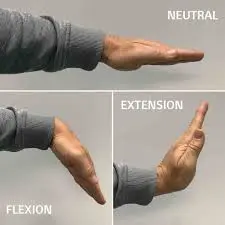
Wrist Flexion and Extension: Gently bend and straighten your wrist, moving through a pain-free range of motion.
Wrist Circles: Rotate your wrist in circular motions to improve flexibility.
Wrist Curls: Hold a lightweight or resistance band and curl your wrist upward and downward.
Isometric Wrist Exercises: Apply gentle resistance with your opposite hand while you push or pull against it in a different direction

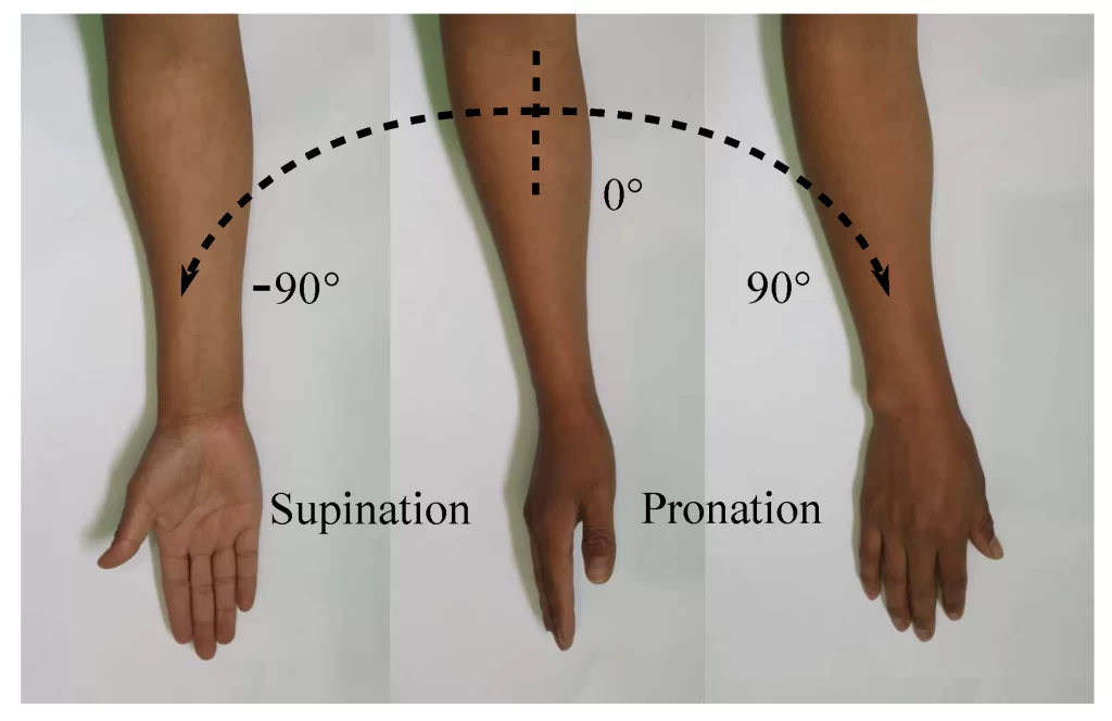
Forearm Strengthening Exercises: Forearm Pronation and Supination: Rotate your forearm to turn your palm facing down (pronation) and then your palm facing up (supination).
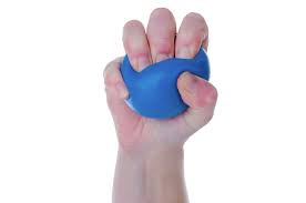
Grip Strengthening: Squeeze a softball or grip a resistance band, holding for a few seconds before releasing.
Ulnar Nerve Gliding Exercises: Ulnar Nerve Glide: Gently move your wrist and fingers in specific patterns to promote mobility of the ulnar nerve.
Stretching Exercises: Wrist Flexor and Extensor Stretches: Gently stretch the muscles on the palm side and back of the forearm.
Forearm Stretch: Hold your arm straight with the palm facing down, then gently pull your fingers towards your body to stretch the forearm muscles.

Finger Exercises: Finger Spread: Spread your fingers apart as far as possible and then bring them back together.
Finger Tapping: Tap each finger to your thumb, working through all fingers.

Functional Activities: Activity-Specific Training: Practice modified versions of activities that may have triggered your symptoms, gradually increasing intensity as tolerated.
Nerve gliding exercises: also known as neural gliding or nerve flossing exercises in wrist joint, are designed to increase the mobility and flexibility of nerves, reducing tension and irritation.
These exercises could be beneficial for the person experiencing symptoms like tingling, numbness, or shooting pain due to nerve compression or entrapment in the wrist joint and hand part.
Before attempting nerve gliding exercises in hand, it’s crucial to consult with a healthcare doctor, as these exercises might not be suitable for every person, especially in certain medical conditions.
Median Nerve Gliding Exercise: Start with standing or sitting with the back straight.
Extend the involved arm in front of the patient, with the palm facing the downside.
Begin by bending patient’s wrist and fingers down towards the floor area, like person making a “stop” gesture with person hand.
Next, slowly extend his wrist and fingers back up, as if his signaling someone to “come here.”
Perform this gliding motion Exercise smoothly and continuously for Ten to Fifteen repetitions.
Ulnar Nerve Gliding Exercise: Sit or stand with your back straight.
Extend the patient’s affected arm out to your side at shoulder level, with the patient’s palm facing upside.
Flex the patient’s elbow slightly and keep it in that position throughout the exercise.
start by moving your wrist, fingers, and thumb in a claw-like motion, curling them downside towards the floor area.
Then, extend the patient’s wrist, fingers, and thumb back up, spreading their wide range.
Repeat this gliding motion for 10-15 repetitions.
Radial Nerve Gliding Exercise: Sit or stand with your back straight.
patient’s injured arm should be extended in front of his, palm upwardside.
Keep your elbow straight throughout the exercise.
Begin by moving your wrist, fingers, and thumb downward, bending the patient’s wrist like you’re pouring out a drink of water from a jug.
Next, extend the patient’s wrist, fingers, and thumb back up, like you’re carrying a tray in your hand.
Perform this gliding motion for 10-15 repetitions.
During these exercises, it’s vital to move gently and avoid any movement that causes pain or discomfort.
If the patient experience worsening symptoms or if nerve gliding exercises exacerbate the patient’s condition, stop immediately and seek guidance from healthcare doctors.
Remember that nerve-sliding exercises are only one part of a comprehensive treatment plan.
If a patient experiences persistent wrist or hand issues, it’s important to consult with a qualified healthcare doctor, such as a physical therapist or an occupational therapist, to receive a exact diagnosis and individualized treatment program.
It’s important to start with gentle, pain-free movements and gradually progress as your wrist improves. Always consult with a physiotherapist or healthcare professional before starting any exercise regimen, especially if you have a medical condition. They can guide you in developing a safe and effective exercise program tailored to your specific needs and goals.
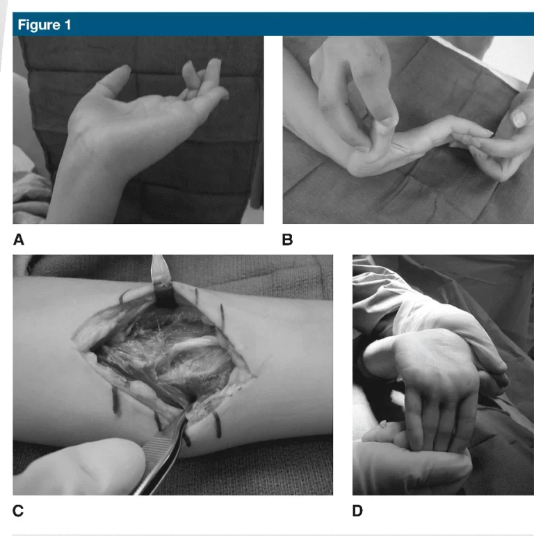
Surgical treatment
Pisiform Excision: In cases where the pisiform bone is causing impingement due to its position or size, surgical removal of the pisiform may be recommended. This procedure is known as pisiform excision.
Pisiform excision aims to eliminate the source of impingement, relieving pressure on surrounding tissues and reducing pain.
Soft Tissue Release or Decompression: Surgical release or decompression of tight or inflamed soft tissues around the pisiform and ulnar nerve can help alleviate compression and improve nerve function.
This procedure aims to create more space and reduce irritation in the affected area.
Arthroscopy: Arthroscopic surgery involves using a small camera and specialized instruments inserted through small incisions to visualize and treat the affected area.
Arthroscopy can be used to assess and address issues within the wrist joint, including the pisiform area.
Open Surgery: In cases where more extensive intervention is required, open surgery may be performed to directly address the impingement and any associated damage.
The specific surgical technique will depend on the underlying cause of the impingement and the individual’s anatomy.
Nerve Decompression: If nerve compression is a significant component of the condition, a surgical procedure may involve decompressing the ulnar nerve to alleviate symptoms.
Tendon Repair or Reconstruction:
If tendons have been affected or damaged due to impingement, surgical repair or reconstruction may be necessary to restore proper wrist function.
FAQ
How do patients treat a pisiform bone sticking out?
The treatment protocol of options is closed for manipulative immobilization and reduction, internal fixation, and open resection and reduction of the pisiform bone
What muscles attach to pisiform?
The pisiform is the only bone carpal bone with insertions and attachments for the abductor digit minimi and the flexor carpi ulnaris.
What type of pain is around the pisiform bone
Arthritis beneath the pisiform bone (pisotrequetral arthritis) causes sharp pain on the outer (ulnar) side of the wrist during movement and is one of the diagnoses that need considering in ulnar wrist pain


One Comment