Brain Aneurysm
What is a Brain aneurysm?
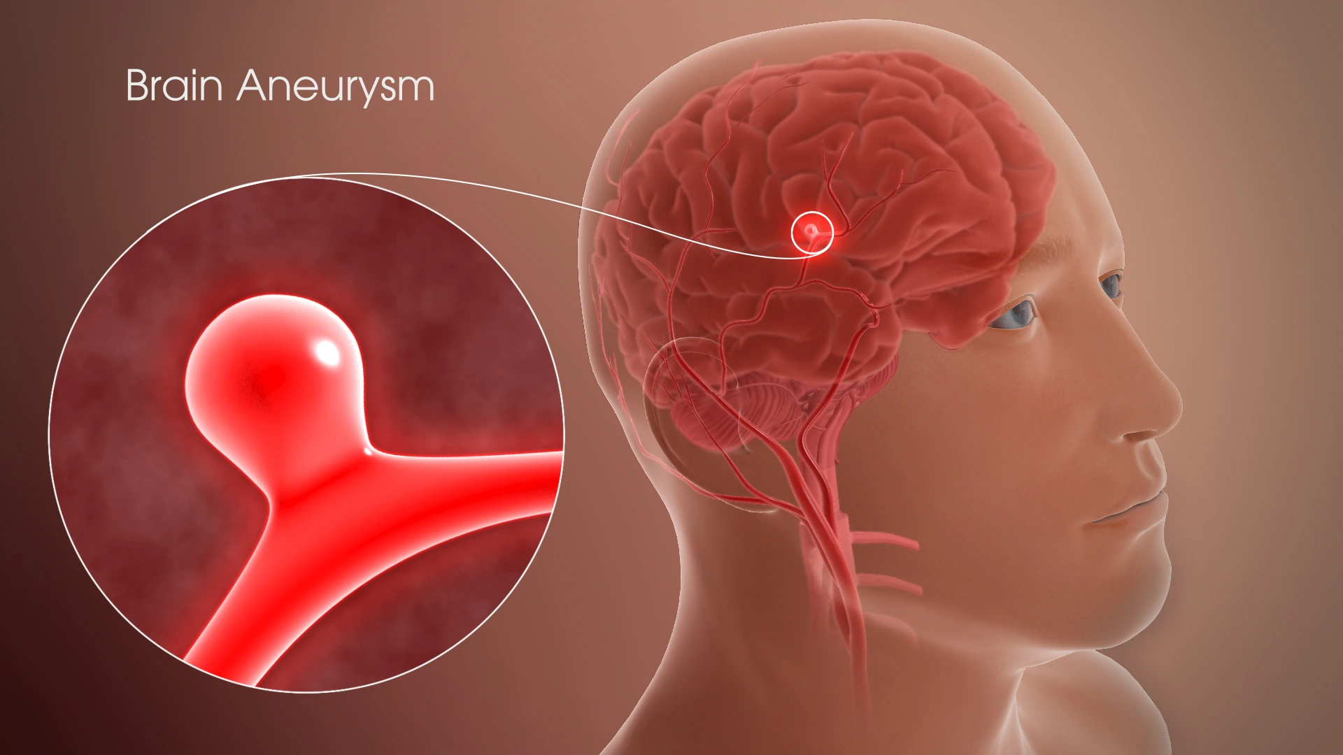
A brain aneurysm, also known as a cerebral aneurysm, is a bulge in a weak region of an artery in or around your brain. The constant pressure of blood flow pushes the weakened area outward, forming a blister-like bump.
When blood flows into this bulge, the aneurysm stretches even distant. It’s equal to how a balloon gets thinner and is more probable to pop as it fills with air.
Brain aneurysms can happen anywhere in your brain, but most of them develop in the main arteries along the bottom of your skull. About 10% to 30% of the person who has a brain aneurysm have multiple aneurysms. The prevalence of brain aneurysms is little and doesn’t cause symptoms.
An aneurysm can generate symptoms if it places pressure on nearby nerves or brain tissue. If the aneurysm leaks or ruptures (bursts open), it generates bleeding in your brain. A ruptured brain aneurysm can be life-threatening and needs emergency medical treatment. As more time passes with a ruptured aneurysm, the possibility of death or disability is raised.
Types of Brain Aneurysm
There are various types:
Saccular aneurysms are the most familiar variety of brain aneurysms. They bulge out in a dome form. They’re linked to the artery by a narrow neck.
Fusiform aneurysms arent is as familiar as saccular aneurysms. They don’t pouch out in a dome shape. Rather, they make an enlarged spot in the blood vessel.
Although brain aneurysms sound alarming, most don’t reason symptoms or health issues. You can enjoy a long life without ever recognizing that you have one.
But in rare patients, aneurysms can grow large, leak, or explode. Bleeding in the brain, called a hemorrhagic stroke, is severe, and you’ll require medical care right away.
What happens when a Brain Aneurysm ruptures?
When it ruptures, blood falls (hemorrhages) into your surrounding brain tissue. The blood can place extra pressure on your brain tissue and make your brain swell. It generally generates an extreme headache known as a thunderclap headache, in extra to further symptoms.
A ruptured brain aneurysm can generate severe health issues like:
Subarachnoid hemorrhage (SAH): Bleeding in the region between your brain and the light tissues that cover and save it (the arachnoid layer). Approximately 90% of SAHs are due to ruptured brain aneurysms.
Hemorrhagic stroke: Bleeding in the area between your skull and brain.
This can result can in permanent brain injury or different complications like:
Vasospasm: This occurs when blood vessels bring narrower or clamp down and less oxygen gets to your brain.
Hydrocephalus: This occurs when a buildup of cerebrospinal fluid or blood near your brain places raised pressure on it.
Seizures: A seizure is a temporary, uncontrolled wave of electrical activity in your brain. It can create brain injury due to a ruptured aneurysm more harmful.
Coma: A form of lengthy unconsciousness. It can stay days to weeks.
Death: Ruptured brain aneurysms result in death in approximately 50% of patients.
Symptoms of Brain Aneurysm
Ruptured aneurysm
A sudden, extreme headache is the essential symptom of a ruptured aneurysm. This headache is frequently defined as the worst headache ever experienced.
In extra to an extreme headache, familiar signs and symptoms of a ruptured aneurysm contain:
- Nausea and vomiting
- Stiff neck
- Blurred or double vision
- Sensitivity to light
- Seizure
- A drooping eyelid
- Loss of consciousness (Coma)
- Confusion
Leaking aneurysm
In some patients, an aneurysm may leak a slight quantity of blood. This leaking may generate only a sudden, intensely extreme headache.
A more extreme rupture frequently follows leaking.
Unruptured aneurysm
An unruptured brain aneurysm may create no symptoms, especially if it’s little. Yet, a bigger unruptured aneurysm may push on brain tissues and nerves, maybe causing:
- Pain above and behind one eye
- A dilated pupil
- A change in vision or double vision
- Numbness on one side of the face
Causes of Brain Aneurysm
Brain aneurysms form when the walls of an artery in your brain evolve thin and weak. They generally start at the branching points of arteries. Occasionally, you can be birth with a brain aneurysm. This is generally due to an abnormality (birth deficiency) in an artery wall. Several further elements can contribute to the weakening of an artery.
The subsequent inherited factors involve the health of your arteries and can raise your risk of creating a brain aneurysm:
- Vascular Ehlers-Danlos syndrome.
- Autosomal dominant polycystic kidney condition.
- Marfan syndrome.
- Fibromuscular dysplasia.
- Arteriovenous malformation.
Having a first-degree relative (biological sibling or parent) with a record of brain aneurysms.
The following states and conditions can weaken your artery walls over time:
- Smoking.
- High blood pressure.
- Substance use, especially cocaine.
- Extreme alcohol use.
Risk factors
Brain aneurysms can involve anyone. Yet, some factors can raise your risk.
There are various risk factors for aneurysm growth and rupture.
Risk factors for aneurysm formation
Several risk factors can raise your risk of creating a brain aneurysm. These contain:
Age. Most aneurysms are analyzed in people who are over the age of 40.
Sex. Females are more probable to obtain aneurysms than males.
Family history. If aneurysms run in your primary family, your risk of having one is more elevated.
High blood pressure. Untreated high blood pressure, or hypertension, can put additional stress on the walls of your arteries.
Smoking. Smoking can raise your blood pressure and cause injury to the blood vessel walls.
Alcohol and drug misuse. Alcohol and drug misuse, especially cocaine or amphetamine use, can raise blood pressure and leads to inflammation in your arteries.
Head injury. In rare circumstances, an extreme head injury can injure blood vessels in your brain, conducting to the construction of an aneurysm.
Genetic conditions. Specific genetic diseases can injure arteries or affect their structure, raising the risk of an aneurysm. Some instances contain:
- autosomal dominant polycystic kidney disease (ADPKD)
- Ehlers-Danlos syndrome
- Marfan syndrome
Congenital conditions. Weaknesses in blood vessels can likely be current from birth. Congenital diseases such as arteriovenous malformations or narrowing of the aorta, known as coarctation, can also increase the risk of aneurysms.
Infections. Some varieties of infections can injure artery walls and raise aneurysm risk. These are known as mycotic aneurysms.
Risk factors for aneurysm rupture
Few aneurysms will ever rupture. Yet, there are also risk factors that can raise the possibility of a ruptured aneurysm.
Few risk factors for rupture are associated with the features of the aneurysm itself. The risk of rupture is increased in brain aneurysms that are:
large
have grown larger over time
found in specific arteries, particularly the posterior communicating arteries and the anterior communicating arteries
Personal factors that raise the risk of rupture contain:
having a personal or family record of ruptured aneurysms
having high blood pressure
smoking cigarettes
Also, some events may promote an aneurysm to rupture. An older 2011 analysis estimated the relative risk of specific events in 250 people who had previously experienced a ruptured aneurysm. It found that the next were associated with the rupture of a current aneurysm:
- extreme exercise
- coffee or soda consumption
- straining during bowel motions
- nose blowing
- experiencing intense anger
- being startled
- sexual intercourse
Brain Aneurysms in Children
Infrequently, kids under 18 can have a brain aneurysm. Boys are eight times more probable to obtain them than girls. Of the few issues in kids, approximately 20% are giant aneurysms (bigger than 2.5 centimeters).
Aneurysms in children can arrive for no cause. But they’re also occasionally related to:
- Head trauma
- Connective tissue diseases
- Infection
- Genetic diseases
- Family record
Diagnosis
If you feel a sudden, extreme headache or further symptoms that could be linked to a ruptured aneurysm, you’ll be given examinations to specify whether you’ve had bleeding into the space between your brain and surrounding tissues (subarachnoid hemorrhage). The examinations can also decide if you’ve had another variety of strokes.
You may also be given examinations if you show symptoms of an unruptured brain aneurysm, like pain behind the eye, modifications in vision, or double vision.
Diagnostic examinations contain:
Computerized tomography (CT). A CT scan, which is a specialized X-ray examination, is generally the first examination used to specify if you have bleeding in the brain or some other variety of strokes. The examination produces pictures that are 2D slices of the brain.
With this examination, you may also obtain an injection of a dye that creates it easier to observe blood flow in the brain and may suggest the presence of an aneurysm. This interpretation of the examination is known as a CT angiogram.
Cerebrospinal fluid test. If you’ve had a subarachnoid hemorrhage, there will most probably be red blood cells in the fluid covering your brain and spine (cerebrospinal fluid). If you have symptoms of a ruptured aneurysm but a CT scan doesn’t show proof of bleeding, an examination of your cerebrospinal fluid can assist create a diagnosis.
The method to draw cerebrospinal fluid from your back with a syringe is known as a lumbar puncture.
Magnetic resonance imaging (MRI). This imaging method uses a magnetic field and radio waves to produce detailed pictures of the brain, either 2D images or 3D pictures.
A variety of MRIs that evaluates the arteries in detail known as MR angiography may see the presence of an aneurysm.
Cerebral angiogram. During this method, a light, flexible tube (catheter) is inserted into a large artery, generally in the groin or the wrist. The catheter lines past your heart to the arteries in your brain. A particular dye injected into the catheter crosses arteries throughout your brain.
A sequence of X-ray pictures can then reveal details regarding the state of your arteries and see an aneurysm. A cerebral angiogram also known as a cerebral arteriogram is generally used when further diagnostic examinations don’t provide sufficient data.
Treatment of Brain Aneurysm
Surgery
There are two familiar treatment choices for a ruptured brain aneurysm.
Surgical clipping is a method to close off an aneurysm. The neurosurgeon extracts an area of your skull to access the aneurysm and finds the blood vessel that feeds the aneurysm. Then the neurosurgeon puts a small metal clip on the neck of the aneurysm to prevent blood flow into it.
Endovascular therapy is a less invasive method than surgical clipping. The surgeon inserts a catheter into an artery, generally in your wrist or groin, and threads it via your body to the aneurysm.
The surgeon then utilizes a machine a flow diverter, an intraluminal flow disrupter, a stent or coils, or different mixtures of various instruments to kill the aneurysm from inside the blood vessel.
Both methods pose potential risks, especially bleeding in the brain or loss of blood flow to the brain. The endovascular coil is slightly invasive and may be originally safer, but it may bring a just higher risk of requiring a repeat method in the future due to the aneurysm reopening.
Flow diverters
Newer therapies available for brain aneurysms contain tubular stent-like implants (flow diverters) that function by shifting blood flow away from an aneurysm sac. The diversion controls blood motion within the aneurysm and facilitates the body to heal the area, facilitating the reconstruction of the parent artery. Flow diverters may be especially helpful in larger aneurysms that can’t be safely managed with further choices.
Your neurosurgeon or interventional neuroradiologist, in association with your neurologist, will make a suggestion based on the size, area, and overall formation of the brain aneurysm, your capability to undergo a method, and further factors.
Different treatments for ruptured aneurysms
Other therapies for ruptured brain aneurysms are purposed at easing symptoms and managing difficulties.
Pain relievers, like acetaminophen (Tylenol, others), may be utilized to manage headache pain.
Calcium channel blockers control calcium from entering cells of the blood vessel walls. These drugs may lessen the risk of having severe symptoms from the irregular narrowing of blood vessels (vasospasm) that may be a difficulty of a ruptured aneurysm.
One of these drugs, nimodipine (Nymalize), has been shown to decrease the risk of delayed brain injury generated by poor blood flow after a subarachnoid hemorrhage from a ruptured aneurysm.
Interventions to control stroke from poor blood flow contain IV injections of medicine to dilate the blood vessels, which promotes blood pressure to overcome the resistance of narrowed blood vessels.
An alternative intervention to control stroke is angioplasty. In this method, a surgeon uses a catheter to inflate a small balloon that grows a narrowed blood vessel in the brain generated by vasospasm. A medicine called a vasodilator also may be used to develop blood vessels in the affected region.
Anti-seizure drugs may be used to manage seizures related to a ruptured aneurysm. These drugs contain phenytoin (Dilantin, Phenytek, others), levetiracetam (Keppra), valproic acid, and others. Their use has been debated by several specialists, and is commonly subject to caregiver control, based on the medical requirements of each person.
Ventricular or lumbar draining catheters and shunt surgery can relieve pressure on the brain from extra cerebrospinal fluid (hydrocephalus) associated with a ruptured aneurysm. A catheter may be put in the areas that are filled with fluid inside the brain or in the region surrounding the brain and spinal cord to drain the extra fluid into an external bag.
Occasionally it may then be required to introduce a shunt system which consists of a stretchy silicone rubber tube and a valve that makes a drainage channel beginning in the brain and ending in the abdominal cavity.
Rehabilitative therapy. Injury to the brain from a subarachnoid hemorrhage may result in the requirement for physical, speech, and occupational therapy to relearn talents.
Treating unruptured brain aneurysms
Aneurysm clip
A surgical clip, an endovascular coil, or a flow diverter can be used to seal off an unruptured brain aneurysm and assist to control a future rupture. Yet, in some unruptured aneurysms, the known risks of the methods may outweigh the potential use.
A neurologist, in association with a neurosurgeon or interventional neuroradiologist, can assist you to specify whether the therapy is proper for you.
Factors to believe in making therapy suggestions contain:
The aneurysm location, size, degree of abnormality of the aneurysm and overall impression of the aneurysm
Your age and general fitness
Family record of a ruptured aneurysm
Congenital diseases that raise the risk of a ruptured aneurysm
If you have high blood pressure, talk to your healthcare provider regarding drugs to control the disease. If you have a brain aneurysm, accurate control of blood pressure may lower the risk of rupture.
In extra, if you smoke cigarettes, speak with your care provider regarding plans to stop smoking since cigarette smoking is a risk factor for the formation, growth, and break of the aneurysm.
Physiotherapy treatment
Physical therapy in the hospital normally begins with comfortable motion in a lying-down or sitting position, transitioning to sitting on the border of the standing, bed, and walking. Later, the regions of focus contain posture, balance, range of movement, and activities of daily living.
Physical therapy continues after the person is released from the hospital. Jointly with your physicians, we can create a customized schedule that permits further healing, with activities at home and/or at our facility. If the therapy was for an unruptured aneurysm, full activity can generally be restarted within a few weeks after surgery.
Aerobic Activity
Aerobic activity raises your heart rate and breathing. Attempt to create up to 150 minutes/week of moderate-intensity exercise (such as light cycling, brisk walking, or water activity). Don’t activity at a high intensity.
Aerobic Activity Cautions
The variety of aneurysms you have can involve how hard and frequently you should work out, so speak to your healthcare provider regarding precise guidelines for you.
Do any medicines you’re carrying, such as beta-blockers, involve your reaction to exercise? Rather than inspecting your heart rate, you may want to concentrate on your feelings of struggle
Strength Training
Strength training, for instance, functioning with weights or resistance bands, creates you stronger and assists your overall health. Plus, strength training can create daily activities such as raising laundry baskets or yardwork more comfortable and securer.
Remember: If you require it, get an asset from a certified activity professional. They can guide you on the right way to do activities and how to breathe correctly.
Strength Training Cautions
Don’t do high-intensity strength training.
Avoid straining or maintaining your breath when raising. This generates your blood pressure to go up.
Different Types of Physical Activity
Aerobic activity and strength training are at the heart of a schedule for those with an aneurysm. But you may want help from these other choices.
Take More Steps
Use a smartphone or exercise tracker to calculate your progress and remain motivated. Measure your everyday steps for the first week or two. Slowly makeup to 7,000-9,000 steps/per day
Tai Chi and Pilates
All encourage flexibility, balance, and strength, and are calming too.
Flexibility Exercises
Stretch big muscle groups for 10-30 seconds (30-60 seconds for more senior grown-ups), 2-3 days/ week. If you have Marfan syndrome, avoid flexibility workouts such as some yoga positions. They may generate joint dislocations or further injuries.
Balance Training
Balance activities may contain standing on one foot, walking in a line, or using a balance board. Be certain to train in an uncluttered region and use a chair or wall for help.
To stay safe and injury free:
Begin with light to medium action.
Slowly raise your pace and time spent being busy. Begin low and go gradually.
Warm-up and cool down at an effortless pace before and after the activity.
Lifestyle modifications to lower your risk
If you have an unruptured brain aneurysm, you may reduce the risk of its rupture by creating these lifestyle modifications:
Don’t smoke or use recreational medicines. If you smoke or use recreational medicines, speak to your healthcare provider regarding plans or a proper treatment schedule to assist you to quit.
Manage your blood pressure if you have high blood pressure.
Eat a nutritious diet and activity. Modifications in diet and activity can assist lower blood pressure. Speak to your healthcare provider regarding modifications that are proper for you.
Complications
When a brain aneurysm ruptures, the bleeding generally stays for only a few seconds. Yet, the blood can cause direct injury to covering cells, and the bleeding can injure or kill further cells. It also raises the pressure inside the skull.
If the pressure evolves too high, it may interrupt the blood and oxygen supply to the brain, and loss of consciousness or even death may happen.
Difficulties that can grow after the rupture of an aneurysm contain:
Re-bleeding. An aneurysm that has ruptured or leaked is a chance of bleeding also. Re-bleeding can cause additional injury to brain cells.
Narrowed blood vessels in the brain. After a brain aneurysm ruptures, blood vessels in the brain may contract, and little is known as vasospasm. This state can generate an ischemic stroke, in which there’s restricted blood flow to brain cells, generating additional cell injury and loss.
A buildup of fluid within the brain is known as hydrocephalus. Most frequently, a ruptured brain aneurysm happens in the space between the brain and the thin tissues surrounding the brain. The blood can block the motion of fluid that covers the brain and spinal cord. As an outcome, an extra fluid places pressure on the brain and can injure tissues.
Alter in sodium level. Bleeding in the brain can interrupt the balance of sodium in the blood. This may happen from injury to the hypothalamus, a region near the bottom of the brain. A decrease in blood sodium levels can conduct in swelling of brain cells and permanent injury.
Brain Aneurysm Prevention
Once your doctor or physician sees an aneurysm, it’s extremely improbable that it’ll heal on its own. The good information is you can bring steps to stop it from expanding or leaking. There are also items you can do to reduce the risk of a new aneurysm:
- Stop smoking.
- Eat a balanced diet.
- Work out daily and avoid regular heavy lifting (stick with moderate activity).
- Don’t use cocaine or different stimulant medicines.
- Get an asset for any alcohol or medicine use issues.
- Control your high blood pressure in control with drug and lifestyle modifications.
FAQ
Can you survive a brain aneurysm?
With rapid, expert therapy, persons can frequently recover completely. An unruptured brain aneurysm may generate zero symptoms. A person can live with them for years before detection. If a brain aneurysm is unruptured, no blood has broken via the blood vessel walls.
Can you live a long life after a brain aneurysm?
Can a person live a prolonged time with a brain aneurysm? Definitely. Many aneurysms generate no symptoms at all. Some people live for years without understanding they have a brain aneurysm.
How can you prevent a brain aneurysm?
You can’t always control brain aneurysms, but you can decrease your risk by not smoking and by lowering high blood pressure.
Do all brain aneurysms cause death?
Ruptured brain aneurysms are fatal in approximately 50% of patients. Of those who stay, approximately 66% suffer some permanent neurological deficiency. About 15% of people with a ruptured aneurysm die before getting to the hospital. Most of the deaths are due to quick and immense brain damage from the initial bleeding.
Does brain aneurysm affect memory?
Memory includes many regions of the brain, and if a brain aneurysm ruptures or therapy damages any of those regions, your memory will be involved. Survivors of ruptured aneurysms generally do not recall the event or much of what occurred in the hospital, and never will.

