Compound Fracture
Introduction
- An open fracture, also known as a compound fracture, is a kind of bone fracture in which the skin around the shattered bone has an open incision. Usually, a skin wound results from a bone piercing the skin’s surface.
- Open fractures are serious medical crises that are frequently brought on by high-energy trauma, such as car crashes, and are linked to significant soft tissue and bone injury.
- A deep infection and/or bleeding could result from an open fracture, which could endanger the patient’s life or cause them to lose a limb.
- Additional concerns include the possibility of a nonunion or malunion of the bone. The degree of open fracture severity can differ.
- The most widely used technique for identifying and categorizing open fractures is the Gustilo-Anderson open fracture classification.
- Additionally, it can be applied to forecast clinical outcomes and guide treatment.
- The first course of treatment for open fractures and the rule-out of other potentially fatal conditions in trauma situations is advanced trauma life support.
- Antibiotics are also given to the patient for a minimum of 24 hours to lower the chance of infection. Generally speaking, cephalosporins are the first class of antibiotics.
- The primary treatments for open fractures include bone repair, early wound closure, wound debridement, and therapeutic irrigation.
- The goal of all of these measures was to lower the chance of infection.
- The tibia is the bone that sustains injuries most frequently, and young men of working age are the population with the highest risk of suffering an open fracture.
- An additional risk factor includes soft-tissue disorders and osteoporosis in older adults.
What is a Compound Fracture?
- A complex fracture, sometimes referred to as an “open fracture,” is a type of bone fracture in which there are skin fractures that allow the shattered ends of the bone to come into touch with the surrounding environment.
- This typically affects the lower leg, though it can happen to any area of the limbs, and is brought on by a broken bone entering the skin following a high-impact trauma.
- The degree of damage to the bone and surrounding soft tissues (muscle, tendon, ligament, etc.), the degree of wound contamination with soil, grease, sand, etc., and whether or not there is any disruption of the major blood vessels to that part of the body are some of the factors that determine how severe a compound fracture is.
What might someone suffer from a complex fracture?
- Due to the significant danger of infection, tissue injury, and difficulties throughout the healing process, compound fractures require immediate attention. Compound fractures and other high-energy fractures are all susceptible to compartment syndrome, a dangerous concomitant ailment.
- Virus Infection
- Comparatively speaking, high-energy open fractures are far more likely to become infected than comparable closed fractures. Compound fractures are not only more likely to require extra surgery to assist healing, but they also carry a higher chance of not healing quickly.
- The Compartment Syndrome
- A medical emergency known as compartment syndrome occurs when damaged muscle tissue grows to the point where it destroys nearby tissues, most frequently nerves, in addition to the damaged muscle itself.
- The enlarged muscle is restrained within a section of Fascia is a type of tissue that is unable to stretch to release pressure.
- Every tissue inside the compartment sustains injury as the pressure increases.
- If compartment syndrome is left untreated, the afflicted muscles and nerves may permanently lose their ability to function due to necrosis, or tissue death.
- A fasciotomy, an urgent surgical procedure that releases pressure from the affected muscle compartment, is the treatment for compartment syndrome.
Symptoms
- If a shattered bone penetrates your skin, it is referred to as a complex fracture. The shattered bone is visible. Severe symptoms are among the others.
- intense agony.
- sensitivity to physical contact.
- Growing.
- bruising.
- Sickened.
- damage to the nerves, which could cause the pain to be less or more intense than usual.
- Since the degree of the injury can vary widely, open fractures can have a variety of characteristics.
- A fractured bone that protrudes from the skin is the typical sign of an open fracture, but it is also possible for a shattered bone to be connected to a tiny “poke-hole”-like skin wound.
- These two wounds are categorized as open fractures. Blood loss from certain open fractures can be quite high.
- Soft tissues like muscles, tendons, blood arteries, and nerves close to and around the bone are severely damaged in the majority of open fractures.
Causes
- Direct hits such as high-energy physical forces (trauma), auto accidents, guns, and falls from heights can result in open fractures.
- Falling from a standing position and twisting (torsional injuries) are examples of indirect processes.
- These processes can present more subtly, such as a little poking hole and an accumulation of clots of blood in the tissues, but they are typically linked to significant degloving of the soft tissues.
- Different kinds of fractures may result from trauma, depending on its nature:
- Typical fractures
- Bone fractures are the outcome of severe bone trauma. A direct hit, axial loading, rotational forces, torque, or a combination of these can all cause this trauma.
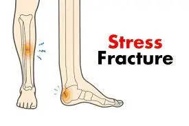
- Different forms of fractures exist, such as stress, closed, open, greenstick, comminuted, simple, and displacement fractures.
- transversal and slanted.
- unstable fractures
- result from a small amount of pathological bone damage. These preexisting conditions include severe osteoporosis, bone cysts, and metastatic lesions, among others.
- Break-dislocations
- severe damage in which a fracture and a dislocation occur at the same time.
- wounds from gunshots
- High-speed projectiles inflict damage to tissue as they pass through it by cavitating and creating a secondary shock wave.
Classification
- Many classification schemes, like the Müller AO Classification of Fractures, the Tscherne classification, and the Gustilo-Anderson open fracture classification, try to classify open fractures.
- On the other hand, the most widely utilized classification scheme is the Gustilo-Anderson open fracture classification.
- The Gustilo system assigns a grade to a fracture based on the degree of contamination, soft tissue damage, energy of injury, and fracture comminution. The outcome of the fracture is worse the higher the grade.
| Gustilo–Grade | Definition |
| I | Compound fracture, clean wound, 1 cm long wound |
| II | Compound fracture wound greater than 1 cm in length but less than 10 cm in length, flaps, avulsions |
| III A | Open fracture of a fractured bone with appropriate soft tissue coverage despite severe soft tissue laceration or flaps, or high-energy trauma (gunshot and agricultural injuries) regardless of wound size |
| III B | Compound fracture with significant soft-tissue loss, periosteal stripping, and bone destruction. Typically connected with widespread pollution. |
| III C | Compound fracture linked with an artery injury needing repair, regardless of the degree of soft-tissue injury, will frequently require additional soft-tissue coverage treatment (i.e. free or rotating flap). |
- The Gustilo system does have certain drawbacks, though. At 50% to 60%, the system’s interobserver dependability is limited.
- The extent of deep soft tissue damage underneath the skin’s surface does not always correspond to the size of the injury.
- Consequently, the only place where Gustilo may be truly graded is in an operating room.
Diagnosis
- Your skin has broken, making a compound/open fracture more noticeable to a medical professional than a simple/closed fracture.
- After performing a physical examination, the medical professional will request X-rays to determine the precise location of the fractures and the necessary alignment of the broken bones.
- To completely evaluate the extent of the fracture’s damage, medical professionals may occasionally need a more sensitive test, such as a computed tomography (CT) scan or magnetic resonance imaging (MRI).
- Your healthcare practitioner will diagnose the compound fracture and also look for any consequences. What they’ll do is as follows:
- Examine the hue and feel of your skin.
- To make sure there isn’t any substantial bleeding, check your blood pressure and pulse.
- Assess for nerve injury by looking at the region surrounding and extending beyond your injury.
Treatment
Non- Surgical Treatment
- To avoid dirt getting inside the open fracture, wrap it with a clean, moist towel before going to the hospital. You should also avoid utilizing any of the joints near the break unless required to seek medical attention.
- Rest: By resting the body parts, you will heal faster and be less likely to cause more injuries.
- Ice: Apply ice to the area above and surrounding the compound fracture as soon as possible after the accident occurs, but be careful not to infect the site.
- Compression: A cast or other device will be used to keep your limb or other body part in place while it heals.
- Elevation: To minimize swelling, elevate (raise) the injury over your heart. You might have to wait till after you’ve been
- To clean the damaged region and reduce the risk of infection, urgent therapies are frequently required.
- These interventions include wound debridement and therapeutic irrigation.
- Long-term consequences, including profound infection, vascular impairment, and total limb loss, are additional hazards associated with delaying intervention.
- To stop bacterial contamination, the wound should be covered with dry or wet gauze after irrigation.
- By taking pictures of the wound, you may be able to avoid the need for several, potentially uncomfortable medical evaluations from different physicians.
- To immobilize a fractured limb, it should be reduced and placed in a well-padded splint. It is important to record pulses both before and after reduction.
- In 22% of pre-debridement and 60% of post-debridement cultures of infected cases, wound cultures are positive.
- Pre-operative cultures are therefore no longer advised. It is unknown what post-operative cultures are worth.
- Regular administration of tetanus prophylaxis strengthens the immune system’s defense against Clostridium tetani. Immunoglobulin against tetanus is only recommended for people with highly infected wounds and a hazy vaccination history.
- To provide protection right away, a single intramuscular injection of 3000–5000 units of tetanus immunoglobulin is administered.
- A crucial therapeutic choice in the acute therapy of open fractures pertains to preventing avoidable amputations when it is evident that functional salvage of the limb is preferable.
- It is important to make sure that the patient, their family, and the care team have fully discussed all of their alternatives and that the decision is not only dependent on the results of an injury severity tool.
Medical Treatment
- To lower the risk of infection, intravenous broad-spectrum antibiotics must be administered as quickly as possible—ideally, within an hour.
- Antibiotics might not be necessary in cases of open finger fractures and injuries from low-velocity weapons, though. When treating open fractures, first-generation cephalosporins like cefazolin are advised as the first line of treatment.
- The antibiotic works well against gram-negative rods and gram-positive cocci, including Klebsiella pneumoniae, Proteus mirabilis, and Escherichia coli.
- A combination of first-generation cephalosporin and an aminoglycoside (gentamicin or tobramycin) or a third-generation cephalosporin is advised to cover against nosocomial gram-negative bacilli like Pseudomonas aeruginosa to increase the coverage of use medicines against more microorganisms in Type III Gustilo fractures.
- Penicillin was added to treat gas gangrene brought on by microorganisms that do not require oxygen The use of Clostridium perfringens is debatable.
- Research has indicated that the usual course of antibiotics may be sufficient to treat Clostridial infections, negating the need for this kind of intervention. Antibiotic-impregnated materials, like antibiotic bone cement and tobramycin-impregnated Poly(methyl methacrylate) (PMMA) beads, can help lower infection rates.
- Another efficient way to give local antibiotics is to use absorbable carriers with implant coatings during surgical fixation.
- The ideal period of antibiotics has not been agreed upon.
- Research has indicated that administering antibiotics for a single day does not confer any extra benefits in terms of infection risk when compared to administering antibiotics for three or five days. But as of right now, there is just low to moderate.
Wound irrigation
- Regarding the best way to irrigate wounds, there is no consensus. Research has revealed that utilizing tap, boiled, distilled, or regular saline water does not affect the rate of infection.
- When irrigating wounds, there is no change in the infection rates between using regular saline with castile soap and regular saline plus bacitracin.
- Research has also demonstrated that when irrigating wounds, low-pressure pulse lavage (LPPL) and high-pressure pulse lavage (HPPL) do not differ in terms of infection rates.
- Additionally, the ideal fluid quantity for irrigation remains undetermined.
- It is advised that the volume of irrigation solution be based on the fracture’s severity, with three liters being the suggested type 6 liters for type II fractures, 9 liters for type III fractures, and type I fractures
Wound debridement
- Debridement of the wound aims to remove all infected and non-viable tissues, such as muscles, bones, subcutaneous fat, and skin.
- The ability of soft tissues and bones to bleed indicates whether or not they are viable. In the meanwhile, muscle viability is defined by color, consistency, contractility, and bleeding ability.
- There is disagreement over the best time to undertake wound debridement and closure; factors include the extent of the injury, the availability of resources and antibiotics, and the needs of the individual.
- Closure times can be quick (less than 72 hours) or delayed (72 hours to up to 3 months), with debridement times ranging from 6 to 72 hours. When surgery is done within six hours of an injury as opposed to waiting until 72 hours later, there is no change in infection rates.
Surgical Management
- The early immobilization and fixing of a fracture helps to minimize future soft tissue injury and facilitates wound and bone healing.
- This is particularly significant in the treatment of intraarticular fractures since early fixation allows for early joint motion, preventing joint stiffness.
- Fracture care is determined by the individual’s overall health, fracture pattern and location, and the extent of soft tissue injury.
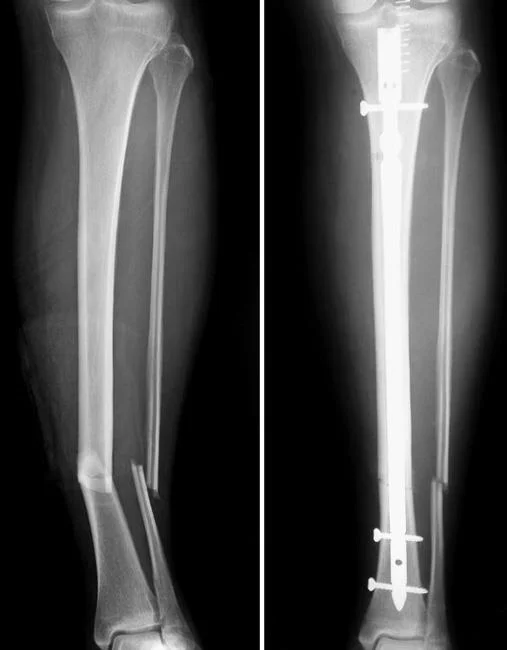
- Both reamed and undreamed intramedullary nailing are commonly used surgical procedures for open tibial fractures. Both methods have comparable rates of postoperative healing, infection, implant failure, and compartment syndrome.
- When compared to external fixation, undreamed intramedullary nailing has a lower incidence of superficial infection and malunion.
- However, undreamed intramedullary nailing might result in a high rate of hardware failure if a person continues to bear weight following the procedure.
- Surgery is not strictly regulated. Unreamed intramedullary nailing shows comparable rates of deep infection, delayed union, and nonunion following surgery to external fixation.
- In children with open tibial fractures, orthopedic casts are increasingly being used instead of external fixation. Bone grafting can also aid in fracture repair.
- Internal fixation using plates and screws, on the other hand, is not recommended because it increases the risk of infection.
- Amputation is a last resort intervention that is influenced by factors such as tissue viability and covering, infection, and the level of circulatory system injury.
- Compound fractures are sent to the operating room for more complete wound cleansing and irrigation, as well as the removal of damaged tissue that may contaminate the site.
- Unless other emergency operations, such as fasciotomy for compartment syndrome, are required, this acute wound care will be followed by surgical realignment of bones.
- At this initial operating room setting, the fracture is either temporarily or permanently stabilized.
- This usually entails open reduction (cutting an incision to manually access and straighten the bone or bones), followed by internal or external fixation.
- At one year, anteroposterior and lateral femur and tibia radiographs show healed distal femur and tibia fractures in excellent alignment.
- X-rays of the corrected femur from the front to the back and the side.
- One year after surgery, and the tibia.
- Fixation is the use of rods, wires, pins, screws, and/or plates to hold the bone in place during healing. extra wound treatment operations (extra washing and/or skin or muscle grafts to cover the wound) may be required depending on the severity of the wound.
Internal Fixation
- Your doctor will install metal implants, such as plates, rods, or screws, on the surface or inside the shattered bone during this treatment.
- The implants will keep the bone in place and hold it together while the fracture heals.
- Internal fixing can be done during the initial surgery after the fracture has been cleansed. If the skin and muscle need time to heal, a subsequent (later) operation may be performed.
- Following internal fixation, your wounded limb will be checked for signs of infection. Antibiotics will be administered to you for a length of time to assist in preventing infection.
- Your doctor will examine the site of the injury to ensure there are no signs of infection during the healing process.
External Fixation
- The doctor inserts metal screws or pins through the skin into the bone above and below the fracture site during this procedure. Pins and screws protrude (stick out) from the skin and are fastened to metal or carbon fiber bars.
- If your wound and damaged bones are not yet suitable for a permanent implant, your doctor may decide to use external fixation to stabilize your injured limb. External fixation is used to stabilize the majority of severe open fractures.
Treatment of More Complex Wounds
- Large wounds with substantial skin and muscle tissue loss can occur with some open fractures. These wounds are frequently too large to close. In this case, the doctor will cover the wound temporarily to reduce the danger of infection and encourage healing.
- Numerous dressings can be applied to provide temporary covering.
- A semi-permeable dressing is frequently used to seal the wound until it can be properly closed.
- This form of dressing allows oxygen and carbon dioxide to pass through while preventing pollutants (such as germs) and water from passing through.
- Antibiotic beads are frequently inserted into the wound before closure to deliver a high concentration of antibiotics directly to the injury.
- The external fixator has the added benefit of supporting the shattered bone while your doctor treats the wound. In some circumstances, further debridement or skin and tissue grafting may be required to cover the fractured bone.
- In most circumstances, an external fixator is only used once until internal fixation may be performed safely. However, an external fixator is sometimes employed to stabilize the bones until healing is complete. When the fracture has healed, it is removed in a further (later) surgery.
- After some time, a permanent method will be utilized to close the wound. Skin grafts are permanent wound-covering procedures.
- If you simply have skin loss, a portion of skin from another part of your body can be extracted and utilized to cover the wound.
- Local disturbance. To cover the defect, muscle tissue from another part of the same leg might be transferred into the lesion.
- The flap is free. Tissue from another section of the body, commonly the thigh, back, flank, or abdomen, can be transferred. A microvascular surgeon who can establish blood circulation to the flap is commonly used in a free flap operation.
Physiotherapy Treatment
- The physiotherapist’s goal is to determine the core cause of the problem and pick appropriate treatment strategies to assist patients in returning to their desired daily activities.
- A patient assessment is essential in any physiotherapy session. The problem-oriented medical record (POMR) system is built on a data-collecting system known as SOAP.
- The treatment delivered is mostly determined by the issues detected during your initial evaluation.
- The following ambulation with assistive aids during various chores such as walking up and down stairs or getting in and out of a car may comprise a combination of the following.
- During these sessions, it is also important to consider the client’s weight-bearing constraints, educating clients on how to ambulate while adhering to the restrictions.
- Because client safety is of the utmost importance, they must be proficient and comfortable utilizing these devices before discharge.
- on the use and removal of slings following upper extremity fractures.
- Massage of the soft tissues, notably to treat edema and swelling
- Scar care after surgery
- Icing therapy
- Exercises for regaining joint range of motion
- Joint manual treatment and mobilizations might help you regain joint mobility.
- The regime of structured and progressive strengthening
- Work on balance and control, as well as gait (walking) re-education, if needed.
- Taping to support the wounded area and aid in edema control
- Return to sports preparation work and advice as needed
Complications
- Compound fractures are complex injuries, although not all of them result in long-term problems other than the shattered bone.
- Your healthcare personnel will look for them when you are in the emergency room, during surgery, and during your recuperation. Following a complex fracture, the following complications may occur:
- Skin injury: When the compound fracture pierces your skin, it causes harm. While your bones recover, your skin will heal.
- The skin under the cast does not require particular care while the cast is on, but it may require some care when the cast is off.
- Joint damage: If your complex fracture is close to a joint, it can impair the ability of that joint to move appropriately.
- The fracture might harm the cartilage at the ends of your bones. When bones are immobile, joints can become stiff as well.
- Physical treatment will be required to get the joint moving properly, and surgery may be required in some cases to restore normal movement.
- Nerve injury: A compound fracture can crush, bruise, tear, or strain your nerves, resulting in some modest nerve harm.
- These can recover entirely, although it can take months (or even years). Some nerve injuries cannot heal, depending on the severity of the injury, but these are less common than milder injuries.
- Problems with healing: In some cases, the bones do not grow back together (this is known as nonunion).
- They can grow back slower than normal (delayed union) or in the wrong location (malunion).
- Your healthcare experts will take X-rays to determine how well you’re recovering and whether additional treatment is required.
- Uneven limbs: Children’s bones continue to grow until they reach their adolescence. Growth plates are areas of the bones toward the ends that are called such.
- This is the stage at which children’s bones grow longer. If a growth plate in your child’s arm or leg is destroyed, the bone may not grow correctly and end up shorter on one side than the other.
- If a growth plate has been injured, your doctor will tell you.
- Shock: When you have a complicated fracture, you may lose a lot of blood, which can be terrifying. The blood loss could be external or inside, which means you could die.
Prevention
- Accidents can happen to anyone at any time. It’s frightening to think that you could shatter a bone by falling off a ladder, being in a vehicle accident, or slipping on a damp floor. You can lower your risk by following basic steps like:
- Keeping a safe distance from heights.
- Yoga or other forms of exercise to increase strength and balance.
- If you have poor balance, hold on to the safety rails when getting in and out of the bathtub.
- Driving defensively and safely.
- Not participating in contact sports such as football.
- Wearing appropriate shoes for the surfaces you’re walking on and be cautious if the ground is slick.
How do compound fractures heal?
- Your bones repair themselves by forming new bone tissue. The new bone is known as an exterior callus.
- This callus appears quickly after the bone is shattered. It’s not like normal bone at first since it’s fragile and doesn’t protect the underlying break.
- However, it becomes stronger as it calcifies and matures into normal bones over weeks to months.
- There are three stages of healing for compound fractures:
- Inflammation.
- Repair.
- Remodeling.
- Stage of inflammation: Your body begins to mend immediately after the fracture. Your immune system’s cells rush to the wounded area immediately.
- They increase blood flow to the area, which can cause the skin around the compound fracture to expand and turn red.
- This enlargement and Redness may persist for some time as your body attempts to mend.
- Stage of repair: Your shattered bone will be kept immobile (immobilized) in a cast throughout this stage, which can take weeks to months.
- Your shattered bones must remain immobile while they recover.
- During this stage, your body will produce new bone tissue. Because the exterior callus (the new bone) is easily destroyed, it must be protected.
- Stage of renovation: The remodeling stage can last months. During this time, the exterior callus thickens and calcifies, making it stronger.
- Your bones grow more normal in shape and less fragile as they rebuild.
Recovery
- Recovery from a compound fracture can take several months. It is critical to remain patient during the mending process.
- Several factors influence how quickly you recover from a complex fracture. Among the factors are:
- The extent of your injury.
- It’s your age. Children, for example, recover faster than adults.
- Complications following treatment.
- Other health concerns. You may heal more slowly if you have a disease that interferes with blood flow. Diabetes and peripheral arterial disease are two examples of such illnesses.
Summary
- This is determined by the nature of the injury. Your compound fracture will still be visible on X-rays after it heals, but you may not be able to tell just by looking at the area.
- The bone tissues frequently blend quite well.
- Depending on the severity of the break, your bones’ capacities to function should return to pre-injury levels.
- You may require extensive physical rehabilitation to return to your typical activities.
FAQ
Is a complex fracture the most serious type of fracture?
However, fractures of many forms can develop even while training for a marathon. A complex fracture is the most serious of them all. They are not only painful, but they are also unappealing to look at.
What are the signs of a complex fracture?
A complex fracture has either penetrated the skin or exposed the bone, resulting in excruciating pain. With a complex fracture, you will experience chronic agony even if you do not attempt to move the affected body part.
Is it possible to repair a complex fracture without surgery?
Almost all patients will believe surgery is necessary. However, surgery will be required depending on the type of fracture. Compound fractures require surgery because they break the skin, whereas compression fractures heal on their own.
Is it possible to heal a complex fracture?
Compound fractures are frequently totally cured through surgical correction of the deformity, as well as care for the shattered bone and the wound created by it.
What is the treatment for a complex fracture?
The primary therapy is surgery, with the primary goal of preventing infection. Internal fixation and external fixation are the two types of procedures used to treat complex fractures. Metal pins, screws, and plates are used in both operations to hold the bone together. External fixation attaches this gear to a metal bar located outside the skin.
References
- Compound Fracture | Open Fracture | HSS Orthopedic Trauma Service. (n.d.). Hospital for Special Surgery. https://www.hss.edu/condition-list_compound-fracture.asp#:~:text=A%20compound%20fracture%20(also%20known,contact%20with%20the%20outside%20environment.
- Open fracture. (2023, December 9). Wikipedia. https://en.wikipedia.org/wiki/Open_fracture
- Professional, C. C. M. (n.d.). Compound Fracture. Cleveland Clinic. https://my.clevelandclinic.org/health/diseases/21843-compound-fracture
- Open Fractures – OrthoInfo – AAOS. (n.d.). https://orthoinfo.aaos.org/en/diseases–conditions/open-fractures/
- Fracture. (n.d.). Physiopedia. https://www.physio-pedia.com/Fracture