
Cuboid Bone Fracture
A cuboid bone fracture refers to a break or crack in the cuboid bone, which is one of the seven tarsal bones located in the foot. The cuboid bone plays a crucial role in maintaining the structural integrity of the foot and facilitating various movements, particularly in the midfoot region.
Introduction
The cuboid bone, located within the midfoot, is one of the seven tarsal bones that make up the anatomical structure of the foot. Cuboid fractures account for an estimated 2-8% of all midfoot fractures, making them much less common than injuries to other tarsal bones like the navicular or talus.
However, due to the important stabilizing role of the cuboid in weight transfer during gait, these fractures can cause significant alterations to foot biomechanics when left untreated.
Anatomy
The cuboid has several distinguishing anatomical features and facets that allow for multiple articulations or joints with neighboring bones:
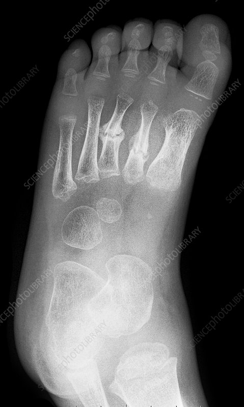
- Location: On the lateral side of the foot, anterior to the calcaneus (heel) and posterior to the 4th and 5th metatarsal bones
- Shape: Irregular, cube-like shape with 6 surfaces—medial, lateral, superior, inferior, anterior, and posterior
- Facets:
- The lateral facet articulates with the calcaneus to form the calcaneocuboid joint
- The medial facet articulates with the lateral cuneiform bone
- Inferior facets articulate with the bases of the 4th and 5th metatarsal bones
- Grooves for tendons of peroneus longus and tibialis posterior muscles
- Borders: Has anterior, posterior, medial, and lateral borders which serve as attachment points for multiple ligaments
Biomechanics
The cuboid plays several essential roles in foot biomechanics and gait:
- Forms part of both the transverse tarsal joint (talocalcaneonavicular) and lateral longitudinal arch of the foot
- Weight-bearing: Transfers loads from the calcaneus to the 4th and 5th metatarsals during the stance phase of gait as lateral column stabilizer
- Lever for propulsion: Helps form rigid lever system needed to progress over the planted foot during toe-off
- Medial longitudinal arch: Helps maintain the structural integrity and height of the arch which controls overall foot flexibility and shock absorption
In summary, the cuboid acts as an important connector between the midfoot and lateral forefoot, facilitating weight transfer and lever action needed for normal foot function in gait. Fractures to this bone can significantly alter foot stability and mechanics.
Causes of Cuboid Bone Fracture
Cuboid fractures have both direct and indirect causes:
- Direct trauma or crush injuries – e.g. the foot is run over by a vehicle causing a cuboid break
- Falls from height – landing directly on the lateral aspect of the foot
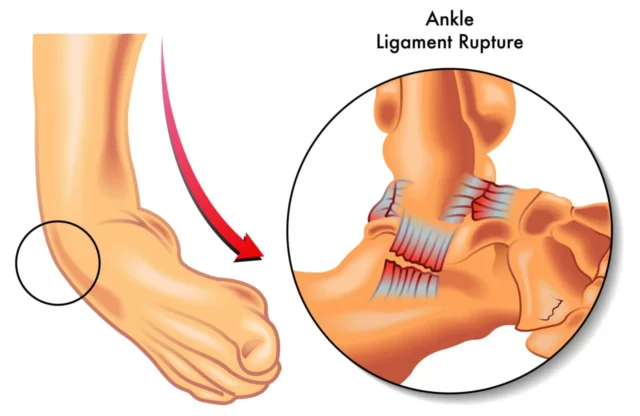
- Inversion ankle sprains – forceful internal rotation of the foot tears ligaments but also avulses (pulls) a piece of the cuboid
- Stress fractures – repetitive overuse and too much pressure on the cuboid leads to microscopic cracks that progress to complete fracture
Types of Cuboid Bone Fracture
Extra-articular Fractures
- Also called isolated cuboid fractures
- The fracture line does not extend into any nearby joints
- Can occur in different parts of the cuboid depending on the force mechanism
- Considered more stable injuries compared to intra-articular fractures
Intra-articular Fractures
- The fracture line extends into a nearby joint
- Most commonly involves the calcaneocuboid joint between the heel and cuboid bone
- Destabilize the lateral column of the foot
- Requires surgery more often due to joint disruption
Avulsion or Chip Fractures
- A small fragment of bone is pulled off by a tendon or ligament
- Common on the foot due to multiple tendon attachments
- Cuboid chip fractures are often caused by forceful inversion ankle sprains
- The bone piece attached to a tendon gets ripped off the main cuboid bone
Comminuted Fractures
- The cuboid bone is shattered into 3 or more pieces
- Can involve both extra and intra-articular fracture lines
- Result from high-energy trauma like falls or crush injuries
- Unstable injury pattern with poor healing rates
Displaced Fractures
- The bone fragments are positioned abnormally out of normal alignment
- Can occur with extra or intra-articular fracture patterns
- Misalignment alters foot mechanics and weight transfer
- More likely to need surgery for reduction and fixation
The severity of a cuboid fracture depends on the type, displacement, intra-articular involvement, and comminution. Proper classification guides the treatment approach.
Signs and Symptoms of Cuboid Bone Fracture
Pain
- Usually described as sudden onset of pain on the outside of the midfoot after an injury
- Can range from mild to severe depending on fracture severity
- Worsens with weight bearing or palpation
- May radiate along the lateral aspect of the foot
- Develops rapidly after the fracture
- Soft tissue inflammation and bleeding into tissues
- Greatest over the lateral midfoot in the area of the cuboid
- Swelling may obscure anatomical contours making deformity harder to detect
Bruising
- Visible ecchymosis or black/blue discoloration
- Indicates blood escaping from damaged vessels
- Usually greatest over cuboid but can extend to forefoot or hindfoot
- May not appear for 48+ hours after initial injury
Tenderness
- Exquisite point tenderness when pushing directly on the cuboid bone
- The patient will display signs of discomfort when the area is squeezed
- Helps distinguish cuboid fracture from general midfoot sprain
Inability to Bear Weight
- Most patients are unable to put full weight on the injured foot
- Too painful because fracture disrupts structural stability
- May progress to walking with crutches or a non-weight-bearing boot
Deformity
- Unnatural positioning or angulation of foot bones
- Sign of displaced fracture fragments
- The deformity is usually visible but can be hard to detect if the swelling is severe
- Requires detailed imaging to characterize fully
These signs and symptoms reflect severity and guide treatment decisions. Sudden onset of pain, swelling, and inability to bear weight after trauma prompts evaluation for a possible cuboid fracture.
Diagnosis
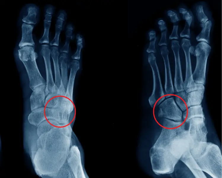
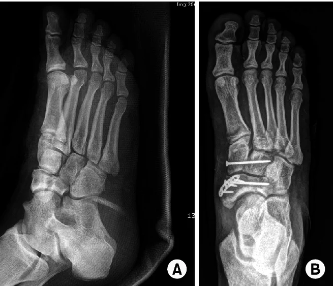
X-Rays
- Initial standard X-rays were taken to evaluate for fracture
- Can detect fracture through visible lines or cortical disruptions
- Not able to provide clear detail on intra-articular fractures, small avulsions
CT Scan
- More advanced imaging with 3D reconstructions
- Better visualizes the shape and size of cuboid fragments
- Identifies intra-articular extension into joints or comminution
- It is helpful when an X-ray fails to show a clear fracture pattern
Physical Exam
- Swelling, ecchymosis, or deformity is assessed first before palpation
- Direct palpation for point tenderness over the cuboid bone
- Special tests: midtarsal adduction/abduction stress tests
Findings are used to characterize the fracture pattern so appropriate treatment can be selected.
Treatment of Cuboid Bone Fracture
Initial Management
For suspected cuboid fractures, the foot is splinted or placed in a post-op shoe immediately after injury to stabilize and offload the area. This limits additional fragment displacement before definitive treatment.
The patient is made strictly non-weight-bearing and likely placed on crutches for 1-2 weeks. Weightbearing too early risks further displacement and damage which can impair long-term outcomes.
Anti-inflammatory medications like NSAIDs are often prescribed to help control pain and swelling. Ice is also frequently applied. Early intervention helps mitigate inflammation that causes secondary injury.
Diagnosis
Once the acute injury settles, standard foot and ankle X-rays are obtained to characterize the location, pattern, and displacement of the cuboid fracture along with ankle/hindfoot alignment. Additional advanced CT or MRI imaging may be ordered for surgical planning purposes.
Based on the imaging and exam, the supervising orthopedist determines optimal management – nonsurgical or surgical. Stable, nondisplaced extra-articular fractures may be treated conservatively in a cast while displaced/comminuted patterns often necessitate surgery.
Nonsurgical Approach
If non-operative:
- Closed reduction is performed if needed to realign fragments
- Short-leg, non-weight-bearing cast or walking boot applied for 6 weeks
- Serial radiographs at follow-ups to ensure maintenance of position while healing
- Begin protected weight bearing at 6 weeks in a walking boot
- Transition to supportive shoes over the next 6 weeks
- Formal physical therapy starts 12 weeks post-injury with an emphasis on stability, mobility, and progressive strengthening
Surgical Approach
- If surgery is indicated:
- Open reduction and internal fixation (ORIF) within 1-2 weeks
- Intra-op fluoroscopy used to manipulate fragments back into anatomic alignment which is then secured with screws/plates
- May utilize bone graft to fill defects
- Post-op non-weight bearing x 6-10 weeks until initial healing occurs
- Similar boot transition, PT schedule as above
Throughout the treatment and rehabilitation process, serial imaging and clinical examination monitor healing progress to ensure the best possible outcome.
Prevention
Properly Fitted Shoes
- Too-tight or too-loose shoes can raise the danger
- Loose shoes allow too much motion, leading to instability
- Tight shoes cause pressure and irritation
- Well-fitted shoes with adequate width and depth are ideal
- Soles should provide cushioning to absorb impact
- Provides support during activity without compressing foot
Ankle Support
- Ankle braces or high-top shoes provide extra stability
- Restrict excess inversion/eversion during activity
- Useful for sports like basketball, tennis, hiking
- Can also tape the ankle in the figure-8 method to limit motion
Avoid Trauma
- Use caution on uneven terrain that risks ankle rolls
- Don’t jump down from heights which causes axial load
- Build strength to prevent falls and trips
- In vehicles, avoid direct impact over the lateral foot
- Take frequent breaks during endurance activities
Implementing good footwear habits, ankle stabilization when needed, and practicing injury prevention during activities can help promote cuboid safety and potentially prevent painful cuboid fractures. Let me know if you have any other questions!
Summary
The cuboid is a cube-shaped tarsal bone located on the lateral side of the midfoot that plays an integral role in stabilizing the foot during gait. Cuboid fractures account for 2-8% of midfoot fractures. These fractures disrupt the transfer of weight from the heel to the lateral metatarsals, often necessitating treatment.
Cuboid fractures have several common injury patterns, including extra-articular (confined to the bone) or complex intra-articular fractures extending into neighboring joints. Avulsion fractures are small chips pulled off by tendons. Symptoms include midfoot pain and swelling, inability to bear weight, and possibly bone deformity.
Diagnosis often starts with X-rays and CT scans to fully characterize the type and displacement of the fracture. Most cuboid fractures necessitate casting/immobilization along with restricted weight bearing to facilitate healing. However, surgical treatment with open reduction and internal fixation is frequently required for displaced or unstable fracture patterns.
While prevention is challenging, properly fitted orthotic shoes and ankle support during vigorous activity can aid stability and minimize trauma that could risk cuboid injury. Caught early, most cuboid fractures heal well, but neglect can result in chronic pain and disability.
FAQs
When is surgery needed?
Surgery is often necessary for displaced, intra-articular, or unstable cuboid fractures to achieve proper realignment and stabilization with internal fixation. Conservative treatment can be successful in mild cuboid fractures.
What are the most common symptoms of a cuboid fracture?
The most common symptoms are pain, swelling, and bruising on the outer midfoot by the cuboid bone. There is also usually tenderness when pushing directly on the cuboid. Many people are unable to bear full weight on the injured foot.
How long does it take a cuboid fracture to heal?
Uncomplicated cuboid fractures can take approximately 6-8 weeks to heal with proper treatment. Intra-articular, comminuted, or displaced fractures often have a longer recovery timeline closer to 12 weeks or more.
What are the treatment options for minimally displaced cuboid fractures?
Minimally displaced extra-articular cuboid fractures are often treated non-operatively with a short leg cast or walking boot for 6 weeks along with restricted weight bearing. Serial imaging checks for maintenance of fracture position.
Can a cuboid fracture heal on its own without treatment?
A minimally displaced cuboid fracture can heal on its own over time. However, the fracture fragments tend to settle into an abnormal position since there is no stabilization, which can negatively impact long-term foot biomechanics. Proper diagnosis and management are recommended.
What can be done to prevent cuboid stress fractures?
To help prevent painful cuboid stress fractures, make sure to wear properly fitting shoes with adequate cushioning and support. Slowly increase the intensity with new activities and weight-bearing exercises. Also strengthen your feet and ankles through regular stretching, massaging, and icing after strenuous activity involving the feet.
References
Cuboid fracture – WikEM. (n.d.). https://wikem.org/wiki/Cuboid_fracture
Lau, H. (2023, August 14). Cuboid Stress Fractures. StatPearls – NCBI Bookshelf. https://www.ncbi.nlm.nih.gov/books/NBK542250/
McCormack, J. (2023, March 27). Cuboid Fracture. James McCormack. https://james-mccormack.com/advice-centre/cuboid-fracture/

One Comment