
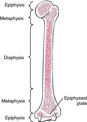
Epiphysis
Overview
When full growth is reached, the extended end of the long bones, known as the epiphysis, fuses with the bone shaft after ossifying independently. The spongy cancellous bone that makes up the epiphysis is covered in a thin layer of solid bone.
The epiphyseal cartilage, also known as the growth plate, connects it to the bone shaft and helps the bone grow longer before being replaced by bone.
Definition
Particularly toward the end of the long bone, it is a portion of the bone that ossifies independently before becoming ankylosed to the main portion of the bone.
It is an important growing region close to the end of a long bone that ossifies to become one with the main bone. More specifically, it is the rounded tip of any long bone where the segment connects to neighboring bones. Only the ends of lengthy bones include this section of bone. To support joints and provide room for the attachment of ligaments and tendons, it is enlarged.
The rounded areas at the ends of a bone that divide the metaphysis from the physis are called epiphyses (singular: epiphysis). Unlike an apophysis, which is a place of tendon or ligament attachment, an epiphysis is a joint component. The metaphysis and epiphysis unite once the growth plate has united.
The spongy bone known as trabecular tissue makes up the epiphysis. The primary functional unit of this kind of bone tissue is the trabecula, which makes up the structural framework of the bone. Between the trabeculae is where the red bone marrow that goes through hematopoiesis is located. It is the job of the osteoblasts covering the epiphysis to transform a spongy bone into a compact bone.
The rounded tip of a long bone is called an epiphysis, and its main job is to link neighboring bones to form joints. Another notable component is the long bone’s diaphysis, or shaft. Between the diaphysis and the epiphysis lies another segment of the long bone that we refer to as metaphysics.
At the joint site, the articular cartilage encircles the epiphysis. It is separated into pieces that are proximal, radial, and distal. It is frequently confused with the brain’s tiny endocrine gland, epiphysis cerebri. Ephephyses is its plural. The most remarkable feature is that the Epiphysis contains red bone marrow, which is used to make erythrocytes or red blood cells.
The metaphysis contains the epiphyseal plate, also known as the growth plate of the epiphysis. Articular cartilage also covers the epiphysis at the joint. Conversely, the bone beneath the articular cartilage and its development plate is known as the subchondral bone.
Its main function is to create joints by joining adjacent bones. The other noteworthy part is the diaphysis, or shaft, of the long bone. The part of the long bone between the diaphysis and the epiphysis is called the metaphysics.
The epiphyseal growth plate or epiphyseal plate is located in the metaphysis. At the joint, articular cartilage also covers the epiphysis. Conversely, the bone underlying the articular cartilage and its growth plate is called the subchondral bone.
Location Of Epiphysis
It exists within the joints. The epiphysis is specifically located near the cartilaginous end of the long bones’ articular surface. It is the ossification’s secondary center.
The rounded end of the bone, situated at the end of the diaphysis and away from the bone’s central point, is referred to as the distal Epiphysis.
Types Of Epiphysis
1) Pressure epiphysis
It is located at the extremities of the long bones.
It’s articular, meaning it plays a part in how joints form.
participates in the weight transfer process.
Examples include the femur’s head and the lower end of the radius.
The part of the long bone that forms the joint, such as the head of the femur, which is a component of the hip joint complex, is called a pressure epiphysis. The parts of the bone under pressure during movement or locomotion are called pressure epiphyses, and they help transfer the body’s weight.
Another example of a pressure epiphysis is the head of the humerus, which is a component of the shoulder complex. The femur of the tibia and the condyles are similarly affected by the pressure epiphysis.
In other words, the secondary center of ossification emerges earlier in pressure epiphyses than in traction epiphyses because pressure epiphyses ossify earlier than traction epiphyses.
2) Traction epiphysis
It offers attachment to the muscle or muscles since it is created as a result of muscular pulling.
It isn’t articulated.
It does not contribute to the transfer of weight.
The mastoid process of the temporal bone, the larger and lesser trochanters of the femur, and the tubercles of the humerus are a few examples.
The long bone’s non-articular areas are not involved in the creation of joints. In contrast to pressure epiphyses, these regions do not help with weight transmission. Because these regions of the bone are close to the pressure epiphysis location, the supporting ligaments and tendons attach to them. Traction epiphysis ossifies after pressure epiphyses.
Two examples of traction epiphyses are the larger and lesser tubercles of the humerus and the trochanters of the femur.
3) Atavistic epiphysis
is a component of another bone in humans, but it is phylogenetically an independent bone in lower species.
Examples are the talus’s posterior tubercle and the scapula’s coracoid process.
a bone joined with another phylogenetically separate bone. An example of atavistically fused bones is the coracoid process of the scapula, which has fused in humans but is separate in four-legged animals. The posterior tubercle of the talus, or os trigonum, is another instance of an atavistic epiphysis.
4) Aberrant epiphysis
The aberrant epiphysis is not always present but may be present in some individuals.
Examples: epiphysis at the head of the first metacarpal and base of other metacarpals.
Normally metacarpals have only one epiphysis i.e. base of 1st metacarpal and the heads of other metacarpals.
These epiphyses diverge from the norm and are not always present. Examples include the epiphysis at the base of other metacarpal bones and the head of the first metacarpal bone.
5) Pseudo Epiphysis
A transverse notch that mimics a growth plate and denotes a pseudo-epiphysis is the epiphysis-looking end of a bone that is not an epiphysis. Conversely, these transverse notches do not affect longitudinal bone formation because they do not include the typical cell columns present in regular growth plates.
At the distal end of the first metacarpal bone and the proximal end of the second metacarpal bone, pseudo-epiphyses are found in 80% of the normal population.
Epiphysis In Bone
Numerous bones have an epiphysis.
- Humerus; It is situated between the elbow and the shoulder.
- Radius; Is one of two bones situated anatomically between the ulna and the hand, the radius lateral to it.
- Ulna; The anatomical location of the ulna is medial to the radius, and it is one of two bones situated between the hand and the elbow.
- Metacarpal; These are the hand’s bones, and they are located closest to the phalanges.
- Phalanges; The bones of the toes and fingers, which are located farther apart from the foot’s metatarsals and the hand’s metacarpals.
- Femur; Located in the thigh, between the hip and the knee, it is the longest bone in the human body.
- Tibia; One of the two lower leg bones, it bears the majority of the body’s weight-bearing duties. It is medial located to the fibula.
- Fibula; One of the two bones in the lower leg, it is smaller than the tibia and located laterally.
- Metatarsal; On the first metatarsal, these are the bones of the foot that are closest to the medial cuneiform; on the other four, they are closest to the phalanges.
Development Of Epiphysis
In babies, the epiphysis is cartilaginous, either entirely or mostly. It is initially made up of growth and articular cartilage, but when a secondary ossification center develops, these cartilage types differentiate.
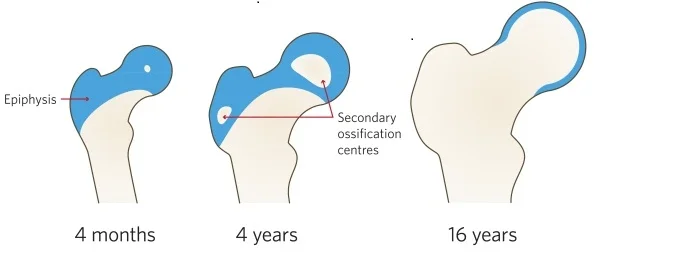
Around four months, the proximal femoral epiphyseal secondary ossification center develops. At skeletal maturity, the cartilaginous (blue) region is nearly entirely replaced by bone as the ossification centers gradually enlarge.
Following the establishment of the secondary ossification center, the lower portion of the epiphyseal cartilage ossifies to create the epiphyseal bone plate. The secondary ossification center first grows in all directions, forming radially oriented spongy bone trabeculae. But when the front approaches the area where the growth plate’s resting cartilage is, the process of ossification significantly alters.
Here, cartilage gives way immediately to compact bone tissue, forming a flat plate with multiple layers of closely spaced bony lamellae that are positioned perpendicular to the bone’s long axis.
Certain unique features distinguish the creation of the epiphyseal bone plate from a typical endochondral ossification process. It is first understood to be a specialized growth of the cortical bone, which is typically created by direct intramembranous ossification. Second, its orientation is uncommon as most bone structures are oriented so that the direction in which they sustain load makes them stronger: perpendicular to the long axis of the bone.
Thirdly, it develops more swiftly than any other area of the bone, allowing cartilage to turn into bone more quickly. Lastly, it forms in the area directly next to the growth plate’s resting cartilage, and its near spatial proximity implies that the latter may have a significant impact on its development.
Notwithstanding the factors as mentioned above and the possibility that the epiphyseal bone plate is a structural element of the growth plate that affects its functionality, this region of the long bones has received little research and is among the least understood. Not much is even known about how disorders linked to anomalies of the bones and growth failure influence it. Based on this, the current study aimed to provide a general review of the physiological processes involved in the formation of the epiphyseal bone plate.
Longitudinal and circumferential bone growth
Blood flow is restricted by the physis, which may be crucial for healing following physical separations. Knowing which physics, at the moment of separation, are susceptible to total disconnection of the blood supply might help predict the likelihood of long-term problems like avascular necrosis (e.g. slipping upper femoral epiphysis).
Because the physis is radiolucent, an x-ray cannot detect it, but the Harris lines can be used to infer indirectly what the physis does. These tiny, radiodense streaks are a typical occurrence in the metaphysis of bone.
Functions Of Epiphysis
Red bone marrow, the primary source of erythrocytes, or red blood cells, makes up this substance. Additionally, it aids in the transfer of weight from locations that experience extreme tension and pressure. To put it simply, its job is to help with improved mobility by evenly distributing pressure throughout the joints. The larger section’s porous nature helps to reduce the bone’s weight.
As new cartilage is pushed to the border of the growth site in the epiphyseal plate, long bones elongate in young infants. Conversely, the diaphysis’s older cartilages undergo a process that results in the formation of new bones. Usually, between the ages of 18 and 25, bone development stops. Typically, this stage is referred to as epiphyseal closure.
Disturbance Of Epiphysis
The knobby growth end is frequently more susceptible to slipping capital or sub-capital femoral epiphysis, a condition in which the femur and hip joint separate. Traumatic forces can cause fractures in the tibia, the terminal portion of the inner and bigger of the lower limb’s two bones that run from the knee to the ankle.
Children frequently sustain fractures to their fibulae epiphyses. Sometimes there is intense pain and limited movement due to inflammation and irritation of the calcaneus, or heel bone. Growth abnormalities are caused by an uncommon malformation called the longitudinal epiphyseal bracket, which affects both the long and short bones of the limbs.
Notably, Multiple Epiphyseal Dysplasia (MED) is a rare hereditary illness that adversely affects the end of the long bones among all other types of epiphyseal abnormalities. The problem is caused by the formation of cartilage, which causes the epiphyseal plate, also known as the growth plate, towards the end of the long bones to expand outward.
Over time, the cartilage that expands outward from its form mineralizes and hardens (a process known as ossification). This type of epiphyseal disease is faulty and can be attributed to multiple causes.
FAQs
What is the epiphysis of the bone?
A long bone’s enlarged, wide end, or epiphysis, is only present on the ends of long bones and is where the bone articulates with other bones at joints. Widening it makes it stronger and makes room for tendons and ligaments to attach.
What is epiphyseal?
A flat bony structure situated between the long bones’ epiphysis and metaphysis is called the epiphyseal bone plate. It contains the growth plate cartilage, which gives strength and stability to the weakest part of the forming bone.
Where is the epiphysis located?
The junction at the end of the bone and the growing plate, or physis, are separated by the epiphysis. In both humans and mice, the majority of tubular bones grow an epiphysis on both ends. This structure is typically formed at one end solely by certain bones, such as the phalanges, metacarpal, and metatarsal bones.
What is epiphysis made of?
The epiphysis is composed of spongy cancellous bone that is covered in a thin layer of solid bone. The epiphyseal cartilage, also known as the growth plate, connects it to the bone shaft and helps the bone grow longer before being replaced by bone.
Does the epiphysis grow?
The mechanism by which bones lengthen at the epiphyseal plate is comparable to endochondral ossification. Through mitosis, the cartilage in the vicinity of the epiphyseal plate next to the epiphysis keeps growing. In the area adjacent to the diaphysis, the chondrocytes deteriorate with age.
Does epiphysis disappear?
In long bones, the epiphyseal growth plate is a layer of cartilage that appears solely during the growth stage and disappears shortly after puberty.
Reference
- Epiphysis | Definition, Anatomy, & Function. (n.d.). Encyclopedia Britannica. https://www.britannica.com/science/epiphysis
- Epiphysis – Explanation, Types, Bones, Similarities and Differences. (n.d.). VEDANTU. https://www.vedantu.com/biology/epiphysis
- Jones, J. (n.d.). Epiphysis | Radiology Reference Article | Radiopaedia.org. Radiopaedia. https://radiopaedia.org/articles/epiphysis
- JANGHU, P. K. (2018, June 29). Epiphyses – Types – Anatomy QA. Epiphyses – Types – Anatomy QA. https://anatomyqa.com/epiphysis-types/
- Fracture Education : Anatomic differences: child vs. adult. (n.d.). Fracture Education : Anatomic Differences: Child Vs. Adult. https://www.rch.org.au/fracture-education/anatomy/Anatomic_differences_child_vs_adult/
- Acharya, M. (2013, February 16). Epiphysis – Definition, Location, Function, and Pictures. Epiphysis – Definition, Location, Function, and Pictures. https://www.knowyourbody.net/epiphysis.html
- Fernández-Iglesias, N., Fuente, R., Gil-Peña, H., Alonso-Durán, L., Santos, F., & López, J. M. (2021, January 18). The Formation of the Epiphyseal Bone Plate Occurs via Combined Endochondral and Intramembranous-Like Ossification. PubMed Central (PMC). https://doi.org/10.3390/ijms22020900

One Comment