
Erector Spinae Muscle
Introduction
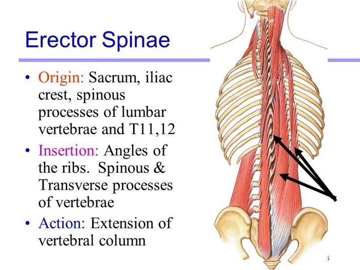
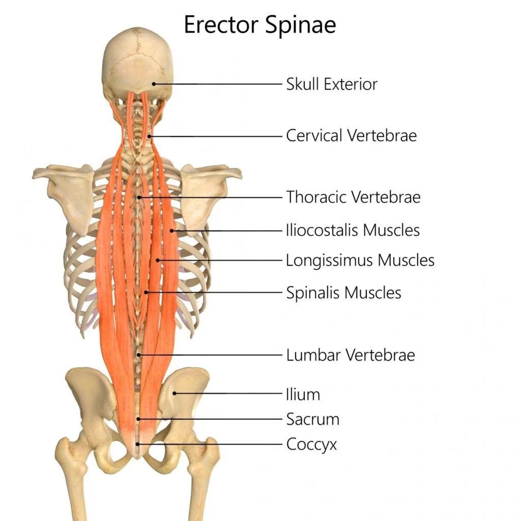
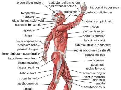
The erector spinae is a series of muscles that extend from the sacrum to the neck and is located within the back, which is the largest muscle group in the body. The muscles of this specific group are divided into three different sections, which are the iliocostalis, longissimus and spinalis, which all have different functions.
The erector spinae muscle, also known as sacrospinalis and extensor spinae in some texts is from the deep muscles of the back.
It lies superficial to the transversospinales muscle group and deep to the intermediate group of back muscles (serratus posterior superior and inferior).
The erector spinae muscle extends the vertebreal column.
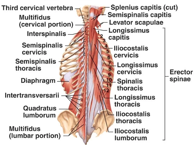
It is formed of 3 muscles and its fibers run more or less vertically throughout the lumbar, thoracic and cervical regions.
It lies in the groove to the side of the vertebral column.
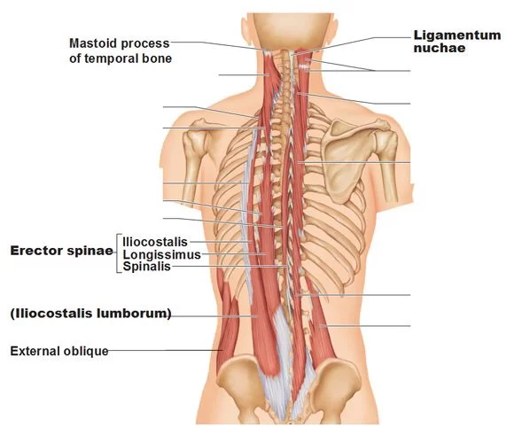
In cervical region it is covered by nuchal ligament and in thoracic and lumber region by thoracolumbar fascia.
The erector spinae are divided into three groups, from medial to lateral:
- Spinalis muscles
- Longissimus muscles
- Iliocostalis muscles
The function of the spinal erectors is to move the vertebral column. Bilateral contraction of these muscles extends the spine, while unilateral contraction causes lateral flexion (ipsilateral).
They also help to maintain posture by steadying the spine on the pelvis during walking.
Anatomy of Erector Spinae Muscle
Spinalis:
- Is the most medial part just next to the spine. It connects the spinous process of the adjacent vertebrae to each other.
- The spinalis muscles are innervated by the lateral branches of the posterior rami of the cervical, thoracic, and lumbar spinal nerves.
- The blood supply to the spinalis muscles comes from the vertebral, deep cervical, occipital, intercostal, and lumbar arteries.
- The function of the spinalis muscles is to extend and laterally flex the cervical and thoracic regions of the spine.
It is divided into 3 parts:
| Muscle | Origin | Insertion |
| Spinalis capitis | Usually blends with semispinalis capitis | With semispinalis capitis |
| Spinalis cervicis | Spinous process of C7 (sometimes T1 to T2) and ligmentum nuchae | Spinous process of C2 and C3-C4 |
| Spinalis thoracis | Spinous process of T11 to L2 | Spinous process of upper thoracic vertebrae |
Longissimus:
- It forms the middle part of the erector spinae muscles, lateral to the spinalis.
- The longissimus muscle forms the main meat of the erector group. It attaches along the transverse process of the vertebrae.
- The longissimus muscles have an identical innervation to the spinalis muscles; from the lateral branches of the posterior rami of the adjacent spinal nerves.
- They receive arterial blood from branches of the vertebral, deep cervical, occipital, transverse cervical, intercostal, and sacral arteries.
- The function of the longissimus muscles is to extend and laterally flex (ipsilateral) the spine. Longissimus capitis also helps to rotate the head ipsilaterally.
It is divided into 3 parts:
| Muscle | Origin | Insertion |
| Longissimus capitis | C4-T4 transverse process | Posterior edge of the mastoid process |
| Longissimus cervicis | T1-T4 transverse process | C2 to C6 transverse process |
| Longissimus thoraci | Transverse process of lumbar vertebra and blends with iliocostalis in the lumbar region | Transverse process of all thoracic vertebrae |
Iliocostalis:
- It is the most lateral part of the erector spinae muscles. It attaches to the ribs.
- Because of it’s lateral position, a tight iliocostalis can bring a hip up, or bring the ribcage down toward the hip.
- Similar to the other erector spinae groups, iliocostalis muscles are innervated by the lateral branches of the posterior rami of the cervical, thoracic, and lumbar spinal nerves.
- Arterial supply is via branches of the occipital, deep cervical, vertebral, posterior intercostal, subcostal, lumbar and lateral sacral arteries.
- The function of the iliocostalis muscle is to extend and laterally flex (ipsilateral) the spine.
It is divided into 3 parts:
| Muscle | Origin | Insertion |
| Iliocostalis cervicis | Angle of ribs 3-6 | Transverse process of C4-C6 |
| Iliocostalis thoracis | Angle of lower six ribs | Angles of upper six ribs and transverse process of c7 |
| Iliocostalis lumborum | Iliac crest | L1-L4 lumbar transverse processes, angle of 4-12 ribs and thoracolumbar fascia |
STRUCTURE OF ERECTOR SPINAE:
- The erector spinae is not just one muscle, but a group of muscles and tendons which run more or less the length of the spine on the left and the right, from the sacrum or sacral region (the bony structure beneath the lower back [lumbar] vertebrae and between your hips/glutes) and hips to the base of the skull.
- They are also known as the sacrospinalis group of muscles. These muscles lie on either side of the vertebral column spinous processes (the bony points up and down the middle of the back) and extend throughout the lumbar, thoracic, and cervical regions (lower, middle, and upper back and the neck).
- The erector spinae is covered in the lumbar and thoracic regions (lower back and lower middle back) by the thoracolumbar fascia, and in the cervical region (neck) by the nuchal ligament.
- This large muscular and tendinous mass varies in size and structure at different parts of the vertebral column. In the sacral region, it is narrow and pointed, and at its origin chiefly tendinous in structure.
- In the lumbar region, it is larger, and forms a thick fleshy mass. Further up, it is subdivided into three columns. They gradually diminish in size as they ascend to be inserted into the vertebrae and ribs.
- Picture a tree trunk branching out left and right.
- The erector spinae is attached to the medial crest of the sacrum to the spinous processes of the lumbar (bony points along your lower back) and the eleventh and twelfth thoracic vertebrae and the supraspinous ligament, to the back part of the inner lip of the iliac crests (the top border of your hips), and to the lateral crests of the sacrum, where it blends with the sacrotuberous and posterior sacroiliac ligaments.
- Some of its fibers are continuous with the fibers of origin of the gluteus maximus muscle.
ACTION:
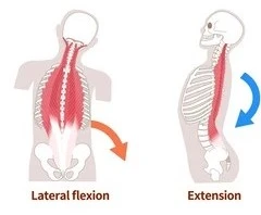
- Bilateral contractions straighten the back and pull the head posteriorly and are involved in the control of the flexion of the vertebral column.
- Unilateral contraction bends the vertebral column laterally and turns the head towards the contracting side.
Nerve Supply:
- Doral rami of spinal nerves
Blood Supply:
- Branches of the vertebral, deep cervical, occipital, transverse cervical, posterior intercostal, subcostal, lumbar and lateral sacral arteries.
Clinical Importance:
- The ESP block is a newer regional anesthetic that can provide thoracic, abdominal, and even some lower extremity analgesia.
- A versatile block, ESP blocking has been used by anesthesiologists to provide analgesia for a myriad of conditions from chronic shoulder pain to pain following hip surgery.
- Much of the information on the efficacy of ESP blocking is from case reports and anecdotal experiences, so formal research is underway to determine if ESP blocks lead to a statistically significant reduction in opioid consumption, lower pain scores, and potential length of hospital stay.
FAQ
What is the erector spinae muscle?
The middle layer of the deep (intrinsic) back muscles is made up of the erector spinae muscles. They stretch between the base of the skull superiorly and the pelvis inferiorly on either side of the spinal column.
What is the main function of the erector muscle?
The erector spinae muscle group has two main purposes: it extends and laterally flexes the spine. To enable aerobic motions like backbends, these muscles contract on various back regions.
What is the action of the erector muscle?
The primary functions of the erector spinae muscles are to maintain proper posture and the curvature of the spinal column, as well as to extend and laterally flex the back. It’s understandable why so many people experience low back discomfort given the abundance of muscles in the lower back!
What do the erector spinae muscles primarily function in?
Stretching from the sacrum to the head, the erector spinae group of muscles is a substantial muscle mass on both sides of the spinal column. The major function of these muscles is to stretch the spinal column in order to preserve an upright posture.
What is the erector spinae also known as?
The term “erector spinae” refers to a collection of muscles and tendons that extend roughly down the left and right sides of the spine, from the hips and sacrum to the base of the skull. The sacrospinalis group of muscles is another name for them.
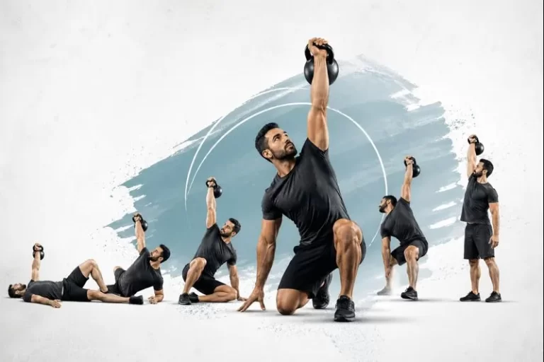
What are the benefits of strong erector spinae muscles?
You are powerful, aligned, and stable thanks to your erectors. Back discomfort and bad posture can result from weak erectors. Increasing your erector strength will improve your range of motion and rotation. For those of you who like to strength train, activating your erectors is an excellent warm-up exercise before a squat or deadlift.
What causes erector spinae pain?
When the lower back’s muscles and connective tissues are overworked or strained beyond what is considered normal, an Erector spinae injury can result. Sometimes, when playing a sport, an individual may trip or fall and cause erector spinae pain.

14 Comments