
Medial Pterygoid muscle
Introduction
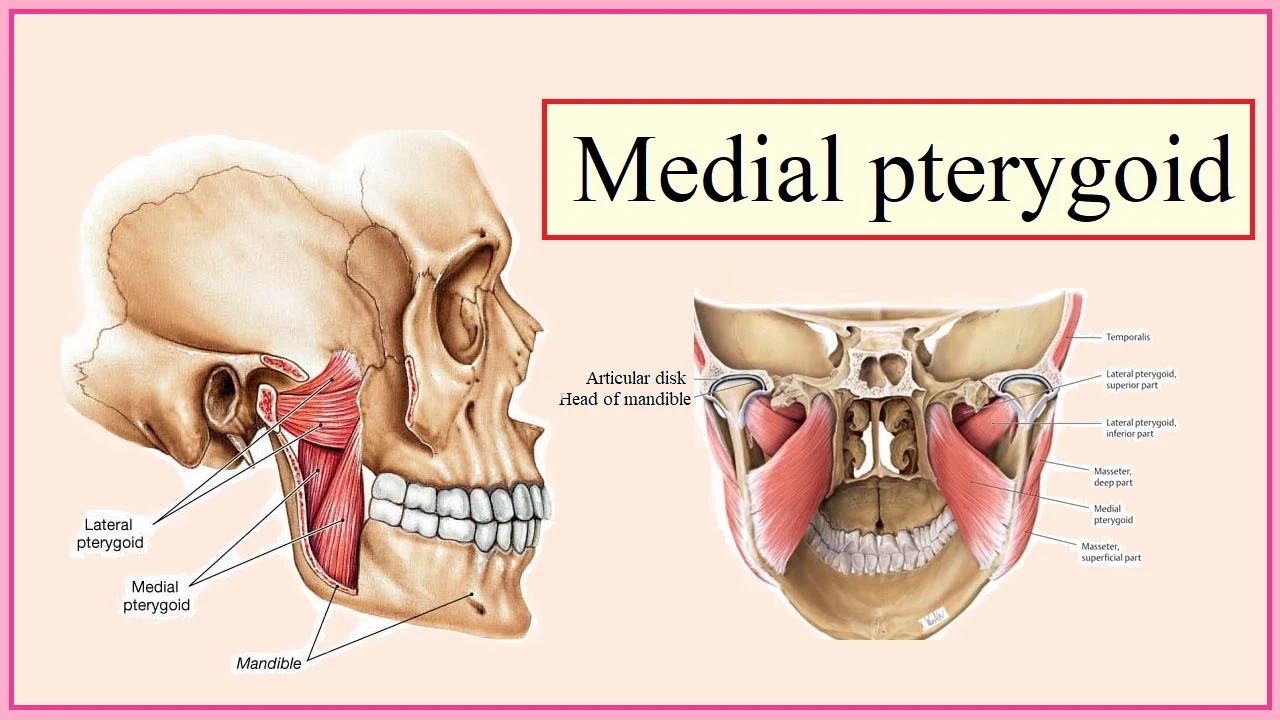
The medial pterygoid muscles, a major elevator of the jaw is a square-shaped masticatory muscle, encountered on the medial part of the lower jaw bilaterally. It is also known as the internal pterygoid muscles. These muscles lie medial to the lateral pterygoid muscles.
The medial pterygoid muscle is a thick quadrilateral muscle that binds the mandible to the maxilla, sphenoid, and palatine bones. It also belongs to the group of masticatory muscles, also along with the lateral pterygoid, masseter, and temporal muscles.
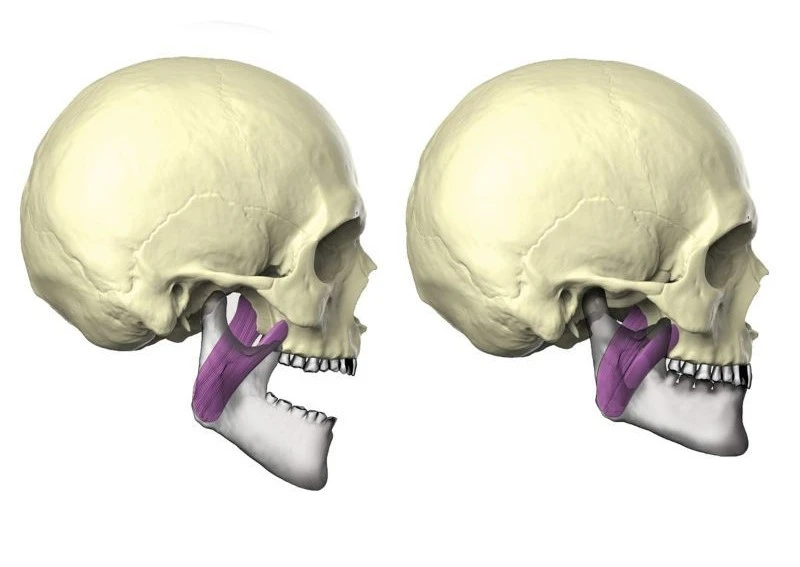
The medial pterygoid muscles consist of two heads; superficial and deep. Although including additional origins, both heads insert on the inner surface of the mandible, assembling an axis for a strong pull of this bone. Unilateral contraction of the medial pterygoid muscles induces rotation of the mandible, while bilateral contraction elevates and protrudes it. In synergy with additional masticatory muscles, these actions facilitate chewing.
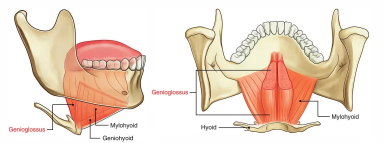
The medial pterygoid muscles have a superficial and deep head that derives from different areas of the jaw. The lateral pterygoid muscles via its lower head divide the two distinct heads. The smaller superficial head has its origin mostly from the maxillary tuberosity with some fibers originating from the pyramidal process of the palatine bone. The larger deep head retains its origin from the medial side of a horseshoe-shaped extension of the sphenoid bone (lateral pterygoid plate) at the bottom of the skull.
Its fibers pass in a downward, lateral, and posterior approach and have their insertion via a strong tendinous lamina, into the roughened area on the lower and posterior part of the medial or inner portion of the ramus and angle of the mandible, are as high as the mandibular foramen.
Origin of Medial Pterygoid muscle
Medial pterygoid muscles consist of two heads; superficial and deep, that are separated by the inferior head of lateral pterygoid muscles at their origin.
The deep portion forms the bulk of the muscle and arises from the medial surface of the lateral pterygoid plate of the sphenoid bone and the pyramidal process of the palatine bone. The smaller, superficial part arises from the maxillary tuberosity and the grooved surface of the pyramidal process of the palatine bone.
Insertion of Medial Pterygoid muscle
As these two sets of fibers descend posterolaterally, they insert into the triangular appearance found on the medial surface of the ramus and the angle of the mandible. The fibers connect via a strong tendinous lamina that expands from the mandibular foramen superiorly, to the mylohyoid groove anteroinferior.
The muscles are separated by the tendinous sheets into three layers recognizable in its upper one-third. The lingual nerve runs in an anteroinferior direction lateral to the belly of the medial pterygoid muscles and medial to the mandible. The area between the medial pterygoid muscles and the superior constrictor muscles is referred to as the prestyloid place, which is filled with fat.
The medial pterygoid muscles are found in the infratemporal fossa lying deep to the masseter and temporalis muscles and the medial to lateral pterygoid muscles.
The outer surface of the muscle lies against the inner surface of the mandible, from which it is divided by the lateral pterygoid muscle, sphenomandibular ligament, maxillary artery, mandibular nerve, and its lingual and inferior alveolar branches. About its insertion, the external surface of the medial pterygoid muscles is corresponding to the process of the parotid gland.
The inner surface of the muscles are concerning tensor veli palatini, styloglossus, and salpingopharyngeus muscles. The latter two muscles diverge the medial pterygoid muscles from the superior pharyngeal constrictor. Near its insertion, the medial pterygoid muscle is medically related to the lateral surface of the submandibular gland, and the facial artery, which descends between these two structures.
Nerve supply
The medial pterygoid muscles are innervated by the medial pterygoid branches of the mandibular nerve (CN V3), one of the three branches of the trigeminal nerve (CN V). This also delivers the tensor tympani muscle and the tensor veli palatini muscle. The medial pterygoid nerve is the main trunk of the mandibular nerve, before the division of the trigeminal nerve – this is unlike the lateral pterygoid muscle and all other muscles of mastication which are provided by the anterior division of the mandibular nerve.
Blood supply
The medial pterygoid muscles receive blood supply from the pterygoid and buccal branches of the maxillary artery. To lower importance, ascending palatine artery and muscular branches of the facial artery also contribute to the blood supply of this muscle. The first muscular branch originates directly from the facial artery near the mandibular angle. The second muscular branch originates from the ascending palatine artery, which arises from the facial artery in the cervical area. The third branch is an anterior muscular branch that originates from either the tonsillar or submental branch of the facial artery. This branch penetrates the muscle from the anteromedial direction.
Occasionally, the medial pterygoid muscle may receive its vascular supply from a direct branch from the external carotid artery that enters the lower part of the muscle posteriorly about 2 cm above the gonion.
Function of Medial Pterygoid muscle
Due to its posteroinferior direction and the reality that its mandibular attachment is always mobile, the medial pterygoid muscles can exhibit several actions:
Unilateral contraction slightly medially rotates the mandible. When this action occurs simultaneously with the contraction of the ipsilateral lateral pterygoid muscle, it results in a noticeable movement; same sided portion of the mandible swings anteriorly and medially. In addition, alternating contraction of the medial pterygoid and lateral pterygoid muscles causes side-to-side movements of the mandible. Bilateral contraction elevates the mandible. This action, when connected with bilateral contraction of the lateral pterygoids protrudes the mandible.
The medial pterygoid usually works together with other masticatory muscles. Therefore, its actions converge with those of other masticators to facilitate chewing and food grinding between the maxillary and mandibular teeth.
Clinical significance
The medial pterygoid muscles can sometimes be injured during inferior alveolar nerve block due to it being close to the nerve. The injury occurs if the anesthetic needle is placed too medially and accidentally injects into the muscles instead of the inferior alveolar nerve. This can cause hemorrhage and then the result of medial pterygoid trismus appears hours to days after the process. This manifests in the incapability to completely open the mouth and significant medial pterygoid muscle pain when attempting to open the mouth outside the restriction.
The medial pterygoid muscles along with the masseter muscles form a ‘pterygomassetric sling’ that suspends the mandible. After orthognathic surgery to treat mandibular prognathism, this sling greatly influences the stability of the two segments of the mandible. Also, the healing process of bone between the two segments (distal and proximal) is subject to impact by the magnitude of force applied by the sling.
Infection in the mandibular teeth more frequently impacts the lower compartment of the masticatory space, which contains medial pterygoid muscle; this could result in pterygoid muscle abscess. Since both the masticator area and infratemporal fossa share the medial pterygoid muscle, there are chances of the potential spread of infection from this space to the infratemporal fossa.
Periodically, occlusal interference may occur due to movement of the accessory medial pterygoid muscle, which is clinically apparent on the buccal cusps of the third molar. This occlusion anomaly may act as a trigger point for a spasm of the muscle, which can be clinically inspected by fixing the mandible under pressure in the most posterior and centric position to create the interfering occlusal contacts evident.
During vertical jaw movements, the lateral pterygoid muscles act as primary muscle that generates horizontal forces. However, the medial pterygoid muscles may also play a role in controlling horizontal jaw positions.
Treatment
Open wider
Begin by gradually opening your mouth as wide as possible. If clicking or popping of the jaw appears, discontinue the stretch immediately. Maintain the stretch for ten seconds then close the jaw. Replicate this stretch ten times, three days a week.
Tongue Stretches
Move the tongue to where it relaxes on the back of the upper front teeth. Next, move the tongue back until it is relaxing on the soft tissue of the roof of the mouth. Open the mouth, making sure to hold the tongue in the same position. Hold for ten seconds and also return to the starting position. Complete three repetitions of these exercises for two duration a week.
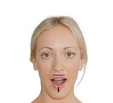
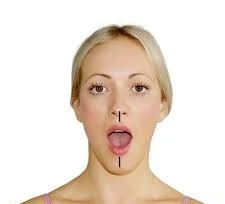
Stretching: The right medial pterygoid muscle, a muscle of mastication, is stretched by depressing and ipsilaterally deviating (deviating to the same side) the mandible at the temporomandibular joints (TMJs).
Myofascial release
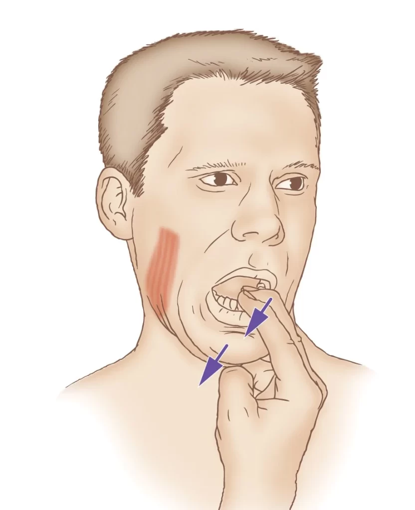
Place index finger, on muscles at the inside of bottom teeth in the mouth. Place the opposite thumb under the jaw line below the ear. Apply pressure to the muscles as if to touch the finger and thumb. Move along the gum line until reach the incisors in front. Hold until relaxes one to two times 1 to 2 per day.
FAQ
What are the origins and insertion of the medial pterygoid muscle?
Located deep in the posterior cheek region, the medial pterygoid muscles maintain two heads that originate from: the medial pterygoid plate; the maxillary tuberosity; and the pyramidal process of the palatine bone. The muscles insert into the ramus and the gonial (or mandibular) angle.
What are the movements of the medial pterygoid muscle?
The medial pterygoid muscle has functions including elevating the mandible (closing the mouth), protruding the mandible, mastication (especially when the maxillary teeth and the mandibular teeth are close together), and excursing the mandible (contralateral expedition occurs with unilateral contraction).
What causes medial pterygoid pain?
Any posture or activity where the head is moved forward moves the lower jaw into a position that places this muscle under tension. In the lengthy run, this can overload the medial pterygoid and activate trigger points.
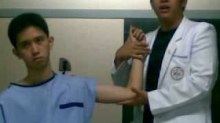
How do you test a medial pterygoid muscle?
Ask the patient to extend about 10-15mm from intercuspal contact. Slide your forefinger posteriorly along the buccal area. Palpate the muscles by pressing medially as well as posteriorly-superiorly. Ask if the patient senses any tension or tenderness.
How do you stretch the medial pterygoid?
Slide your finger between your teeth until it reaches the bone at the back. Slide your finger around it going further inside. The pad of your finger is on the medial pterygoid muscle. Curl your fingertip against it to expert pressure.
One Comment