How Many Bones In The Skull
Introduction
The cranium, or skull, is made up of 22 bones, of which 8 are cranial and the remaining 14 are facial.
Protection and structure are the primary purposes of the skull’s bones and the meninges that encircle them. Defense of the eye orbits and brain (cerebellum, cerebrum, brainstem). It serves as a structural anchor for the facial and scalp muscles’ tendinous and muscular attachments. The numerous nerves and veins that supply and innervate the skin, facial muscles, and brain are likewise shielded by the skull.
Anatomy
Parts of the skull
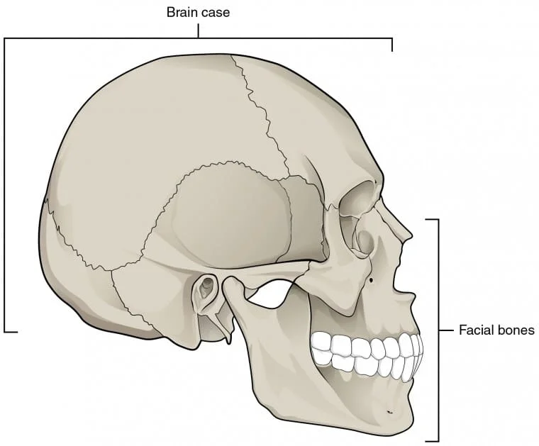
There are two major sections of the human skull. The component that surrounds the brain is called the neurocranium, or braincase, while the part that creates the facial skeleton is called the viscerocranium.
The two divisions of the skull are the cranial base, also known as the skull base, and the calvaria, also known as the skullcap or skull vault.
Covering the cranial cavity, the calvaria is the upper portion of the neurocranium. The floor of the cranial cavity and the inferior region of the viscerocranium are components of the cranial base.
Neurocranium
The craniofacial cavity is surrounded by the neuron. The brain’s entire structure is safeguarded, encompassing the brainstem, cerebellum, cerebrum, and nervous system’s associated sensory organs. The nose cavity’s olfactory receptors, the eyes, the ears, and the tongue—which has taste buds—are among these organs.
The neural surrounds the craniofacial cavity. The entire structure of the brain is protected, including the brainstem, cerebellum, cerebrum, and the sensory organs of the nervous system that are related to it. These organs include the tongue, which possesses taste buds, the eyes, the hearing, and the olfactory receptors in the nose cavity.
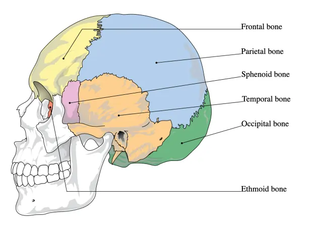
Bones of neurocranium
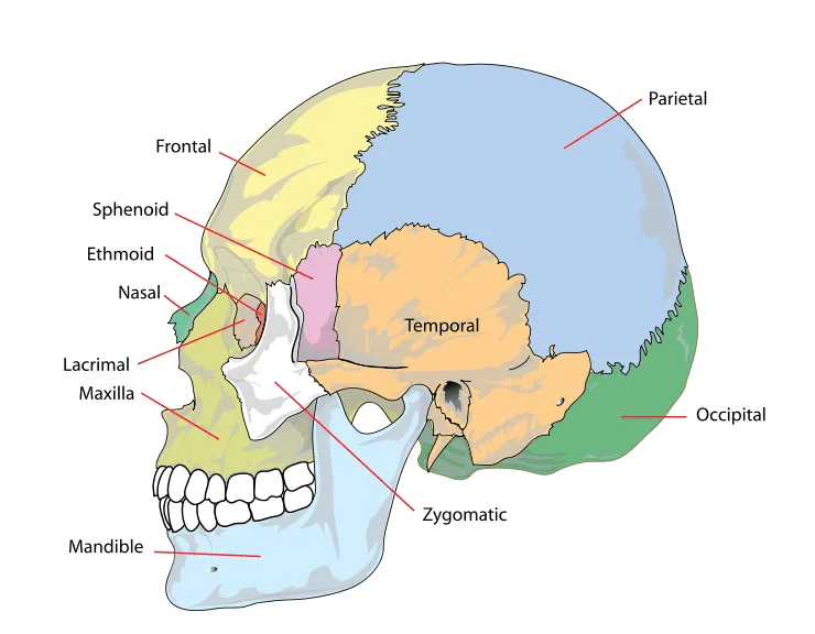
Two paired and four unpaired (single) bones make up the neurocranium. The following bones are not yet paired:
- Sphenoid bone -It forms the back of the orbit and is positioned in the center of the skull toward the front;
- Frontal bone– builds the forehead’s bones;
- Occiput bone– forms the base of the skull and its back;
- Ethmoid bone– situated in front of the skull, dividing the brain and cranial cavity from the nasal cavity.
The paired bones of the neurocranium are:
- Temporal bones– situated at the skull’s sides and base;
- Parietal bones– comprise the sides and roof of the skull, and are positioned above the temporal bones.
The paranasal sinusus is a collection of air-filled cavities found only in the sphenoid, frontal, and ethmoid bones.
Viscerocranium
The region directly in front of the neurocranium is called the viscerocranium. It shapes the skeleton of the jaw and the face. The face’s soft tissue is also supported by the viscerocranium.
Bones of viscerocranium
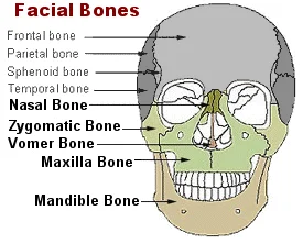
Six paired and two unpaired bones make up the viscerocranium. The following are the paired bones:
- The tiniest bones in the face, the lacrimal bones make up the orbit’s medial wall;
- Nasal bones: these bones create the nose bridge and are positioned in the midline of the face;
- The nasal cavity’s lateral wall contains the inferior nasal conchae.
- Zygomatic bones: these contribute to the orbits and form the cheekbones;
- Maxillae: These two bones combine to form a single bone in the midline of the face, the maxilla, which forms the upper jaw and helps create the hard palate;
- The floor of the nasal cavity and the roof of the oral cavity are formed by the hard palate, which is formed by the midline fusion of the palatine bone.
And the unpaired bones of the viscerocranium are:
- The nasal septum’s posterior portion is formed by the vomer;
- The mandible, which is made up of the lower jaw, is joined to the skull via the temporomandibular joint.
There is only one paranasal sinus (maxillary sinus) in the viscerocranium, which is found in the maxilla bone. The maxilla is the most noticeable immobile bone in the bones of the face.
The unpaired hyoid bone, albeit it is in the upper neck area, is occasionally also categorized as a viscerocranium bone. Ligaments serve as a link between the hyoid bone and the skull.
Calvaria
The calvaria and the cranial cavity are two other divisions of the skull previously mentioned. The top portion of the neurocranium is called the calvaria. It envelops the brain-containing cerebral cavity. It is sometimes referred to as the cranial roof or skullcap. The brain is encircled and shielded by the sturdy calvaria. However, the cranial base, which is mostly made up of thin-walled bones, is more sensitive.
The parietal, frontal, and occipital bones are sutured together to form the calvaria. To be more specific, it is made up of the larger wing of the sphenoid bone, the parietal bones, the squamous part of the temporal bone, the squamous part of the frontal bone, and the squamous section of the occipital bone (above the superior nuchal line).
The cranial base and calvaria are separated by the following structures:
- The frontal bone’s supraorbital border A process of the front bone called zygomatic Infratemporal crest on the sphenoid’s larger wing Zone of the temporal bone zygomatic process The temporal bone’s mastoid process The occipital bone’s superior nuchal line Occipital protuberance on the outside
- The frontal bone’s supraorbital border
- A process of the front bone called zygomatic
- Infratemporal crest on the sphenoid’s larger wing
- Zone of the temporal bone zygomatic process
- The temporal bone’s mastoid process
- The occipital bone’s superior nuchal line
- Occipital protuberance on the outside
Cranial cavity
The space inside the skull is referred to as the “cranial cavity” or “intracranial space”. The cranial cavity contains the brain, meninges, and intracranial segments of the cranial nerve. It also contains arteries that supply the brain with arterial blood as well as veins and venous sinuses, which gather venous blood.
Embryology Of The Human Skull
Development of the Skull:
Typically, the mesenchyme surrounding the growing brain gives rise to the skull or cranium. There are two sections to it:
The neurocranium surrounds the brain like a protective shell.
The pharynx, upper respiratory tubes, and oral cavity are surrounded by the viscerocranium.
Neurocranium
The neurocranium is made up of two sections: the cartilaginous (chondrocranium), which forms the bone of the skull, and the membranous (cranial vault) component, which is made up of flat bones that surround the brain.
Membranous Neurocranium:
It develops from paraxial mesoderm and neural crest cells. This mesoderm undergoes intramembranous ossification after investing in the brain. Many flat, membranous bones then begin to develop.
The bone spicules on these bones resemble needles. Though the individual plates do not fuse, these spicules expand outward from the central ossification core toward the periphery. These flat bones are joined at birth by thin, dense connective tissue seams known as sutures of the calvaria, which are derived from paraxial mesoderm (coronal suture) and neural crest cells (sagittal suture). There are six such areas, known as fontanels, where more than two sutures converge:
- A single fontanel anterior
- A single posterior fontanel
- Posterolateral fontanels on two
- Anterolateral fontanels on two
The largest and most visible fontanel is the anterior one. The fontanels and sutures aid in the skull’s shaping during birth, allowing the skull to fit through the birth canal. The sutures here begin to close.
After birth, the posterior fontanel closes one to two months, but the anterior fontanel closes after 18 months. Even as adults, some of the sutures are still open. Because of this, we experience an intracranial pressure vibration in the anterior fontanel during the first year.
Cartilaginous Neurocranium or Chondrocranium:
It is made up of the cartilaginous base of the growing skull, which develops from neural crest cells as well as sclerotomes. The cartilages are made of neural crest cells that are located in front of the notochord’s rostral limit and terminate in the sella tunica’s center.
They form the prechondral chondrocranium. While those that lie posterior to the rostral limit are formed from occipital sclerotomes that are formed by paraxial mesoderm. They form chordal chondrocranium.
- When the cartilage from the occipital sclerotomes fuses with the basal plate, the occipital bone is created.
- The hypophysial cartilage fuses to form the sphenoid bone, while its lesser wing is formed from the fusing of ala orbitalis.
- The trabeculae carnie fuses to the ethmoid bone.
- The otic capsules form the temporal bone’s petrous and mastoid portions.
- In the end, all these bones fuse by endochondral ossification to form the base of the skull except for one part called the foramen magnum.
Viscerocranium:
Membranous and Cartilaginous portions make up the other division of the viscerocranium.
Membranous Viscerocranium:
Membranous ossification creates the zygomatic, maxillary, and squamous temporal bones in the dorsal region of the first pharyngeal arch. Subsequently, the squamous temporal bone integrates with the neurocranium.
The mandible is formed by intramembranous ossification, which condenses and ossifies the mesenchyme surrounding the Meckel cartilage in the ventral region. Later on, this Mecklel vanishes, with the sphenomandibular ligament being the only exception.
Cartilaginous Viscerocranium:
In the head area, neural crest cells often compose the majority of the mesenchyme. These cells proliferate into the pharyngeal arches, particularly the initial two, which give rise to the facial bones and tissues. While the other arches solely contain ventral sections, the first and second arches have both dorsal and ventral parts.
The malleus and incus bones are formed by the dorsal tip of the first pharyngeal arch cartilage, whereas the stapes and the styloid process of the temporal bone are formed by the dorsal end of the second.
The lesser horn and superior portion of the hyoid bone are formed by the ossification of the ventral end of the second pharyngeal arch cartilage, whereas the greater horn and inferior portion of the hyoid bone are formed by the third.
All of the laryngeal cartilages—aside from the epiglottis—are formed by the fusion of the fourth pharyngeal arches.
Views Of The Human Skull
The adult skull is made up of 22 separate bones, 21 of which are joined into a single, immobile unit. The mandible, or lower jaw, is the 22nd bone in the skull and the only one that can move.
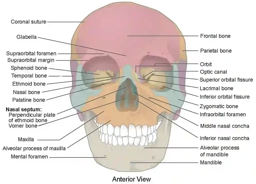
Anterior View of Skull
The skeletal support for the eyes and other facial structures is provided by the facial bones that make up the anterior skull. The main features of this skull picture are the orbital and nasal cavity openings. The lower and upper jaws, along with their corresponding teeth, are also visible.
The orbit is the bony socket in which the eyeball and the muscles that move it or lift the top lid are housed. The anterior orbit’s upper border is known as the supraorbital margin. The supraorbital foramen is a tiny aperture that is situated close to the supraorbital margin’s midpoint.
This makes it possible for a sensory nerve to reach the forehead’s skin. The anterior face below the orbit is supplied by a sensory nerve that emerges at the infraorbital foramen, located below the orbit.
The nasal septum splits the nasal cavity in half inside the nasal region of the skull. The perpendicular plate of the ethmoid bone forms the upper part of the nasal septum, whereas the volmer bone forms the lower part. The nasal cavity is triangular, with a wide inferior area that narrows superiorly on each side.
Two bony plates can protrude from each lateral wall of the nasal cavity when viewed from the front of the skull. The inferior nasal concha, a separate skull bone, is the bigger. Situated in the location of the middle nasal concha A portion of the ethmoid bone is the inferior concha. Part of the ethmoid bone, the superior nasal concha is a third bony plate. Above the middle concha, it is much smaller and hidden. The superior nasal concha is lateral to the perpendicular plate in the upper nasal cavity.
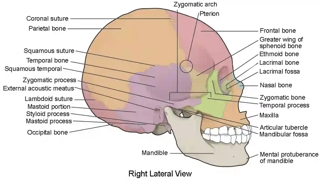
Lateral View of Skull
The broad, spherical brain case above and the top and lower jaws with their teeth below dominate a view of the lateral skull. The zygomatic arch, a bone bridge, divides these regions. The bony arch on the side of the skull that extends from the cheek region to slightly above the ear canal is known as the zygomatic arch.
The temporal process of the zygomatic bone, or cheekbone, is the short anterior component that joins the longer posterior zygomatic process of the temporal bone, which extends forward from the temporal bone, to form this structure.
The zygomatic arch is thus formed by the anterior temporal process and the posterior zygomatic process joining together like the two ends of a drawbridge. The zygomatic arch gives rise to one of the main muscles that pull the mandible upward during biting and eating.
The temporal fossa is a shallow area located above the level of the zygomatic arch on the lateral side of the braincase. The infratemporal fossa is another area that is deep within the jaw, below the level of the zygomatic arch. The muscles in the temporal and infratemporal fossas work on the mandible when chewing.
Arterial Supply Of The Human Skull
The ocular artery, a branch of the internal carotid artery, and the external carotid artery provide a strong circulatory supply to the scalp. The external carotid artery has three branches that are affected:
- Superficial temporal – provides for the temporal and frontal areas
- Posterior auricular – provides the region that is both superior and posterior to the ear.
- Occipital – supplies the scalp’s back
The supraorbital and supratrochlear arteries, two branches of the ophthalmic artery, supply the scalp with extra blood both superiorly and anteriorly. The supraorbital and supratrochlear nerves are accompanied, respectively, by these veins.
Venous Drainage Of The Human Skull
There are two categories of the scalp’s venous drainage system: superficial and deep.
The superficial temporal, occipital, posterior auricular, supraorbital, and supratrochlear veins are the arteries that supply the superficial drainage.
The Pterygoid Venous Plexus drains the deep (temporal) part of the skull. This extensive vein plexus empties into the maxillary vein and is located between the lateral pterygoid and temporalis muscles.
Crucially, valveless emissary veins allow the scalp’s veins to join the skull’s diploic veins. This creates a link between the dural venous sinuses and the scalp.
Innervation Of The Human Skull
The roots of the cervical nerve or branches of the trigeminal nerve innervate the scalp cutaneously.
Trigeminal Nerve
- Supratrochlear nerve – anteromedial forehead supplied by a branch of the ophthalmic nerve.
- Supraorbital nerve – a part of the scalp between the anterolateral forehead and the vertex that is supplied by a branch of the ophthalmic nerve.
- Zygomaticotemporal nerve – The maxillary nerve branch that supplies the temple.
- Auriculotemporal nerve – a mandibular nerve branch that provides skin anterosuperior to the ear.
Cervical Nerves
- Lesser occipital nerve – originates from the C2’s anterior ramus (division), supplying the skin behind the ear.
- Greater occipital nerve – originates from the C2 posterior ramus (division) and supplies the occipital skin area.
- Great auricular nerve – originates from the C2 and C3 anterior rami and provides the skin above the mandibular angle and posterior to the ear.
- Third occipital nerve – originates from C3’s posterior ramus and supplies the inferior occipital region’s skin.
Muscles Of The Human Skull
The majority of the striated muscles in the face and scalp are innervated voluntarily. They are essential for mastication, language, vision, and facial expression. The facial, oculomotor, or trigeminal nerves innervate most of these muscles, whereas the tongue is innervated by the hypoglossal nerve.
To produce aqueous humor and accommodating lenses, the little smooth muscles of the eye are controlled by the sympathetic or parasympathetic nervous system.
Oculomotor Nerve (CN III)
The superior rectus muscle and the striated muscle component of the levator palpebrae superior muscle are innervated by the superior division of CN III. The medial and inferior rectus muscles as well as the inferior oblique muscle are innervated by the inferior division of CN III.
Trigeminal Nerve (CNV)
Four of the eight muscles that are innervated by the mandibular nerve (V3) of the trigeminal nerve are involved in mastication. During chewing, the lateral pterygoid protrudes from the mandible, while the masseter and temporalis muscles raise it. The mandible can move laterally, rocking it back and forth, thanks to the medial pterygoid.
The tensor tympani, which dampens sound, the mylohyoid muscles, the anterior belly of the digastric muscle, and the tensor veli palatini, which tenses the soft palate, are also innervated by V3 of CN V.
Facial Nerve (CN VII)
The posterior belly of the digastric muscles, the stylohyoid muscle, and the following facial expression muscles are innervated by the motor root of CN VII:
- Buccinator
- Corrugator supercilii
- Depressor anguli oris
- Frontalis (part of occipitofrontalis)
- Levator labii superioris alaque nasi
- Mentalis
- Orbicularis oculi
- Orbicularis oris
- Platysma
- Procerus
- Risorius
- Zygomaticus major
- Zygomaticus minor
Glossopharyngeal Nerve (CN IX)
CN IX innervates the stylopharyngeus muscle, which elevates and dilates the pharynx.
Vagus Nerve (CN X)
Derived from CN X, the pharyngeal plexus innervates numerous soft palate muscles, including the levator veli palatini and musculus uvulae. The superior, middle, and inferior pharyngeal constrictors, as well as the salpingopharyngeus muscle, are also innervated by the pharyngeal plexus.
The external laryngeal branch of the superior laryngeal nerve, a division of CN X, innervates the cricothyroid muscle. Along with being a branch of CN X, the motor component of the recurrent laryngeal nerve supplies the oblique and transverse arytenoids, lateral and posterior cricoarytenoids, thyroarytenoids, and vocalis muscles.
Spinal Accessory Nerve (CN XI)
The muscles of the sternocleidomastoid and trapezius are innervated by CN XI.
Hypoglossal Nerve (CN XII)
The intrinsic muscles of the tongue are mostly innervated by the hypoglossal nerve (CN XII). The muscles of the hyoglossus, genioglossus, and styloglossus are also innervated by its motor component. A C1 branch innervates the geniohyoid muscle by traveling with CN XII. Interestingly, CN X innervates the palatoglossus muscle, which elevates the tongue.
FAQs
Are there 22 or 29 bones in the skull?
There are 29 bones in an adult human skull. Eight cranial bones, fourteen facial bones, one hyoid bone, and six auditory bones are among them. Thus, ’29’ is the right response.
Which 14 facial bones make up the skull?
Two nasal bones, two palatine bones, two lacrymal bones, two zygomatic bones, two maxilla bones, two inferior nasal conchae, one vomer bone, and the mandible are among the fourteen facial bones.
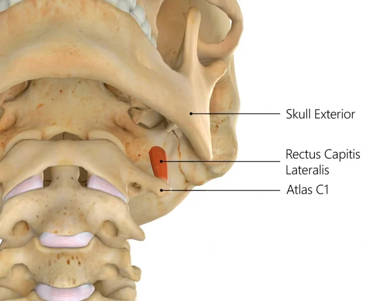
What are the 6 bones in the human skull?
The frontal, sphenoid, ethmoid, occipital, parietal, and temporal bones make up the cranial base. These bones articulate with the mandible (jaw), the facial bones, and the first cervical vertebra (atlas).
How does the skull primarily function?
The meninges that surround the skull bones serve primarily as structural and protective elements. The brain (cerebrum, cerebellum, and brainstem) and the orbits around the eyes are shielded by the skull. It provides a structural anchor for the muscles and tendons that attach to the facial and scalp muscles.
How many bones does the brain contain?
One frontal bone, two parietal bones, two temporal bones, one occipital bone, one sphenoid bone, and one ethmoid bone make up the eight bones that round your brain. The skull is composed of these eight bones.
Reference
- Skull. (n.d.). Physiopedia. https://www.physio-pedia.com/Skull
- Next, A. (n.d.). %s | Anatomy.app | Learn anatomy | 3D models, articles, and quizzes. https://anatomy.app/encyclopedia/skull
- Brennan, V. A. P. B. E. (2020, December 23). 25 Days of Skeletal Facts: Day 24 – The Neurocranium. GRAVE THOUGHTS. https://gravethoughtsblog.com/2020/12/24/25-days-of-skeletal-facts-day-24-the-neurocranium/
- Advocate, W. (n.d.). Viscerocranium (Facial Skeleton) < Skull Axial Skeleton Division << Axial Skeleton System <<< Bones by Region @ Wellness Advocate . com. https://wellnessadvocate.com/?dgl=2853
- The Skull | Anatomy and Physiology I. (n.d.). https://courses.lumenlearning.com/suny-ap1/chapter/the-skull/
- The Scalp – Layers – Innervation – Blood Supply – TeachMeAnatomy. (2020, November 22). TeachMeAnatomy. https://teachmeanatomy.info/head/areas/scalp/
- Anderson, B. W. (2023, November 9). Anatomy, Head and Neck, Skull. StatPearls – NCBI Bookshelf. https://www.ncbi.nlm.nih.gov/books/NBK499834/