
Levator Anguli Oris Muscle
Introduction
A muscle of facial expression, the levator anguli oris, helps to elevate both corners of the mouth. Along with the zygomaticus major and minor, the levator labii superioris, and the levator labii superioris alaeque nasi, it is accountable for providing upper dental show, primarily during smiling, and for sustaining the resting tone and position of the upper lip. While it is not the most significant of the smile muscles, unilateral dysfunction of the levator anguli oris can lead to noticeable asymmetry of the smile, which can also have a noticeable influence on a person’s quality of life.
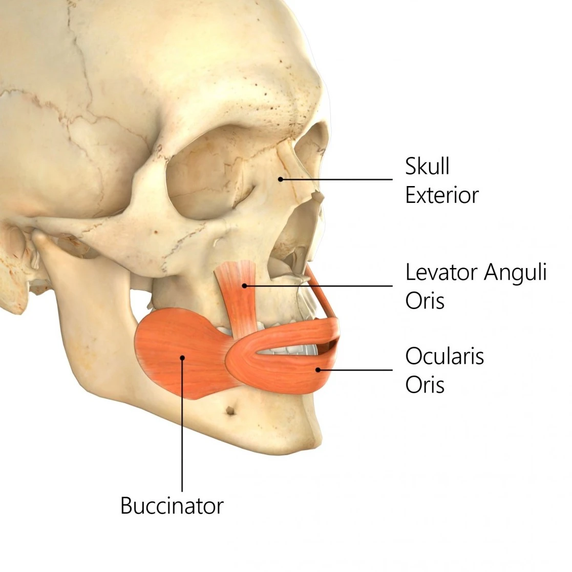
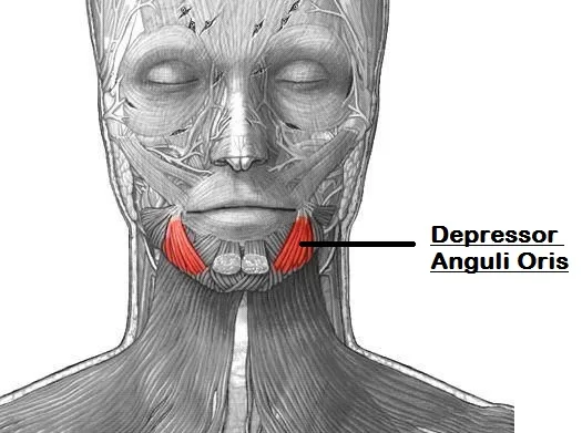
Levator anguli oris muscle is a paired strap-like muscle of the face, located beyond the angles of the lips. It belongs to an extensive group of muscles of facial expression named the buccolabial group. Besides levator anguli oris muscles, this grouping also contains levator labii superioris alaeque nasi, levator labii superioris, zygomaticus major, zygomaticus minor, risorius, depressor labii inferioris, depressor anguli oris, mentalis, orbicularis oris, incisive superior and inferior, and buccinator muscles.
Like all the buccolabial muscles, the levator anguli, or function contributes to producing facial expressions by maintaining the shape, position, and movements of the lips. Especially, it accomplishes so by elevating the angle of the mouth and deepening the nasolabial lines, encouraging smiling.
Embryology
The levator anguli oris muscles develop alongside the other mimicking muscles of the face, beginning in the third to fourth week of development. The muscles of facial expression arise from the second branchial arch, which also creates the facial nerve, the stapedial artery (which subsequently involutes in most people), and Reichert’s cartilage, which turn develops the stylohyoid ligament, the lesser cornu of the hyoid bone, and the styloid process of the temporal bone.
Origin of Levator anguli oris muscle
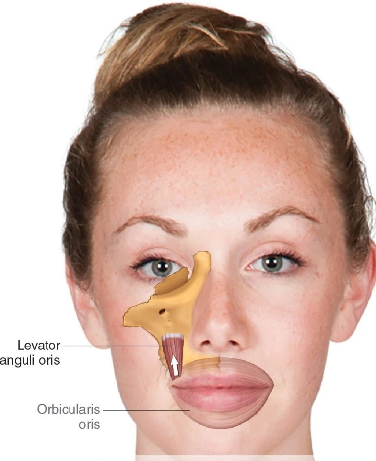
Levator anguli oris muscle originates from the canine fossa of the maxilla, inferior to the infraorbital foramen. After a short inferior course, it connects with the modiolus at the angle of the lips.
Insertion
Modiolus is a fibromuscular nodule at the angle of the mouth created by at least nine muscles. It serves as an attachment hub for most of the buccolabial muscle, thus integrating the motions of the lips with those of the jaw and cheeks. Its exact description varies across various textbooks but, surely, the direct tractors of the lips donate to modiolus; zygomaticus major, depressor anguli oris, and orbicularis oris.
The Levator anguli oris muscle lies deep, and lateral, to the levator labii superioris. These two muscles are attached to the infraorbital tissue space (canine space), which is clinically influential as odontogenic infections may spread into it. Infraorbital nerve and vessels pass between the bony attachments of levator anguli oris and levator labii superioris muscles on their way to the face.
Nerve supply
Zygomatic and buccal branches of the facial nerve (CN VII) innervate / or supply the levator anguli oris muscle.
The facial nerve, penned of approximately 10,000 axons, provides motor control of the muscles of facial expression, in addition to the posterior belly of the digastric, the stylohyoid, and the stapedius muscles. Upon emerging from the temporal bone, the facial nerve travels through the parotid gland, separating the deep and superficial lobes, and then continues distally underneath the superficial musculoaponeurotic system (SMAS) of the face. Because the emulative muscles are contiguous with the SMAS layer, the majority of the muscles of facial expression are innervated from their deep surfaces; the peculiarities are the buccinator, the mentalis, and the levator anguli oris muscles, which lie deeper within the face and are accordingly innervated from their superficial surfaces. Their innervation reference is a terminal buccal branch of the facial nerve that innervates the levator anguli oris muscle.
Blood supply
Blood supply to the depressor labii inferioris muscle arrives from the superior labial branch of the facial artery and the infraorbital branch of the maxillary artery.
Function of Levator anguli oris muscle
The levator anguli oris lifts the angle of the mouth, thus participating in producing a smile. Contractions of these muscles produce a facial expression associated with self-confidence. Medially drags the angle of the mouth upwards. It contributes to the widening of the mouth. It also supports the zygomaticus major muscle in producing a smile.
Clinical significance
The levator anguli oris is most important for its critical role in producing a natural-appearing, dentate, or “Duchenne” smile, without which people have difficulty expressing emotion and maintaining human social connection.
Excision is the most influential and safe method for treating tumors of the nasal skin. However, excision surgery can impair the appearance of the patient and cause unnecessary psychological trauma. Therefore, reconstructive surgery is often required behind resection. The levator anguli oris muscles can be used as a surgical flap for nasal reconstruction following the excision of nasal tumors.
Facial reanimation surgery desires to repair the inability to smile in patients suffering from facial nerve palsy. New techniques for measuring the physiological consequences of these types of surgeries provide surgeons with specific muscle targets for reanimation. Specifically, levator anguli oris and zygomaticus major muscles have a measurable effect on the perception of appropriate smile formation.
Exercise of Levator anguli oris muscle
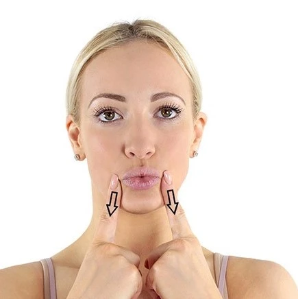
When performed regularly, facial exercises can reduce tension and even improve the smoothness of your face. To focus on levator anguli oris which is responsible for lifting the corners of your mouth, first stand in front of a mirror then position your index fingers on your cheeks near the corners of your mouth. While applying gentle pressure with your fingers, and then smile broadly. Hold the smile while opposing the pressure from your fingers for ten seconds then return to a neutral position. Repeat this exercise ten times and also recognize to smile frequently throughout the day.
FAQ
Where is the levator Anguli located?
The levator anguli oris muscles, located along with the buccinator and mentalis muscles in the deepest layer of the mimetic muscles, originate roughly 1 cm inferior to the infraorbital foramen within the canine fossa of the maxilla.
What does the levator anguli oris muscle do?
Like all the buccolabial muscles, the levator anguli oris function contributes to producing facial expressions by controlling the shape, position, and movements of the lips. Specifically, it does so by elevating the angle of the mouth and deepening the nasolabial lines, encouraging smiling.
How do you test the levator function?
Levator function is estimated by having the patient look down, and with a hand on the patient’s forehead to prevent any brow action, asking the patient to look upward as far as potential without a change in head position. The distance of the upper lid margin elevates in millimeters is the levator muscle function.
Is the nerve to levator anguli oris a branch of the facial nerve?
Specifically, the levator anguli oris muscle is innervated by the buccal branches of the facial nerve.
Is the levator anguli oris involved in smiling?
The levator anguli oris muscle raises the mouth’s angle in smiling and contributes to the depth and contour of the nasolabial fold. This muscle lies deep and lateral to the levator labii superiors muscle. It arises in the canine fossa of the maxilla and connects the modiolus of the lips.


One Comment