
Occipitofrontalis muscle
What is the Occipitofrontalis Muscle?
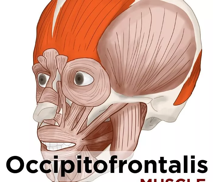
Occipitofrontalis is a scalp muscle that is long and wide muscle. This muscle and the temporoparietalis are both the epicranial group of the facial expression muscles.
The muscles of facial expression enable the face to perform different unique movements, from smiling and frowning to raising one or both eyebrows in excitement or surprise. The occipitofrontalis represents mainly these muscles of facial expression. From wrinkling the forehead to facilitating the communication of different emotions, the occipitofrontalis contributes to a different of facial expressions. Stretching from the back of the head to the forehead, the occipitofrontalis is also called the frontalis muscle, even though the frontalis represents one of the muscle bellies comprising also the occipitofrontalis.
While the human body sometimes relies upon the cooperation or combination of several muscles to produce motion, the occipitofrontalis is different because it is the only muscle that can raise eyebrows. In this article, the anatomy and blood supply. also, the innervation and function of the occipitofrontalis muscle will be explored in detail.
Anatomy
Occipitofrontalis Muscle Structure
The occipitofrontalis possesses two muscle bellies: 1)the frontalis and 2) the occipitalis. These two muscle bellies are connective tissues, that are often found in large, sheetlike muscles such as the occipitofrontalis.
Frontal Belly of the Occipitofrontalis Muscle
The frontal belly of the occipitofrontalis is located directly over the forehead on the human skull bone, extending down from the general area of the hairline to the eyebrows. In appearance, the frontal belly, sometimes also referred to as the frontalis, looks like a pair of fans, known as the left and right frontalis bellies. These ”fans” possess vertical striations that correspond to the muscle fibers that comprise each muscle belly.
Occipital Belly of the Occipitofrontalis Muscle
The occipital belly is located at the back of the head and forms a rectangular band stretching from ear to ear at the base of the skull. Sometimes also referred to as the occipitalis, the occipital belly is also named for the skull bone it overlays: the occipital bone. Like the frontalis muscle, the occipitalis muscle is characterized by vertical striations, which reflect the direction of the muscle fibers found in this muscle.
Origin
The frontal belly of the occipitofrontalis possesses no direct attachments to the bone. Instead, the frontal belly originates at the anterior portion of the galea aponeurotica, the aponeurosis connecting the two bellies at the scalp. The galea aponeurotica is located roughly from the hairline on the forehead.
The occipital belly also originates at the superior nuchal line, one of four curved lines found each on the occipital bone. Another point of origin for an occipital belly is the temporal bone.
Insertion
The occipitofrontalis muscle consists of two parts or bellies:1) the occipital belly, near the occipital bone. also, It originates on the lateral two-thirds of the highest nuchal line, and from the mastoid process of the temporal bone. It inserts into mainly epicranial aponeurosis. At the frontal belly, the frontal bone.
Nerve Supply
- Frontal belly: Temporal branches of the facial nerve (CN VII)
- Occipital belly: Posterior auricular nerve (branch of the facial nerve (CN VII))
Blood Supply
Blood supply to both parts of occipitofrontalis comes from many branches;
- The frontal belly is supplied mainly by the ophthalmic artery and the frontal branch of the superficial temporal artery.
- The occipital belly receives blood from the occipital branch of the posterior auricular and also a descending branch of the occipital artery.
- All the arteries supplying this muscle are the branches of the external carotid artery, except for the ophthalmic artery which arises from the internal carotid artery.
Function
Occipitofrontalis muscle has many actions depending on which of its attachments is fixed;
When its aponeurotic attachments are fixed,
- the frontal belly elevates the eyebrows and skin of the forehead, producing a facial expression of shock or surprise.
- When its forehead attachment is fixed, the frontal belly aids in the procerus, orbicularis oculi, and corrugator supercilii muscles to frown the eyebrows by pulling the scalp forwards and wrinkling the forehead. Since the muscles and heads of the frontal belly are separated, it is possible to perform these movements on only one of the halves of the face side.
The occipital belly retracts the scalp when its nuchal part is fixed and also moves it forwards when the aponeurotic attachment is fixed. These movements are insignificant on their own, but also in case they occur simultaneously with both contractions of the frontal belly, they help move the entire scalp backward and forwards same respectively.
Clinically myofascial trigger points in this muscle can mimic headaches. Pain is usually experienced on the forehead and the top of the head.
Clinical Importance:
Damage to the facial nerve (CN VII) can cause atony of the occipitofrontalis muscle.
What is the attachment for occipitofrontalis?
The occipitofrontalis muscle attaches to the occiput and mastoid part of a temporal bone, the epicranial aponeurosis, and the temporal fascia attachment to a zygomatic arch. These attachments limit the potential posterior and lateral spread of infections from the scalp.
Exercise
1)Eyebrow lift

(Lift your eyebrows as high as possible. Then relax and repeat.)
2)Self-massage
For the self-massage of these muscles, firstly I recommend using your fingers or a massage ball. As a massage technique, firstly you can choose from ischemic compression, precise massage strokes, or the pressure-motion technique. On this page, I will also show how to massage the muscle with your two fingers at the site and the precisely given massage strokes.
Note: Try to relax during the massage and maintain deep and even continue normal breathing.
1)Self-massage of the anterior fibers of the occipitofrontalis
- Shape your hand like a shovel.
- Place two to three fingertips on the muscle in the area of your forehead at the scalp area.
- Examine the entire area of your forehead and search for the painful tension side.
- As soon as you find one, apply stroke over it a few times. Do not slide over your skin but gently pull the skin over the muscle.
Frontalis muscle self-massage video:
2)Self-massage of the posterior fibers of the occipitofrontalis
- You have already felt the occipitalis – at the posterior area, this is exactly the area you massage, in the same way as the area of the frontalis given.
- However, there is still a tiny spot in the muscle which you probably will miss without a detailed guide. also, We don’t want this to happen. This is why I’ll give information you about where it is situated and all.
- Place a finger in the center on the back of your head at the posterior and move your chin a few times towards your chest side.
- Feel the line protruding with each at the “forward tilt” of the head.
- Feel the hollow next to this line.
- From here, move your finger two to three centimeters sideways and look for a very mild, yet noticeable hollow.
- Take your time – this small hollow is no at easy to find.
- occipitalis-palpation-1
- In this hollow, there is the spot I just spoke about at the location.
- Press into this hollow and check the sensitivity of pressure.
- To massage this small spot and press into it, and perform some very small gentle circular movements.
Trigger Point Release Therapy for Headache of Occipital Muscle video
Take Home
Myofascial release/soft tissue techniques that work the forehead and the top of the head can often relieve headache pain, this is useful when treating patients with post-concussion syndrome.
FAQ
What is the function of the occipitofrontalis?
The occipital part of the occipitofrontalis muscle moves the scalp forwards, and the frontalis portion lifts the brows and moves the anterior scalp backward. When the frontalis muscles contract, the vertical fibers pull the skin of the eyebrows upward
Is frontalis same as occipitofrontalis?
The occipitofrontalis is an interesting muscle. It is comprised of three divisions: the frontalis, the occipitalis, and the galea aponeurotica. Each division is responsible for a separate action involving the scalp, forehead, or brows
What muscle is also known as the occipitofrontalis?
The occipitofrontalis muscle (epicranius muscle) is a muscle that covers parts of the skull. It consists of two components or bellies: the occipital belly, near the occipital bone, and the frontal belly, near the frontal bone.
Is occipitofrontalis a flat muscle?
Flat and quadrilateral in form, it is one of the facial muscles. Along with the occipitalis muscle, it forms the occipitofrontalis muscles due to a common tendon sheet attachment; the galea aponeurotic.
How do you strengthen the occipitofrontalis muscle?
To activate the occipital muscles, lie on a table, on your back. Start with the back of your head against the table. Gradually press the back of the head into the table, creating a double chin. Complete eight to twelve repetitions, one to three duration per day.


One Comment