Adductor longus muscle
What is Adductor longus muscle?
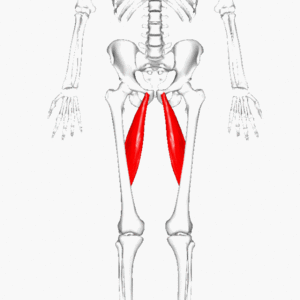
The large, fan-shaped adductor longus muscle is in the medial part of the thigh. It is a member of the adductors of the thigh, along with the adductor brevis, adductor magnus, pectineus, and gracilis muscles. The obturator nerve’s anterior division (L2-L4) innervates the adductor longus.
The adducting of the thigh at the hip joint is the primary function of this muscle group. It is accepted that both the extensor and flexor muscles in this area were formed.
Origin
A strong tendon that connects the pubic crest to the anterior aspect of the body.
Insertion
Forms a fan shape and widely joins the linea aspera, which is located in the middle third of the femur.
This insertion point is between the adductor magnus muscle’s insertion and the vastus medialis muscle’s origin, and it is inferior to the adductor brevis’ insertion.
Relations
Before the adductor magnus muscle, the adductor brevis muscle, the front of the obturator nerve, and the deep femoral vessels are the adductor longus muscles. The pectineus muscle is lateral to the gracilis muscle, which is located medially. Anterior relations to its upper part are the spermatic cord and fascia lata, while the femoral artery and vein are accessible before it in the lower part near its association.
Innervation
The obturator nerve‘s anterior division supplies the adductor longus. The obturator nerve, which is made up of the anterior divisions of the anterior rami of spinal nerves L2-L4, supplies all three adductors (except the hamstring portion of the adductor magnus) and the gracilis.
Through a branch of the lumbar plexus that descends medially to the psoas major muscle, the obturator nerve enters the pelvis. It arrives behind the lateral to the internal iliac artery and ureter and runs behind the normal iliac vessels. After that, it will go through the lateral wall of the pelvis and eventually reach the obturator foramen’s highest point. It isolates into a posterior and anterior branch after going through the foramen and entering the obturator canal. The obturator externus and adductor brevis separate these branches.
The posterior branch pierces the obturator externus and enters the medial compartment to supply the adductor brevis and adductor magnus. The nerve also sends a sensation to the upper medial thigh. The anterior branch connects the adductor longus, which it supplies, and the pectineus, which is supplied by the femoral nerve, to the obturator externus posteriorly.
The nerve also serves as an articular branch for the joint as it travels through the hip. It supplies the adductor longus, gracilis, and brevis.
Blood supply
The blood supply to the adductor longus comes from two courses, the deep femoral artery (a piece of the femoral artery) and the obturator vein (a piece of the internal iliac vein).
The medial circumflex artery, which is a part of the deep femoral artery, supplies the muscle’s proximal area. Tributaries are received by the artery’s deep femoral vein-corresponding branches.
Function
Adducting the thigh at the hip joint is the main work of the adductor group of muscles. Additionally, thigh flexion and external/lateral rotation are both performed by the adductor longus muscle.
When standing and walking, the adductors are essential for maintaining a stable stance and balancing the body on the lower limb.
Clinical relations
The medial line of the femoral triangle is formed by the adductor longus muscle. The inguinal tendon forms the superior boundary, while the sartorius forms the lateral boundary. In this triangular area are the femoral nerve, artery, and vein. The nerve runs horizontally under the inguinal tendon and closes to the anterior superior iliac spine (ASIS). Around here, the nerve supplies the iliacus and descends to supply the anterior (extensor) thigh compartment.
The lateral nerve, the supply route, the vein, and lastly the y-front, otherwise called the pubic area, are the designs in this space that are organized from lateral to medial. This order is simple to remember thanks to the NAVY acronym. Some space between the femoral vein and the femoral artery allows it to expand.
Adductor longus muscle stretching
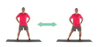
Standing Adductor Stretch
Start by standing approximately three feet apart.
Bent your knee and move the load to the side.
Maintain one knee straight and sense a stretch on the inside of the thigh.
Keep stretching for 15 to 30 seconds.
Repeat on the further side.
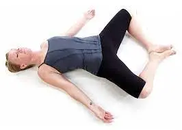
Reclining angle bound pose
This is the best stretch if you sit most of the day.
On the ground, rests in an inclined position.
Twisting the two knees so the knees are contacting to the floor.
Drop the knees down toward the floor with the objective that you can feel the groin muscles stretch.
Maintain this position and take a deep breath every 15 to 30 seconds.
four or three times. Try to bring the feet closer to the buttocks with each stretch.
Adductor longus muscle strengthening exercise
Cossack Squat
The Cossack Squat exercises move the body from side to side in the frontal plane.
Flexibility in the hip, knee, and ankle can be improved by working the lower limb at this angle.
You will strengthen your stability by stretching and strengthening your hip adductors through this activity.
You must drop your hips backward while standing with your feet in a wide stance and your toes pointing outside for this exercise.
With the other leg extended, toes facing up, and heels on the floor, lunge over to that side and shift your weight.
With your flexed leg, push through the ground and bring your back to the starting position.
By shifting the weight and lowering it into a squat position on the opposite side, you can perform this exercise again on the opposite leg.
Note: Because the Cossack Squat requires a great deal of flexibility, if you are unable to immediately descend, try to improve with each exercise by going as far as you are able. Your back can be protected in a neutral location throughout the activity.

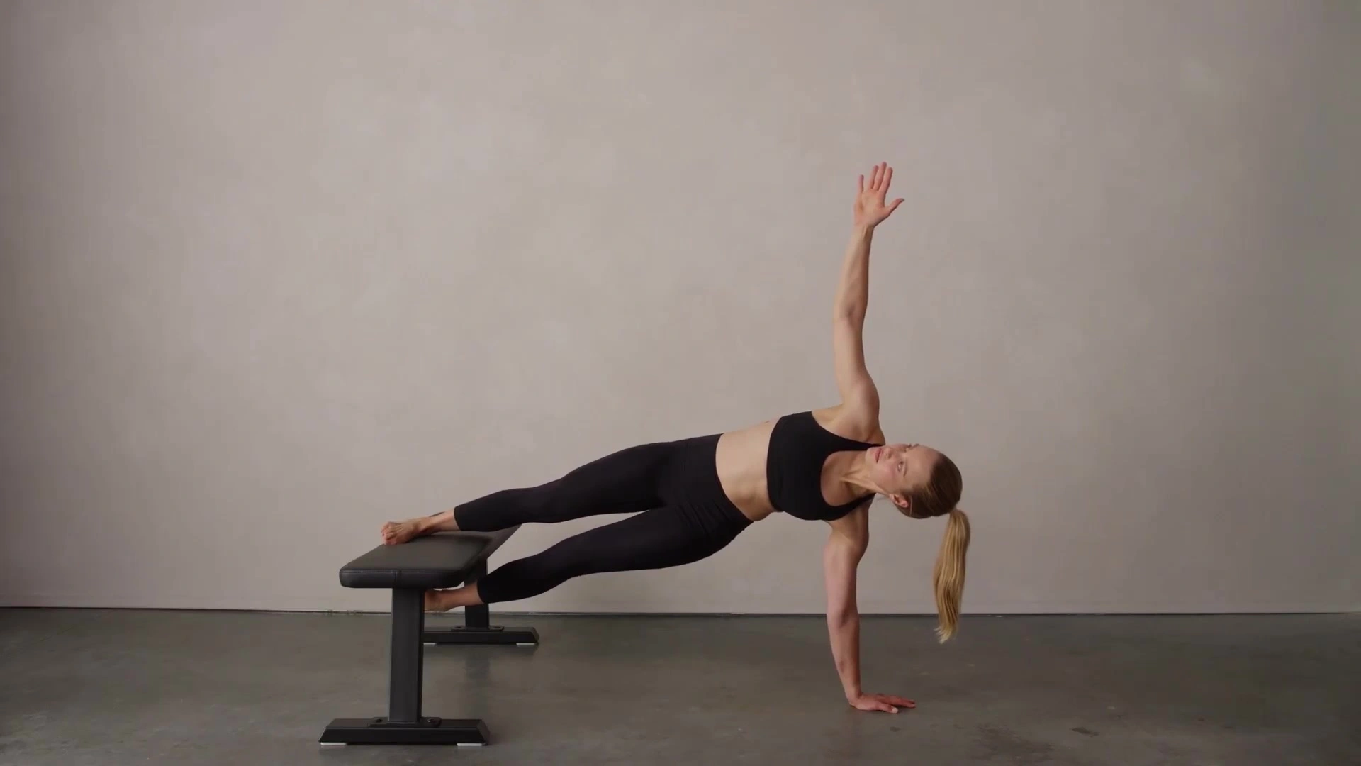
Copenhagen Side Plank
The Copenhagen Side Plank is the most difficult plank exercise because it strengthens the hip adductors while also working the core muscles. The Copenhagen Side Plank will help strengthen the muscles outside the hip to get back into balance.
For this exercise, you must lie on the floor perpendicular to the bench and straighten your elbows and forearms.
Lift with your flexed knee lift your top leg and put it on the seat.
Keep this posture.
Switch sides and repeat after 20 to 25 repetitions in two sets.
Note: To make the exercise more difficult, perform it with your legs extended and only your ankle on the bench.
Cable Hip Adductor
You could see ladies at the gym center doing this activity while the men complete it.
Everybody might be participating in this movement to boost the adductors and diminish the risk of injury.
Perform some dynamic stretches as a warm-up before beginning this exercise. Then, until you feel confident lifting more weight, start with as little weight as possible and as many repetitions as possible.
You can use a connection to lash onto the lower leg that is closest to the pulley.
Check to see that the pulley is at the calf’s level. Your back ought to be to the pulley. Prepare yourself by squeezing your hand against the machine in a protected place where your fingers will not be squeezed. Your active leg may be approaching the pulley from below.
Pull your leg away from the pulley and toward the center of your body.
completed the ideal number of counts or steps by allowing the leg to slowly return to its starting position. Note: You can practice by connecting an opposition band to the right anchor point using the same method.
FAQ
Why does my adductor longus hurt?
A common cause of groin pain and injury in athletes is adductor strain. Age, weak adductors, muscle fatigue, restricted range of motion, insufficient adductor muscle complex stretching, and previous hip or groin injuries are all risk factors.
How do you heal the adductor longus muscle?
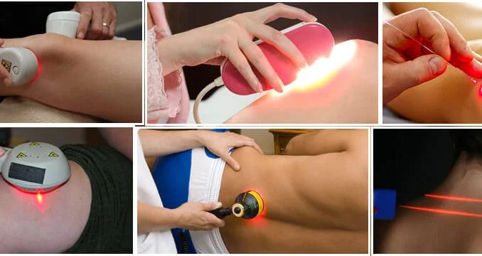
The majority of adductor muscle types can be treated moderately. Moving toward change at the beginning of treatment can quickly merge upholds. When severe muscle strains occur, ice and a sedative are ideal. Fragile expansions are appropriate for reinforcing practices as secondary effects decline.
Where is adductor longus pain?
In acute adductor longus injuries, tendon rupture or avulsion can occur, typically at the proximal insertions. Clinical signs of an adductor strain include pain in the inner thigh and tenderness along the muscle belly, tendon, or insertion.
What are the signs and symptoms of adductor longus?
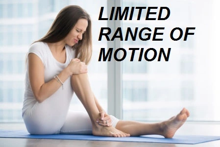
Pain on the inside of your thigh, pain when bringing your legs together, and pain when lifting your knee are all symptoms of an adductor strain. Moreover, there might be a popping sensation in the thigh and some limping. There may similarly be broadening and expansion.
What nerve controls the adductor longus?
The obturator nerve (L2L4) supplies the pectineus; longus, brevis, and magnus adductor; gracilis; and this nerve controls the external obturator muscles’ adduction and thigh rotation.