
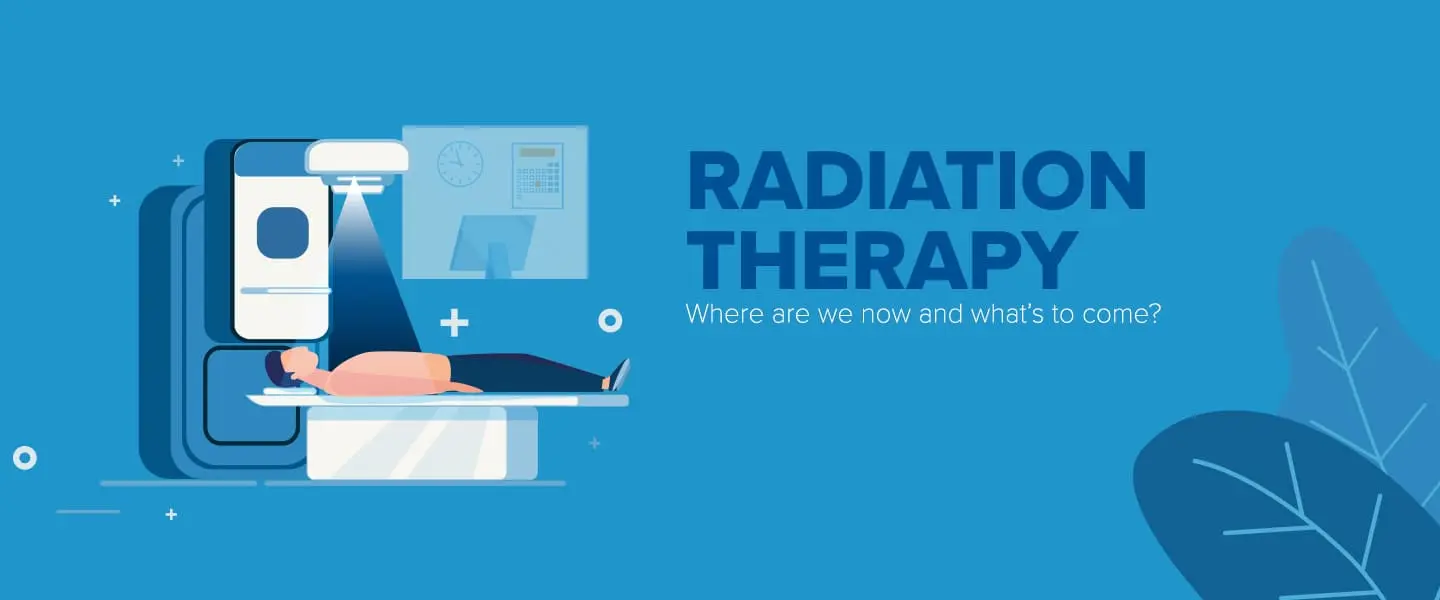
Radiation Therapy
Introduction
Ionizing radiation therapy, often known as radiotherapy (RT, RTx, or XRT), is a medical procedure used to treat cancer. Its goal is to either destroy or stop the proliferation of cancerous cells. Usually, a linear particle accelerator delivers it.
Many cancers can be cured with radiation therapy if they only affect one portion of the body and haven’t spread to other areas.
Additionally, it can be used as adjuvant therapy to stop tumor recurrence following primary tumor removal surgery. malignant tumor (e.g., breast cancer in its early stages).
In tumors that are susceptible, radiation treatment has been employed before, during, and following chemotherapy because it works in concert with the drug. Radiation oncology is the name of the specialization of oncology that deals with radiation therapy.
Radiation oncologists are doctors who specialize in this field of medicine. Because radiation therapy can restrict cell proliferation, it is frequently used to treat malignant tumors. Ionizing radiation causes cellular death by causing damage to the DNA of malignant tissue.
Shaped radiation beams are focused from many angles of exposure to intersect at the tumor, producing a significantly bigger absorbed dose there than in the surrounding healthy tissue, thus sparing normal tissues (such as organs or skin that need to be exposed to radiation in order to treat the tumor).
If the draining lymph nodes are clinically or radiologically implicated, they may also be included in the radiation fields in addition to the tumor itself. with the tumor, or if a subclinical malignant spread risk is believed to exist.
To account for errors in daily setup and internal tumor mobility, a margin of normal tissue must be included around the tumor.
Both internal movement (breathing, for example, and bladder filling) and exterior skin mark movement in relation to the tumor site can contribute to these issues.
Different from radiology, which uses radiation for medical imaging and diagnosis, radiation oncology is the medical profession that deals with prescription radiation. A radiation oncologist may prescribe radiation as adjuvant therapy or with the goal of curing cancer.
It can also be used as therapeutic treatment (when the therapy has survival benefits and can be curative) or palliative treatment (where a cure is not possible and the purpose is for local disease control or symptomatic alleviation).
Additionally, radiation therapy is frequently used in conjunction with immunotherapy, hormone therapy, chemotherapy, surgery, or a combination of the four. Radiation therapy is a viable treatment option for the majority of prevalent cancer types.
The exact purpose of the treatment—curative, adjuvant, neoadjuvant therapeutic, or palliative—will depend on the kind, location, and stage of the tumor as well as the patient’s overall condition.
A radiation therapy procedure called total body irradiation is performed to get the body ready for a bone marrow transplant. An additional kind of radiation therapy is used to lessen the amount of healthy tissue exposed during procedures to treat breast cancer.
Prostate and other organs are called brachytherapy, in which a radioactive source is positioned inside or adjacent to the area that has to be treated. There are various uses for radiation treatment in non-cancerous diseases, including pterygium.
Pigmented villonodular synovitis, auditory neuromas, severe thyroid eye disease, trigeminal neuralgia, and the avoidance of heterotopic ossification, keloid scar formation, and vascular restenosis.
Concerns regarding the possibility of radiation-induced malignancies restrict the use of radiation therapy in non-malignant illnesses.
Medical Uses
Radiation therapy was reportedly a part of the treatment plans for half of the 1.2 million invasive cancer cases in the US that were identified in 2022. Radiation therapy has varying effects on different malignancies.
Radiosensitivity is a term used to characterize how a cancer reacts to radiation. Mild radiation dosages quickly destroy highly radiosensitive cancer cells. These comprise germ cell malignancies, leukemias, and the majority of lymphomas.
Since most epithelial tumors are only mildly radiosensitive, much higher radiation dosage is needed to produce a drastic cure. Certain cancers stand out for being radioresistant, meaning that far greater doses are needed to provide a drastic cure than could be appropriate in real-world settings.
Carcinoma of the renal cells and Although radiation therapy is still a palliative option for many patients with metastatic melanoma, melanoma is generally thought to be radioresistant. Research on the potential benefits of combining radiation therapy and immunotherapy for melanoma and other malignancies is ongoing.
It’s critical to distinguish between a tumor’s radiosensitivity, which is largely a lab measurement, and a cancer’s radiation “curability” in real-world clinical settings. For instance, because leukemias spread throughout the body, radiation treatment is typically not effective in curing them.
If lymphoma is restricted to a single part of the body, it might be drastically treatable. Likewise, a large number of common cancers that respond moderately to radiation are frequently treated with radiation therapy at curative doses if detected early.
Skin cancer, cancer of the head and neck, breast cancer, non-small cell lung cancer, cervical cancer, anal cancer, and prostate cancer are a few examples. Because radiation therapy cannot treat the entire body.
Metastatic tumors are incurable, with the exception of oligometastatic illness.In order to create a sophisticated radiation treatment plan, modern radiation therapy uses a CT scan to detect the tumor and surrounding normal structures and to calculate doses.
To help with the positioning of the treatment fields, the patient is given tiny skin markings. At this point, patient placement is quite important because the patient needs to be in the same position for every treatment.
For this reason, numerous patient positioning tools have been created, such as masks and cushions that may be shaped to fit the patient. A technique called image-guided radiation therapy makes use of imaging to adjust for positioning inaccuracies that arise during each treatment session.
The size of a tumor also affects how well it responds to radiation treatment. Very large tumors react to radiation less successfully than smaller tumors or microscopic illnesses because of complex radiobiology.
Several tactics are employed to counteract this impact. Before receiving radiation therapy, surgical resection is the most often used method. This is most frequently observed in the course of adjuvant radiation therapy after broad local excision or mastectomy for breast cancer.
Another approach is to use neoadjuvant chemotherapy to reduce the tumor size before undergoing intense radiation treatment.
A third method involves administering specific medications to the malignancy during a radiation therapy session in order to increase its radiosensitivity. Medications that sensitize the body to radiation include cetuximab, nimorazole, and cisplatin.
The effects of radiation therapy varied for various cancer kinds and populations. For instance, it has been discovered that radiation therapy for breast cancer after breast-conserving surgery reduces the disease’s recurrence rate by half. Radiation therapy has extended the survival periods of inoperable tumors in pancreatic cancer.
Side Effects
In and of itself, radiation therapy (RT) is painless. Numerous low-dose palliative treatments (such as radiation therapy to bony metastases) have little or no side effects, but because edema compresses the nerves in the treated area.
There may be a brief pain flare-up in the days that follow treatment. Increased dosages may result in different side effects during therapy (acute side effects), months or years after treatment (long-term side effects), or cumulative side effects with repeat treatment.
The organs that receive the medication determine the type, intensity, and duration of any side effects. radiation, the patient, and the actual treatment (radiation type, dose, fractionation, and concurrent chemotherapy). In 5% of RT patients, serious radiation problems could arise.
50 Gy RT dosage may result in acute (near-immediate) or sub-acute (two to three months post-RT) radiation adverse effects. After 65 Gy, late or delayed radiation harm (many months to decades) may manifest.
Most adverse consequences are anticipated and foreseeable. Radiation side effects are often confined to the treated area of the patient’s body. The side effects of radiation to the head and neck are dose-dependent; for instance.
Increased radiation doses have been linked to thyroid and pituitary axis malfunction, cardiovascular problems, and other disorders. The goal of contemporary radiation therapy is to minimize adverse effects while assisting the patient in managing those that cannot be avoided.
Fatigue and skin irritation, like mild to moderate sunburn, are the most common adverse effects mentioned. The tiredness might persist for weeks after therapy concludes and usually begins in the middle of the course of treatment. Although the inflamed skin will recover, its elasticity can decrease.
Acute Side Effects
Nausea and Vomiting
This is not a common side effect of radiation therapy; instead, it is primarily related to treatment of the stomach or abdomen (which typically reacts a few hours after treatment) or to radiation therapy administered to specific head structures that cause nausea when treating specific head and neck tumors, most commonly the inner ear vestibules.
Some people vomit during radiation therapy, or even before, as is the case with any upsetting treatment; nonetheless, this is thought to be a psychological reaction. Antiemetics can be used to treat nausea for any reason.
Damage to The Epithelial Surfaces
Radiation therapy may cause damage to epithelial surfaces. This can include the skin, pharynx, intestine mucosa, ureter, and oral mucosa, depending on the location being treated. The rate at which damage occurs and heals is determined by the rate at which epithelial cells turnover.
Usually, a few weeks into therapy, the skin starts to get pink and hurt. Throughout the course of treatment, the reaction could get worse and last up to It’s possible for the skin to break down approximately one week after radiation therapy ends.
Even though this moist desquamation hurts, healing normally happens quickly. Skin reactions are often more severe in places where the skin naturally folds, such as the groin, behind the ear, and beneath the female breast.
Mouth, Throat, and Stomach Sores
Mouth and throat ulcers and transient pain are frequent if the head and neck region is treated. If severe, this may interfere with swallowing, necessitating the use of medications food supplements, or nutritional support.
Additionally, receiving direct treatment or, more frequently, receiving a dosage of collateral radiation during lung cancer treatment might cause soreness in the esophagus.
Gastric, stomach, or duodenal ulcers may result from collateral radiation used to treat liver cancers and their metastases. The non-targeted delivery of the radioactive agents being infused, also known as reflux, is frequently the origin of this collateral radiation.
There are tools, procedures, and strategies available to reduce the likelihood of this kind of unfavorable side effect.
Intestinal Discomfort
Radiation therapy can be used to treat rectal or anal cancer directly, or it can be used to expose the lower bowel to radiation while also treating other pelvic tissues such as the bladder, female genital tract, and prostate.
Nausea, diarrhea, and pain are common complaints. Nutritional therapies could be able to assist in cases of radiation-induced diarrhea.
Changes in dietary fat, fiber, and lactose during radiation therapy have been shown to minimize diarrhea at the conclusion of treatment in individuals receiving pelvic radiotherapy as part of anticancer treatment for primary pelvic cancer.
Swelling
Swelling of soft tissues as a result of the overall inflammation that happens could be problematic during radiation therapy. When treating brain tumors and brain metastases, this is a worry, particularly if there is a history of elevated intracranial pressure or if the tumor is obstructing a lumen almost completely (e.g., trachea or major bronchus).
Radiation therapy may not always be administered before surgical surgery. Steroids may be given to the patient during radiation therapy in order to minimize edema if surgery is determined to be unneeded or inappropriate.
Infertility
The testicles and ovaries, or gonads, are extremely radiation-sensitive organs. After direct exposure to the majority of standard treatment radiation dosages, they might not be able to develop gametes. If the gonads are not the main site of treatment, treatment planning for all body sites is made to reduce, if not fully eliminate, dosage to them.
Late Side Effects
Late side effects usually only affect the area that was treated and manifest months or years after therapy. They are frequently brought on by vascular and connective tissue cell injury. Partially fractionating a treatment reduces many of the late effects.
Fibrosis
Radiation-exposed tissues gradually lose their elasticity as a result of a diffuse scarring process.
Epilation
With dosages over 1 Gy, epilation (hair loss) may happen on any skin that has hair. It only takes place in the radiation field or fields. A single dosage of 10 Gy may result in permanent hair loss; however, if the dose is fractionated, irreversible hair loss might not happen until the dose reaches 45 Gy.
Dryness
The majority of aggressive head and neck cancer treatments exceed the radiation tolerance of the salivary and tear glands, which is around 30 Gy in 2 Gy fractions. Dry eyes (xerophthalmia) and dry mouth (xerostomia) can worsen with time and significantly lower a patient’s quality of life.
Similar to this, after pelvic irradiation, the normally moist vaginal mucosa frequently becomes dry, and sweat glands on irradiated skin (such as the armpit) frequently cease to function.
Chronic Sinus Drainage
Treatments for soft tissue, palate, or bone cancer with radiation therapy to the head and neck region can result in fistulae from the bone and chronic sinus tract drainage.
Lymphedema
Radiation therapy can cause damage to the lymphatic system, which can lead to lymphedema, a disorder characterized by localized fluid retention and tissue swelling.
When patients with breast radiation therapy undergo adjuvant axillary irradiation after axillary lymph node clearance surgery, this is the most often reported side effect.
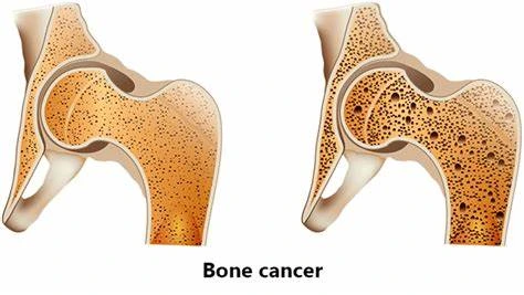
Cancer
Some people experience secondary malignancies, and radiation is a possible cause of cancer. Because of a variety of factors, such as genetics, prior radiation therapy, and lifestyle choices, cancer survivors are already at a higher risk of developing malignancies than the general population.
The incidence of these secondary malignancies from any one etiology is hard to pin down. Only a small percentage of patients acquire secondary cancers linked to radiation therapy, according to studies.
Novel methods like carbon ion radiation and proton beam therapy, which Lower the dosage to healthy tissues will help to reduce these hazards. It begins 4-6 years after therapy, while certain hematological malignancies might appear in as little as 3 years.
Even with pediatric cancers, which have a greater incidence of secondary cancers, this danger is typically considerably balanced by the risk reduction that results from treating the underlying disease.
Cardiovascular disease
As seen in earlier breast cancer RT regimens, radiation can raise the risk of heart disease and death. With aggravating factors taken into account, therapeutic radiation raises a person’s chance of a subsequent cardiovascular event (heart attack or stroke) by 1.5 to 4 times their normal rate.
The strength, volume, and placement of the RT’s dose all affect the rise, which is dose-dependent. Concomitant treatment with anthracyclines raises the risk of worsening the disease.
An estimated 10% to 30% of cases of cardiovascular disease are thought to be caused by radiation therapy. Radiation-induced heart disease (RIHD) and radiation-induced cardiovascular disease (RIVD) are terms used to describe late-stage cardiovascular adverse effects.
Cardiomyopathy, myocardial fibrosis, valvular heart disease, coronary artery disease, cardiac arrhythmia, and peripheral artery disease are among the dose-dependent symptoms. These and other late side effect symptoms might result from oxidative stress, radiation-induced fibrosis, and damage to vascular cells.
Determining the etiology of radiation-induced cardiovascular illnesses can be challenging because the majority manifest ten years or more after treatment.
Cognitive Decline
Radiation therapy may result in cognitive loss in situations where the head is exposed to radiation. Early children, those between the ages of five and eleven, showed the greatest signs of cognitive deterioration. For instance, research revealed that 5-year-old children’s IQ dropped by several IQ points a year following therapy.
Radiation Enteropathy
After receiving radiation therapy to the abdomen and pelvis, the gastrointestinal tract may sustain damage. In cases when the ileum is involved, atrophy, fibrosis, and vascular alterations result in malabsorption, bile acid diarrhea, steatorrhea, and bleeding.
In addition to radiation proctitis, which causes bleeding, diarrhea, and urgency, pelvic radiation illness can also result in radiation cystitis when the bladder is impacted.
Radiation-Induced Polyneuropathy
Due to the radiosensitivity of nerve tissue, radiation therapies may cause damage to nerves in the vicinity of the target area or along the delivery line. Ionizing radiation-induced nerve damage develops gradually; the first stage is caused by microvascular damage, capillary damage, and nerve demyelination.
Radiation-induced fibrous tissue growth that is out of control causes nerve compression and vascular constriction, which both cause more harm. ICD-10-CM Code G62.82, or radiation-induced polyneuropathy, affects 0.5–1% of patients undergoing radiation therapy.
Late-impact neuropathy can affect the peripheral nervous system (PNS) or the central nervous system (CNS), depending on the irradiated zone. For instance, cranial nerve damage in the CNS usually manifests as a loss of visual acuity 1–14 years after therapy.
Radiation-induced brachial plexopathy or radiation-induced lumbosacral plexopathy, which can manifest up to three decades after treatment, are examples of plexus nerve injury in the PNS.
Radiation Necrosis
The loss of healthy tissue close to the radiation location is known as radiation necrosis. This form of coagulative necrosis arises from radiation-induced blood vessel damage, either directly or indirectly.
This diminishes the blood flow to the remaining healthy tissue, leading to ischemia-induced death, akin to an ischemic stroke. This treatment-related side effect manifests months to decades after radiation exposure.
Osteoradionecrosis, vaginal radionecrosis, soft tissue radionecrosis, and laryngeal radionecrosis are the most prevalent presentations of radiation necrosis.
Cumulative Side Effects
Reirradiation can still be dangerous even if short-term effects have subsided and long-term effects are subclinical. Cumulative effects from this approach should not be confused with long-term consequences. These doses are calculated by the radiation oncologist and many factors are taken into account before the subsequent radiation takes place.
Effects on Reproduction
Radiation therapy is deadly but not teratogenic in the first two weeks following fertilization. Excessive radiation exposure during pregnancy can cause birth defects, stunted growth, intellectual incapacity, and a higher chance of leukemia and other cancers in the progeny.
There seems to be no increase in genetic flaws or congenital malformations in the offspring conceived after radiotherapy in guys who have previously had the treatment. Micromanipulation methods and the use of assisted reproductive technologies, however, may raise this danger.
Effects on The Pituitary System
Commonly, hypopituitarism arises during whole-body irradiation for systemic malignancies, extracellular brain tumors, head and neck cancers, and radiation therapy for sellar and parasellar neoplasms.
About 40–50% of kids with pediatric cancer who get treatment experience endocrine adverse effects. Growth hormone and gonadal hormones are the principal hormones affected by radiation-induced hypopituitarism.
On the other hand, individuals with radiation-induced hypopituitarism are least likely to have deficiencies in thyroid stimulating hormone (TSH) and adrenocorticotrophic hormone (ACTH). Prolactin secretion changes are typically minor, and vasopressin insufficiency as a result of radiation seems to be quite uncommon.
Effects on subsequent surgery
Doses exceeding 65 Gy frequently result in delayed tissue damage with reduced wound healing capacity. The external beam radiation’s holographic iso-dosing results in a diffuse damage pattern.
The majority of the radiation is directed towards the targeted tumor, but because of beam divergence, healthy tissue at progressively greater distances from the tumor’s center is also exposed to radiation in a diffuse pattern.
The inflammatory artery linings in these lesions show signs of proliferative, progressive endarteritis, which interrupts the blood supply to the affected tissue. This type of tissue becomes fibrotic, hypoxic, and lacks a sufficient amount of oxygen and nutrients.
Surgery on previously exposed tissue has a very high failure probability. For example, women who get radiation therapy for breast cancer experience hypervascularity and late-effect tissue fibrosis in the chest wall, which makes healing and reconstruction extremely challenging, if not impossible.
Radiation Therapy Accidents
Strict protocols are in place to reduce the possibility of patients unintentionally receiving too much radiation treatment. Errors do happen, though.
Between 1985 and 1987, the radiation therapy machine Therac-25 was involved in at least six incidents in which patients received doses up to 100 times higher than intended; two of these patients died as a direct result of the radiation overdoses.
A Missouri hospital overexposed 76 patients (the majority of whom had brain tumors) over the course of five years in 2005–2010 as a result of improper setup of new radiation equipment.
Radiation oncologists, medical physicists, and other members of the radiation therapy treatment team are striving to eradicate medical errors, despite the fact that they are incredibly uncommon.
Target Safely, a safety program started by the American Society for Radiation Oncology (ASTRO) in 2010, sought to, among other things, document errors across the country so that medical professionals may learn from each and every mishap and keep them from happening again.
In order to make sure that every treatment is as safe as possible, ASTRO also publishes a list of questions that patients should pose to their doctors regarding radiation safety.
Use in Non-Cancerous Diseases
Ledderhose disease and the early stages of Dupuytren’s disease are treated with radiation therapy. Radiation therapy is utilized to stop the illness’s progression when Dupuytren’s disease reaches the nodules and cords stage or when the deformity in the fingers is less than 10 degrees.
In certain situations, radiation therapy is also used after surgery to stop the disease from getting worse. Low radiation dosages are usually administered for five days at a rate of three gray radiation, followed by a three-month hiatus and then another five-day period of three gray radiation.
Technique
Mechanism of Action
Radiation therapy causes cancer cells to experience mitotic catastrophe by causing damage to their DNA. Either a charged particle or a photon is the energy source causing this damage to DNA.
The atoms that comprise the DNA chain are either directly or indirectly ionized as a result of this damage. When water ionizes, free radicals, particularly hydroxyl radicals, are created. These radicals cause indirect ionization and damage DNA.
Free radicals are primarily responsible for the radiation action in photon therapy. Both single-stranded and double-stranded DNA damage can be repaired by cells. On the other hand, double-stranded DNA breaks are far more difficult to fix and can result in significant genetic losses and chromosomal abnormalities.
The likelihood that cells will die increases when double-strand breaks are targeted. Generally speaking, cancer cells are less differentiated and more like stem cells. They also divide more frequently than the majority of healthy, differentiated cells and are less able to repair damage that is not fatal.
Cell division subsequently perpetuates single-strand DNA damage, which eventually leads to the accumulation of damaged DNA in cancer cells and their slower rate of death or reproduction. The oxygen deficiency that photon radiation therapy causes in solid tumor cells is one of its main drawbacks.
When solid tumors grow larger than their blood supply, hypoxia—a low oxygen state—occurs. Because oxygen creates free radicals that damage DNA, it is a powerful radiosensitizer that can increase the effectiveness of a given radiation exposure.
Compared to tumor cells in a normal oxygen environment, those in a hypoxic environment may be up to three times more resistant to radiation damage. Overcoming hypoxia has been the subject of much research.
This has included the use of high-pressure oxygen tanks, hypoxic cell treatment, hyperthermia therapy (heat therapy that dilates blood arteries to the tumor site), and blood substitutes that deliver higher oxygen.
medications that act as radiosensitizers, such as metronidazole and misonidazole, and hypoxic cytotoxins, or tissue poisons, like tirapazamine.
Preclinical and clinical studies on the use of an oxygen diffusion-enhancing substance like trans sodium crocetin as a radiosensitizer are among the more recent research methodologies being examined.
Protons, boron, carbon, and neon ions are examples of charged particles that can directly damage the DNA of cancer cells through high-LET (linear energy transfer).
These particles also have an antitumor effect that is independent of the oxygen supply to the tumor because they primarily act through direct energy transfer, which typically results in double-stranded DNA breaks.
Protons and other charged particles have low lateral side dispersion in the tissue because of their relatively large mass; as a result, the beam does not broaden much, remains focused on the shape of the tumor, and only little affects the surrounding tissue.
They also use the Bragg peak effect to more accurately target the tumor. For a good illustration of the various impacts of intensity-modulated radiation therapy, see proton therapy. Charged particle treatment vs (IMRT).
This process establishes a limited range for tissue damage after the tumor has been reached and lessens harm to healthy tissue between the charged particle radiation source and the tumor.
On the other hand, because IMRT uses uncharged particles, their energy leaves the body and damages healthy cells. This residual damage has little therapeutic value, raises the risk of adverse drug reactions, and raises the possibility of developing cancer again.
This distinction is crucial when other organs are close by, making any stray ionization extremely harmful (such as malignancies of the head and neck). Because of their developing bodies, children are more vulnerable to the harmful effects of X-ray exposure.
Depending on a number of conditions, children are approximately ten times more likely than adults to acquire secondary malignancies following radiation therapy.
Dose
Depending on the kind and stage of cancer being treated, the radiation dosage for photon radiation treatment is expressed in grays (Gy). For solid epithelial tumors, the usual dose for curable patients is 60–80 Gy; for lymphomas, the usual dose is 20–40 Gy.
In general, preventive (adjuvant) dosages for breast, head, and neck malignancies range from 45 to 60 Gy in 1.8 to 2 Gy portions.
When choosing a dose, radiation oncologists take into account a number of additional criteria, such as the patient’s comorbidities, whether chemotherapy is being received before or after surgery, and the surgical success rate.
Treatment planning establishes the delivery characteristics of a specific dose (part of dosimetry). Treatment planning is typically carried out with specialized treatment planning software on dedicated computers.
The total dose required can be calculated using many angles or sources, depending on the radiation delivery mechanism. The planner will make an effort to create a plan that minimizes dosage to nearby healthy tissues while providing the tumor with a consistent prescription dose.
With radiation therapy, gel dosimetry is a dosimetry technique that can be used to examine three-dimensional dose distributions.
Fractionation
There are numerous key reasons why the entire dose is fractionated, or spread out over time. Normal cells can recuperate more slowly due to fractionation, however, tumor cells are typically less adept at repairing themselves in between fractions.
Additionally, fractionation enables tumor cells in a relatively radio-resistant phase of the cell cycle during one treatment to transition into a sensitive phase before the administration of the subsequent fraction.
Similarly, tumor cells may reoxygenate between fractions, increasing the tumor cell death, if they were hypoxic either acutely or chronically (and hence more radioresistant). Individualized fractionation regimens are used by various radiation therapy facilities and even by physicians.
The standard adult fractionation schedule in North America, Australia, and Europe is 1.8 to 2 Gy per day, five days a week. It is preferable to finish radiation treatment for some cancer types, such as head-and-neck and cervical squamous cell cancers.
Within a specific time frame prolonging the dose schedule too much can allow the tumor to start repopulating. A common fraction size for children might be 1.5–1.8 Gy per day in normal tissues.
Smaller fraction sizes are linked to a lower incidence and severity of late-onset adverse effects. When a treatment course is coming to a close, two fractions per day may be administered in certain situations.
This regimen, sometimes referred to as hyperfractionation or a concurrent boost regimen, is applied to cancers that proliferate more rapidly at smaller sizes. This tendency is especially seen in head and neck malignancies.
Not more than one fraction of radiation should be given to patients receiving palliative radiation therapy for simple, painful bone metastases.
A single treatment is ideal for improving patient comfort in patients with low life expectancy, as it offers comparable pain reduction and morbidity outcomes to multiple-fraction treatments.
Schedules For Fractionation
Hypofractionation is one fractionation schedule that is being used more and more and is still being researched. This is a radiation therapy where the overall radiation dose is broken up into big doses.
The standard dosages for different types of cancer range from 2.2 Gy/fraction to 20 Gy/fraction; the latter is more common for treatments using stereotactic techniques such as stereotactic ablative body radiation (SABR) for subcranial lesions (also called SBRT, or stereotactic body radiotherapy) or stereotactic radiosurgery (SRS) for intracranial lesions.
Hypofractionation is justified by the desire to lower the likelihood of a local recurrence by preventing clonogenic cells from having the time to proliferate and taking advantage of some cancers’ radiosensitivity.
The goal of stereotactic therapies is specifically to eliminate clonogenic cells using a procedure called ablation, which involves administering a dose that will directly destroy the cells instead of continually interrupting the process of clonogenic cell division (apoptosis), as is the case with standard radiotherapy.
Estimation of Dose Based on Target Sensitivity
The sensitivity of various cancer forms to radiation varies. Genomic signals of intrinsic cellular radiosensitivity have been demonstrated to correlate with clinical results when used to predict radiation effects on individual patients, notwithstanding the difficulty of forecasting sensitivity based on proteomic or genomic analysis of biopsy samples.
The finding that non-enzymatic compounds of manganese and tiny organic molecules provide radiation protection in bacteria provided an alternate way to genomes and proteomics.
Manganese’s content and variety were discovered to be reliable indicators of radiosensitivity (measured by electron paramagnetic resonance), and this conclusion also applies to human cells.
A correlation was verified between the overall amounts of manganese in cells and their fluctuations, as well as clinically deduced radio responsiveness in various tumor cells. This discovery could have applications in more accurate radiodosing and better cancer patient care.
Types
Brachytherapy, also known as sealed source radiation therapy, external beam radiation treatment (EBRT or XRT), or teletherapy; and systemic radioisotope therapy, also known as unsealed source radiotherapy.
The variations pertain to the location of the radiation source: external radiation is administered externally, brachytherapy employs sealed radioactive devices precisely positioned in the treated area, and systemic radioisotopes are administered orally or by infusion.
Radioactive sources can be positioned either permanently or temporarily during brachytherapy. The afterloading technique is typically used to place the temporary sources. Afterloading involves surgically implanting a hollow tube, or applicator, into the organ to be treated.
The sources are then loaded into the applicator. By doing this, medical staff members’ exposure to radiation is reduced. Protons or heavier ions are used in particle therapy, a specific type of external beam radiation therapy.
A review of randomized clinical trials (RCTs) in radiation therapy conducted between 2018 and 2021 revealed a wealth of new concepts and practice-changing data.
These RCTs also identified techniques that lead to more tailored treatments and improve the therapeutic ratio, emphasized the significance of patient satisfaction, and identified areas that warrant further investigation.
External Beam Radiation Therapy
The X-ray treatment is discussed in the next three parts.
Conventional External Beam Radiation Therapy
Historically, medical linear accelerators that produce high-energy X-rays, kilovoltage therapy X-ray units, or devices that resembled linear accelerators in appearance but used a sealed radioactive source like the one above were used to deliver conventional external beam radiation therapy (2DXRT).
A single radiation beam is primarily used in 2DXRT, and it is generally administered to the patient from the front, back, and both sides. Traditional refers to the planning and simulation of the treatment using a specially calibrated diagnostic X-ray machine called a simulator.
Which replicates the actions of a linear accelerator (or sometimes by eye) and the generally well-established configurations of the radiation beams to accomplish a desired plan. Accurately targeting or localizing the volume that has to be treated is the goal of the simulation.
This method is tried-and-true, usually fast and dependable. The concern is that the radiation toxicity of nearby healthy tissues may restrict the effectiveness of some high-dose treatments. An illustration of this issue can be found in the radiation treatment of the prostate gland.
Where the sensitivity of the nearby rectum restricted the safe dose that could be prescribed using 2DXRT planning to the point where tumor control might not be easily attained.
Doctors and physicists knew very little about the actual radiation dose that was applied to both malignant and healthy tissue before the CT was invented. Because of this, practically all tumor locations now receive 3-dimensional conformal radiation therapy as the standard of care.
Ultrasound, MRI, PET, SPECT, and other modalities of imaging have been used more recently.
Stereotactic Radiation
Article focus: radiosurgery One particular kind of external beam radiation therapy is called stereotactic radiation. It makes use of precise imaging scans to target a precisely defined tumor with concentrated radiation beams.
For cancers in the brain or spine, radiation oncologists use stereotactic therapies; frequently, a neurosurgeon assists them. Stereotactic radiation comes in two varieties.
When clinicians apply one or more stereotactic radiation treatments to the brain or spine, it is known as stereotactic radiosurgery (SRS). One or more stereotactic radiation treatments to the body, including the lungs, is referred to as stereotactic body radiation therapy, or SBRT.
Compared to standard treatments, which can typically take six to eleven weeks, some doctors claim that one benefit of stereotactic treatments is that they can deliver the appropriate quantity of less time to provide radiation to the malignancy.
Furthermore, treatments are administered with a high degree of precision, which should reduce the radiation’s impact on healthy tissues. The fact that stereotactic therapies are limited to specific tiny tumors presents a challenge.
Because many institutions refer to stereotactic treatments by the manufacturer’s name rather than SRS or SBRT, it can be confusing. Axesse, Primatom, Novalis, Cyberknife, Gamma Knife, Synergy, X-Knife, and TomoTherapy.
Trilogy and Truebeam are some of the brands that are associated with these therapies. As equipment makers continue to create new, specialized technologies to treat cancer, this list is subject to change.
Virtual Simulation, and 3-Dimensional Conformal Radiation Therapy
The capacity to use specialist CT and/or MRI scanners along with planning software to create three-dimensional images of tumors and surrounding normal structures has transformed the planning of radiation therapy treatments.
The simplest type of planning, virtual simulation, places radiation beams more precisely than conventional X-rays, which frequently makes it difficult to protect normal tissues and difficult to assess soft-tissue structures.
3-dimensional conformal radiation therapy (3DCRT) is a virtual simulation enhancing technique that uses a multileaf collimator (MLC) and a variable number of beams to mold each radiation beam’s profile to fit the target’s profile from a beam’s eye view (BEV).
A higher dose of radiation can be administered to the tumor than would be possible with conventional procedures when the treatment volume is shaped to fit the shape of the tumor. This is because the radiation’s relative toxicity to the surrounding normal tissues is decreased.
Intensity-Modulated Radiation Therapy (IMRT)
The next phase of 3DCRT is intensity-modulated radiation therapy (IMRT), which is a sophisticated form of high-precision radiation. Additionally, IMRT enhances the capacity to tailor the treatment volume to concave tumor geometries, such as those that encircle a major organ, blood vessel, or the spinal cord, which are all susceptible to tumor growth.
Accurate radiation dosages are applied to malignant tumors or certain regions within the tumor by means of computer-controlled X-ray accelerators.
The radiation delivery pattern is established using extremely customized computer programs for treatment simulation and optimization (Treatment Planning). By adjusting the strength of the radiation beam, the dose of radiation is in accordance with the three-dimensional shape of the tumor.
While radiation exposure to nearby normal tissues is either entirely avoided or reduced, the radiation dose intensity is increased close to the gross tumor volume. Compared to even 3DCRT, this leads to better tumor targeting, fewer side effects, and better treatment outcomes.
While IMRT is increasingly being employed in more complex body sites such as the CNS, head and neck, prostate, breast, and lung, 3DCRT is still widely used for many body locations.
Regrettably, IMRT is constrained by the fact that skilled medical practitioners must devote more time to it. This is because, in contrast to 3DCRT preparation, doctors have to manually outline each tumor along the entire disease location, one CT picture at a time.
Then, in order to develop a workable treatment plan, medical physicists and dosimetrists need to be consulted. Furthermore, IMRT technology has only been applied in a commercial capacity.
Radiation oncologists who did not learn it as part of their residency programs must find extra sources of knowledge before deploying IMRT, as it has been around since the late 1990s even at the most sophisticated cancer facilities.
For many tumor sites, there is increasing evidence that any of these two approaches offers a superior survival advantage over traditional radiation therapy (2DXRT); yet, the capacity to lower toxicity is widely acknowledged.
This is especially true, according to a number of critical trials conducted by Professor Christopher Nutting of the Royal Marsden Hospital, for head and neck cancers. Dose escalation is possible with both methods, which may increase utility.
There has been some worry regarding the increased radiation exposure of normal tissue and the possibility of secondary cancer that results from this, especially with IMRT.
Excessive faith in the precision of imaging can raise the likelihood of overlooking lesions that are not evident on the planning scans and hence not contained in the treatment plan) or that change during a treatment or in between (due to insufficient patient immobilization or respiration, for example).
New methods, including real-time imaging in conjunction with real-time therapeutic beam modification, are being developed to better control this ambiguity. Image-guided radiation therapy.
Often known as four-dimensional radiation therapy, is the name of this revolutionary method. One or more tiny implantable electric devices placed inside or close to the tumor can be tracked and localized in real time using this method.
This is accomplished by using a variety of implantable medical devices. In order to determine the location, it might be a magnetic transponder that measures the magnetic field produced by many transmitting coils and relays the results back to the positioning system.
An additional option for the implantable device is a tiny wireless transmitter that emits an RF signal that is picked up by a sensor array and utilized for tumor localization and real-time tracking.
The “tongue and groove effect”—a well-researched problem with IMRT—causes undesired underdosing when radiation passes between the extended tongues and grooves of overlapping MLC (multileaf collimator) leaves.
Although methods have been found to either totally eliminate or drastically diminish the TG impact, their effectiveness is dependent on the particular IMRT technique employed, and some of them come with additional expenditures.
Certain texts differentiate between “tongue and groove error” and “tongue or groove error” based on whether one or both sides of the aperture are blocked.
Volumetric Modulated Arc Therapy (VMAT)
A radiation approach called volumetric modulated arc treatment (VMAT) was introduced in 2007 and has the ability to create highly conformal dose distributions on target volume coverage while sparing normal tissues.
This technique’s specificity lies in adjusting three parameters while the patient is receiving therapy.
Via the use of a rotating gantry (often 360° rotating fields with one or more arcs), a multileaf collimator (MLC) or “sliding window” device for shifting the beam, and the fluence output rate (or dose rate) of a medical linear accelerator, VMAT delivers radiation.
VMAT is beneficial while treating patients. shorter radiation delivery times when compared to traditional static field intensity modulated radiation treatment (IMRT).
Depending on the type of cancer, comparisons between VMAT and traditional IMRT are made about how well they spare healthy tissues and Organs at Risk (OAR). When it comes to treating nasopharyngeal, oropharyngeal, and hypopharyngeal carcinomas.
Vacuum mask administration (VMAT) offers comparable or superior organ at risk (OAR) protection. The results of OAR protection in the treatment of prostate cancer are inconsistent, with some research supporting VMAT and others supporting IMRT.
Temporally Feathered Radiation Therapy (TFRT)
In 2018, the radiation treatment known as temporally feathered radiation therapy (TFRT) was established. It seeks to spare these tissues without changing the dose given to the tumor by taking advantage of the natural non-linearities in tissue repair.
This procedure has not yet been automated, but its application has been thoroughly explained to improve departments’ capacity to carry it out. In 2021, a small clinical experiment showed that the technique was feasible,[86] while its effectiveness has not yet been properly investigated.
Automated Planning
Radiation therapy treatment planning now incorporates automated treatment planning. Generally speaking, there are two methods for automated planning. 1) Knowledge-based planning, in which the treatment planning algorithm predicts the target and dose-volume histogram of the organ at risk by drawing from a library of excellent plans.
The alternative strategy is known as protocol-based planning, in which the treatment planning system attempts to imitate a skilled treatment planner and assesses the plan quality iteratively based on the protocol.
Particle Therapy
Protons or carbon ions are examples of energetic ionizing particles that are directed toward the target tumor in particle therapy. As the particle enters the tissue, the dose rises to a maximum (the Bragg peak) that appears close to the particle’s range, after which it falls to (almost) zero. This energy deposition profile has the benefit of depositing less energy into the surrounding healthy tissue of the target tissue.
Auger Therapy
Auger treatment (AT) uses an extremely high in-situ dose of ionizing radiation to produce atomic-scale molecular changes. In comparison to conventional radiation therapy, accelerated thermotherapy (AT) is different in a few ways.
Firstly, it does not rely on radioactive nuclei to cause radiation damage to cells at the cellular level. Secondly, it does not use multiple external pencil beams pointing in different directions to deliver a dose to the targeted area at a reduced dose outside the targeted tissue/organ locations.
Rather, in situ, molecular modifications, such as molecular breakages and re-arrangements like a change in stacking structures, as well as cellular metabolic functions associated with the aforementioned molecule structures, are the goals of the in situ delivery of a very high dose at the molecular level using AT.
Motion Compensation
Motion can cause target tissue to move out of the intended beam path or introduce other healthy tissue into it, which can have a negative effect on the delivery of treatment in many forms of external beam radiation.
It is usual practice to immobilize patients in some way to limit their ability to move their bodies in substantial ways while receiving treatment, although this cannot stop all motion, such as breathing.
Various methods have been devised to accommodate such movements. When it’s crucial to prevent radiation to the heart during breast treatments, deep inspiration breath-holds, or DIBHs, are frequently employed.
In order to provide a stable position for the therapy beam to be turned on, the patient in DIBH holds their breath after inhaling. Using an external monitoring system, this can be completed automatically.
Systems like a camera, markers, or a spirometer. For respiratory-gated treatment, in which the patient breathes freely and the beam is only activated at specific times during the breathing cycle, the same monitoring techniques as well as 4DCT imaging can be used.
Additional methods involve active movement of the treatment couch, or beam, to follow motion, and 4DCT imaging to schedule treatments with motion-accounting margins.
Contact X-Ray Brachytherapy
In order to treat rectal cancer, contact X-ray brachytherapy, also known as “CXB,” “electronic brachytherapy,” or the “Papillon Technique,” is a form of radiation therapy that applies low energy (50 kVp) kilovoltage X-rays directly to the tumor.
First, an endoscopic examination is used to locate the tumor in the rectum. Next, a treatment applicator is inserted into the rectum through the anus and positioned against the diseased tissue.
Lastly, a treatment tube is put into the applicator to apply high-dose radiation (30 Gy) directly onto the tumor twice a week for three weeks.over a span of four weeks. It is usually used to treat patients with early-stage rectal cancer who might not be surgical candidates.
According to a 2015 NICE study, the most common side effects were ulcers brought on by radiation, which happened in 27% of instances, and bleeding, which happened in roughly 38% of cases.
Brachytherapy (Sealed Source Radiotherapy)
The delivery of brachytherapy involves putting a radiation source(s) inside or close to the area that is to be treated.
In addition to being a popular and successful treatment for tumors in many different body regions, brachytherapy is also useful in the treatment of skin, breast, prostate, and cervical cancer. In brachytherapy, radiation sources are positioned exactly where the malignant tumor is located.
This indicates that the radiation only impacts a very small area and that healthy tissues farther from the sources are exposed to less radiation.
These benefits of brachytherapy over external beam radiation therapy include the possibility of treating the tumor with extremely high doses of targeted radiation while lowering the likelihood of avoidable harm to the nearby healthy tissues.
Compared to other radiation therapy methods, brachytherapy frequently requires less time to finish a course. This can lessen the likelihood that cancer cells that survive radiation therapy will divide and proliferate in the time between doses.
One instance of the confined character of breast brachytherapy is the way the radiation dose is delivered via the SAVI device, which has numerous individually controllable catheters.
This technique, when compared to prior forms of breast brachytherapy and external beam radiation therapy, reduces the exposure of healthy tissue and the associated adverse effects.
Radionuclide Therapy
Targeted therapy includes radionuclide therapy, commonly referred to as systemic radioisotope therapy, radiopharmaceutical therapy, or molecular radiotherapy. The chemical characteristics of the isotope, such as radioiodine.
The thyroid gland specifically absorbs 1,000 times better than other body organs, which may be the reason for targeting. In order to direct the radioisotope to the intended tissue, targeting can alternatively be accomplished by binding it to a different molecule or antibody.
One way to administer the radioisotopes is either ingestion or infusion (into the bloodstream). Examples include the use of oral iodine-131 to treat thyroid cancer or thyrotoxicosis, the infusion of metaiodobenzylguanidine (MIBG) to treat neuroblastoma, and the use of hormone-bound lutetium-177 and yttrium-90 to treat neuroendocrine tumors (peptide receptor radionuclide treatment).
In order to radio embolize liver tumors or liver metastases, another example is the injection of radioactive yttrium-90 or holmium-166 microspheres into the hepatic artery. Selective internal radiation therapy is a therapeutic method that makes use of these microspheres.
The microspheres, which have a diameter of roughly 30 µm, or one-third of a human hair, are inserted straight into the artery that supplies blood to the tumors.
A catheter is first inserted into the leg through the femoral artery in order to reach the intended target spot and start the therapy process. Unlike conventional systemic chemotherapy, the blood supplying the tumor will transport the microspheres straight to the tumor, allowing for a more targeted approach.
SIR-Spheres, Thera Spheres, and Quirem Spheres are the three types of microspheres that exist at the moment. The treatment of cancerous bone metastases is one of the main applications for systemic radioisotope therapy.
Radioisotopes target specific regions of diseased bone while avoiding healthy, undamaged bone. Often used isotopes for treating bone metastases include radium-223, strontium-89, and lexidronam samarium (153Sm).
The anti-CD20 monoclonal antibody ibritumomab tiuxetan (Zevalin) was approved by the US Food and Drug Administration (FDA) in 2002. It is conjugated to yttrium-90.
The tositumomab/iodine tositumomab regimen (Bexxar), which consists of an unlabeled and an iodine-131 labeled anti-CD20 monoclonal antibody, was approved by the FDA in 2003.
Approved for the treatment of refractory non-Hodgkin’s lymphoma, these drugs were the pioneers of what is now known as radioimmunotherapy.
Intraoperative radiotherapy
Applying therapeutic doses of radiation to a target location, like a cancerous tumor, while the area is exposed during surgery is known as intraoperative radiation therapy or IORT.
Rationale
The idea behind IORT is to accurately target a high radiation dose while exposing the surrounding tissues—which are protected or relocated during the procedure—to as little radiation as possible.
After the tumor is surgically removed, conventional radiation treatments like external beam radiation therapy (EBRT) have a number of disadvantages: Even with contemporary radiotherapy planning, the difficult localization of the wound cavity sometimes results in the missed tumor bed where the highest dosage should be delivered.
Furthermore, the customary interval between the tumor’s surgical excision and EBRT may Allow the tumor cells to repopulate.
By accurately targeting the targeted organs with radiation, it is possible to prevent these potentially detrimental effects and immediately sterilize any remaining tumor cells. An additional feature is that wound fluid stimulates tumor cells. It was discovered that IORT prevents wound fluid’s stimulating effects.
History
Radiation therapy has been used for more than a century in medicine to treat cancer, with its origins dating back to Wilhelm Röntgen’s discovery of X-rays in 1895.
Starting in 1896, Emil Grubbe of Chicago may have been the first doctor in the United States to treat cancer with X-rays. Radiation therapy has been used in medicine to treat cancer for almost a century; its origins may be traced to Wilhelm Röntgen’s discovery of X-rays in 1895.
Possibly the first American doctor to utilize X-rays to treat cancer was Emil Grubbe of Chicago, who started doing so in 1896.
Thanks in large part to the pioneering work of Nobel Prize–winning physicist Marie Curie (1867–1934), who discovered the radioactive elements polonium and radium in 1898, the field of radiation treatment started to expand in the early 1900s.
This ushered in a new phase of medical research and treatment. Little protection was employed throughout the 1920s because the risks of radiation exposure were not recognized. It was thought that radium had broad therapeutic benefits.
Radiotherapy was used to treat a variety of illnesses. Radium and its “emanation”, radon gas, and the X-ray tube were the only effective sources of radiation for radiotherapy prior to World War 2.
Beginning around the turn of the century, external beam radiation therapy (teletherapy) used X-ray machines with relatively low voltage (<150 kV). It was discovered that whereas low-voltage X-rays might be used to treat superficial cancers, higher energy.
More penetrating voltages were needed in order for beams to reach inside malignancies. The 1920s saw the introduction of orthovoltage X-rays, which employed tube voltages between 200 and 500 kV.
“Megavolt” radiation, or radiation with an energy of at least one megavolt, is needed to reach the deepest-buried cancers without subjecting surrounding skin and tissue to hazardous radiation dosages.
Megavolt X-rays require 3 to 5 million volts on the X-ray tube to be produced, necessitating large, costly installations. The first megavoltage X-ray machines were constructed in the late 1930s, but only a small number were produced due to expense.
One of the first had a 30-foot-long X-ray tube and weighed 10 tons. It was installed at St. Bartholomew’s Hospital in London in 1937 and was in operation until 1960. Due to its low occurrence in ores, radium was highly uncommon and expensive, even though it produced megavolt gamma rays.
The total amount of radium available for use in radiotherapy in the globe in 1937 was 50 grams, which was worth £800,000 (about $50 million in 2005 USD). The creation of artificial radioisotopes for radiotherapy was made possible by the development of the nuclear reactor during the Second World War’s Manhattan Project.
Between the 1950s and the beginning of the 1980s, cobalt therapy was revolutionized by teletherapy equipment that used megavolt gamma rays emitted by cobalt-60, a radioisotope created by irradiating regular cobalt metal in a reactor.
Cobalt machines were comparatively inexpensive, sturdy, and easy to use, but they required replacement of the cobalt approximately every five years because of its 5.27-year half-life.
Since the 1980s, medical linear particle accelerators—which have been used since the 1940s—have been supplanting X-ray and cobalt units, which are becoming less effective.
In 1953, the London hospital Hammersmith used the first medical linear accelerator. Higher energy and more collimated beams can be produced using linear accelerators, which also don’t produce radioactive waste and the associated disposal issues associated with radioisotope therapy.
The development of computed tomography (CT) by Godfrey Hounsfield in 1971 made three-dimensional planning feasible and led to a transition in radiation administration from two-dimensional to three-dimensional.
With CT-based planning, doctors can use axial tomographic scans of the patient’s anatomy to more precisely calculate the dosage distribution.
The introduction of new imaging technologies, such as positron emission tomography (PET) and magnetic resonance imaging (MRI) in the 1970s.
Intensity-modulated radiation treatment (IMRT) and image-guided radiation therapy to tomotherapy were the advancements in radiation therapy that occurred in the 1980s due to the introduction of PET.
Radiation oncologists are now able to detect and target malignancies more accurately because of these advancements, which have improved treatment outcomes, preserved more organs, and reduced side effects.
Even though radiation access is expanding worldwide, as of 2017, over half of patients in low- and middle-income nations lacked access to the treatment.
FAQ
What are the side effects of radiation therapy?
Radiation therapy may cause skin changes in certain individuals, such as reddening, lightening, or darkening of the skin tone, or itching and drying of the skin. It may peel and blister occasionally. Usually, this starts one to two weeks following the commencement of treatment. Inform your healthcare staff if you have any skin changes or pain.
What is done in radiation therapy?
High radiation doses are used in radiation therapy, commonly known as radiotherapy, to destroy cancer cells and reduce tumor size. Radiation is utilized in small doses for internal body imaging, such as X-rays of teeth or fractured bones.
Is radiation treatment safe?
Radiation therapy safety is a concern for certain patients. Radiation therapy has been safely treating cancer for more than a century, despite exposing patients to dangerous radioactive particles. Numerous developments have resulted in safety guidelines and treatment-related milestones.
What is the success rate of radiation therapy?
“In fact, the reviewed literature suggests that external-beam radiation therapy may be a more effective treatment in certain cases.”Individuals receiving modern external beam radiation therapy had an overall survival rate of 93.3%. cure rate and a 96.9% metastasis-free survival rate after five years.
How long is radiation therapy?
What is the duration of radiation therapy? An average radiation therapy session lasts for ten minutes. For roughly five to eight weeks, radiation therapy is typically administered daily, Monday through Friday, in an attempt to treat cancer. Normal cells can heal over the weekend.
How serious is radiation treatment?
Physicians have long been aware that radiation exposure can result in cancer. Furthermore, studies have demonstrated that receiving radiation therapy for one cancer may increase the chance of getting another cancer in the future. The area that was treated and the amount of radiation used are two variables that can impact that risk.
What is next after radiation?
Regular follow-up visits are when the majority of patients see their radiation oncologist. Some are directed to return to their general practitioner, a surgeon, or a medical oncologist—a physician skilled in administering chemotherapy, or treatment with anticancer medications.

5 Comments