
CT Scan (Computerized Tomography Scan)
Introduction of the CT Scan
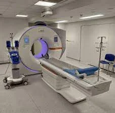
A CT Scan (computerized tomography scan) utilizes a tube with multiple X-ray apparatuses to form a three-dimensional image from two-dimensional X-ray photographs from multiple cross-section ways. Radiolucent things must be nicely caught on computerized Tomography scans rather than X-rays. Since Computerized Tomography is a different angle of x-rays, the radiation exposure is excessively greater.
A computerized tomography scan is a useful diagnostic instrument for catching diseases and injuries. It uses a sequence of X-rays and attaches a computer to generate a three-dimensional photograph of soft tissues and bones. computerized tomography is a painless, noninvasive path for healthcare providers to diagnose conditions. the person might have a computerized tomography scan at a hospital or imaging center.
A computed tomography scan is typically compressed to a Computerized Tomography scan; earlier named computed axial tomography scan which is a medical imaging procedure that is utilized to get thorough internal photographs of the body. The person that performs Computerized Tomography scans is named radiographers or radiology technologists.
Computerized Tomography scanners execute a spinning X-ray tube and a row of sensors positioned in the surround to consider X-ray views weakness by further tissues inside the body. The numerous X-ray measurements would carry out from multiple angles of view like anterolateral, superior, etc. A Computerized Tomography scan should be utilized on patients with metallic implants or pacemakers, for whom magnetic resonance imaging is contraindicated.
Medical professionals utilize computed tomography, even named as computerized tomography scan, to analyze designs inside the body. A computerized tomography scan brings images that display very thin “pieces” of the bones, muscles, organs, and blood vessels so that healthcare providers must notify the body at significant points.
Traditional X-ray devices utilize a set tube to point X-rays at a single spot. As X-rays run through the body, rays should be soaked in different amounts by different tissues. elevated density tissue creates a whiter photographes rather than other tissues against the black background of the X-ray film. X-rays make two-dimensional images. computerized tomography scans have a doughnut-shaped tube that turns the X-ray 360 degrees surround you. The data captured in the X-ray delivers a precise three-dimensional view of the inside of the body.
Types of Computerized Tomography Scan
Spiral Computerized Tomography
A spinning tube, commonly known as spiral Computerized Tomography is an imaging approach in which a full X-ray tube is turned near the central axis of the region would be examined. Computerized tomographies are the major type of scanners on the market because spiral Computerized Tomography should manufacture longer and offers a lower cost of the display and buy. The primary limitation of spiral Computerized Tomography will be the size and inactivity of the equipment X-ray tube body and the detector will display on the contrasting side of the circle which slows the speed at which the equipment should circle. Some designs utilize two X-ray sources and sensor arrays compensated by an angle, as a method to improve temporal resolution
Electron beam tomography
Electron beam tomography is a specific form of Computerized Tomography in which a large adequate X-ray tube is created so that just the direction of the electrons, touring in the middle of the cathode and anode of the X-ray tube, should utilizing using deflection coils. This type of electron beam tomography should have a significant benefit since sweep speeds must be much faster, give granting for slightly blurry imaging of moving formats, like the heart, and arteries. More occasional scanners of blurry design must be constructed when compared with twisting tube types, specifically because of the higher cost linked with making a considerably larger x-ray tube and sensor array and limited anatomical range of motion.
Dual-source Computerized Tomography
Dual-source Computerized Tomography is a progressive scanner with a two X-ray tube detector design, unlike conventional single-tube designs. These two detector systems should ascend on a single platform at 90 degrees in the same plane. Dual-source computerized tomography scanners give grant fast scanning with a higher temporal resolution by designing a full computerized tomography piece in just half a spin. Fast imaging lessens the movement blurring at high heart rates and potentially gives grants shorter breath-hold time. This is specifically helpful for ill patients who keep difficulty holding their breath or are untrained to take heart-rate-lowering medication.
Computerized Tomography perfusion imaging
Computerized Tomography perfusion imaging is a precise form of computerized tomography to estimate flow via blood vessels whilst infiltrating a different mechanism. Blood flow, blood transit time, and organ blood volume must all be calculated with moderate sensitivity and specificity. This type of computerized tomography must be operated on the heart, although sensitivity and carefulness for catching abnormalities should constantly be more down than for other formats of Computerized Tomography. perfusion imaging should again be used on the brain, whereas Computerized Tomography perfusion imaging should typically see poor brain perfusion well before it would be caught using a conventional spiral Computerized Tomography scan.
Mechanism of computerized tomography
Computed tomography generally works by operating an X-ray generator that turns surrounding the thing/object; X-ray sensors must be organized on the contrasting side of the circle from the X-ray origin. As the X-rays pass via the patient, they are reduced differently by different tissues regarding tissue density. A visible representation of the basic data obtained is named a sinogram, yet the sinogram is not adequate for interpretation. Once the scan data should be collected the collected data must be processed utilizing a format of tomographic reconstruction, which delivers a sequence of cross-sectional photographs. These cross-sectional pictures should be made up of small units of pixels or voxels.
Pixels in an appearance got by computerized tomography scanning should be shown in terms of relative radiodensity. The pixel itself is shown regarding the mean reduction of the tissue that it coordinates to on a scale from plus 3,071 most reducing to minus1,024 least reducing on the Hounsfield scale. A pixel is a two-dimensional unit depending on the matrix size and the field of outlook. When the computerized tomography slice consistency is also factored in, the unit, therefore, is known as a voxel, which is a three-dimensional unit.
Water has a reduction of 0 Hounsfield units, while air should be 1,000 Hounsfield units, cancellous bone is normally plus 400 Hounsfield units and cranial bone must achieve 2,000 Hounsfield units or more os temporale and can cause damage. The lessen of metallic implants depends on the atomic number of the part utilized: Titanium normally has an amount of plus 1000 Hounsfield and iron steel should destroy the X-ray and therefore, responsible for well-known line destruction in computed tomograms. these destroy should be caused by short transitions in the middle of low and high-density fabrics, which results in data worths that overreach the dynamic range of the processing electronics. Two-dimensional computerized tomography
images should be consistently affected so that the view is as though looking up at the image side from the patient’s feet. Thus, the left side of the image is to the patient’s right and vice versa, while the anterior side in the image again is the patient’s anterior and vice versa. This left-right interchange coordinates with the thought that doctors typically have facts when placed in front of patients.
Originally, the photos produced in computerized tomography scans should be in the transverse axial anatomical plane, crosswise to the long axis of the body. Modern scanners give grants to the scan data to be reformatted as pictures in other planes. Digital geometry clarification must produce a three-dimensional concept of a thing/object inside the body from a sequence of two-dimensional radiographic pictures got by the spin surrounding a fixed adjusted axis. These cross-sectional images should be broadly utilized for medical diagnosis and physical therapy
Preparations for the CT scan machine
The healthcare provider will provide instructions on how to prepare for the computerized tomography scan. On the day of the examination, the person should pay concentration to:
Depending on which part of the body is being scanned, the person might be asked to:
- Coming: the person should plan to reach early, depending on the healthcare provider’s instructions. Reaching early helps the testing visit on time.
- Diet: Avoid eating and drinking for four hours before the examination.
- Medicines: Ask the healthcare provider if you should take the person’s regular medicines before the computerized tomography scan.
- Comfort: You should wear comfortable clothes. she might require to change clothes before the examination and remove the watch and jewelry, involving any piercings she should remove. he/she might require to remove dentures and hearing aids, too. Zippers and metal things should block the scan.
- remove some or all of the clothing and wear a hospital gown which is provided by the staff
- Remove metal objects, such as a belt, jewelry, and eyeglasses, which might interrupt photo outcomes
- Blood test: the person might require a blood test before the planned computerized tomography scan. The blood test will make sure the healthcare provider chooses the right pigment.
- Diet restrictions: the person will require to observe what he/she eats and drink for four hours before the computerized tomography scan. Drinking only clear liquids helps control nausea when receiving the contrast pigment. he/she should normally drink tea or black coffee, strained fruit juices, simple gelatin, and soft drinks, like ginger ale, etc.
- Allergy medicine: If she/he is allergic to the contrast agent utilized in computerized tomography which comprises iodine, the person might require to take a steroid pill before the night and the morning of the procedure along with an antihistamine, such as Benedryl, before the examination. Be sure to inspect with the healthcare provider and have them request these medicines for the person if required. Contrast agents for magnetic resonance imaging and computerized tomography are different; being allergic to one doesn’t mean being to allergic the other.
- Preparation of the liquid solution: the person should drink the contrast solution orally by mouth as guided by the healthcare provider or physicians.
Contrast material for CT scan
A special pigment named contrast material is required for some computerized tomography scans to support highlighting the places of the body being scanned. The contrast material agent should obstruct the X-rays and reveals the white film, which should assist in the highlighting of blood vessels, intestines, or other structures of the body.
Contrast material might be given to the person:
- By mouth: If the person’s esophagus or stomach should be scanned, the person might require to consume a liquid that includes contrast material. That drink might taste distasteful.
- By injection: Contrast material agents should be infiltrated via a vein in the arm to support the gallbladder, urinary tract, liver, or blood vessels to stand out in the photos. the person might experience a sensation of heat during the injection or a gold taste in the mouth.
- By enema: Contrast material agents might be inserted in the rectum to support imagine the intestines. This technique should create a feeling of puffy and uncomfortable.
Reactions to contrast material
In certain cases, the doctor might suggest that the person obtain a special pigment named contrast material. This pigment is something like the doctor requested to drink before the computerized tomography scan, or something that is given via a vein in the arm or inserted into the rectum. Although infrequent, the contrast material should cause medical problems or allergic reactions.
Most reactions are mild and outcome in a rash or itchiness. In rare examples, an allergic reaction should be serious, even life-threatening. Tell the doctor if the person ever had a reaction to contrast material.
Preparing the child for a computerized tomography scan
If the infant or toddler is keeping a computerized tomography scan, the doctor might suggest sedative medicines to preserve the child peaceful and motionless. Motion blurs the pictures and might conduct in incorrect consequences. Ask the doctor how to prepare the child for a CT scan and follow the terms and conditions prescribed by the doctor.
During the procedure
computerized tomography scanners are shaped like a large doughnut food standing on its side. the person lies on a narrow, motorized table that glides via the opening into a tunnel. Straps and pillows might be utilized to support the person to survive in position. During a head scan, the table might be fitted with a special hotbed that holds the head always.
While the table thrusts the person into the scanner, sensors, and the X-ray tube spin surround the person. Individually spinning produces several photos of slim portions of the body. the person might listen to whizzing noises.
A technologist in an individual room should be able to see and listen to the person’s voice. the person will be able to speak with the technologist through the intercom. The technologist might ask the person to keep his/her breath at specific moments to avoid blurring the photographs.
After the procedure of CT scan
After the examination, the person should return to his/her normal way. If he/she were provided a contrast material, he/she might accept special instructions. In some cases, the person might be requested to stay for a short time before leaving to assure that he/she feels okay after the examination. After the computerized tomography scan, he/she will probably be exposed to drinking lots of fluids to support the kidneys and release the contrast material from the body.
Results of the CT scan
computerized tomography scan photographs should be reserved as electronic data files and normally examined on a computer screen. A radiologist analyzes these photographs and sends a report to the doctor.
Medical use of the Computerized Tomography scan
Computerized tomography should become an essential tool in medical imaging to increase traditional X-ray imaging and medical ultrasonography. It should more recently be utilized for preventative drugs or screening for disease, computerized tomography colonography for people with a high risk of colon cancer, or full movement heart scans for individuals with an elevated chance of heart disease. Several organizations deliver full-body scans for the general people although the technique goes against the direction and authorized function of many professional organizations in the area mainly because of the radiation dose used.
The uses of computerized tomography scans should improve dramatically over the last two decades in many nations’
head
computerized tomography scanning of the head is simply analyzed to carry out data regarding infarction (stroke), tumors, calcifications of bones, hemorrhage, and traumatic bone injury. Of the above-mentioned hypodense dark parts should reveal edema and infarction, hyperdense bright parts reveal calcifications and hemorrhage, and bone trauma should be seen as disconnection in bone windows. Tumors should be caught by the swelling and anatomical distortion that parts cause, or by enclosing edema. computerized tomography scanning of the head is also utilized in computerized tomography-directed stereotactic surgery and radiosurgery for the therapy of intracranial tumors, arteriovenous malformations, and accessory surgically treatable problems utilizing a device termed the N-localizer.
Neck
computerized tomography is commonly the initial choice for neck masses in adults. computerized tomography of the thyroid performs an essential role in the evaluation of thyroid cancer. computerized tomography scan frequently incidentally discovers thyroid abnormalities, and so the CT scan is frequently preferred experimentation modality for thyroid abnormalities.
Lungs
A computerized tomography scan must be utilized for catching both acute and chronic differences in the lung parenchyma, the tissue of the lungs. A computerized tomography scan is significantly appropriate in the lungs case because normal two-dimensional X-rays should not show such deficiencies. A variety of procedures should be utilized, depending on the presumed abnormality. For evaluation of chronic interstitial methods like emphysema, and fibrosis, thin units with high spatial frequency reconstructions should be utilized; frequently computerized tomography scans should be executed both on inhalation and exhalation. This unique method is known as high-resolution CT that generates a piece of the lung, and not constant pictures.
bronchial wall thickening must be seen on lung CTs and normally but not always indicates inflammation of the bronchi.
An incidentally discovered nodule in the absence of symptoms sometimes referred to as an incidental coma might increase problems that might represent a tumor, either benign or malignant. Possibly influenced by fear of patients and doctors sometimes conform to an intensive program of CT scans, sometimes up to every three months and exceeding the instructed guidelines, in trying to do management on the nodules. However, selected guidelines suggest that patients without an initial history of cancer and whose solid nodules have not grown beyond a period of two years should be doubtful to have any malignant cancer risks related to having computerized tomography scans, patients should not obtain screening in the quantity of those recommended by specified guidelines.
Angiography
Computed tomography angiography is a type of contrast CT to imagine the arteries and veins all over the body. This ranges from arteries giving the brain to those carrying blood to the lungs, kidneys, arms, and legs. this type of CT pulmonary angiogram is utilized to diagnose pulmonary embolism. It utilizes computed tomography and an iodine-based difference agent to get an idea of the pulmonary arteries.
Cardiac CT scan
A CT scan of the heart should be conducted to achieve details regarding cardiac or coronary anatomy. Traditionally, cardiac CT scans should be utilized to catch, diagnose, or follow up for coronary artery disease. More newly computerized tomography has played an essential role in the fast-developed area of transcatheter structural heart interventions, more particularly in the transcatheter repair and replacement of heart valves.
The main formats of cardiac CT scanning should
Coronary CT angiography (CCTA): the use of computerized tomography to estimate the coronary arteries of the heart. The cardiac CT scan obtains an intravenous injection of radiocontrast, and then the heart is monitored on the computer by utilizing a high-speed CT scanner, giving a grant to radiologists to calculate the capacity of occlusion in the coronary arteries, normally to diagnose coronary artery disease.
Coronary CT calcium scan
coronary CT calcium scan will also utilize to check the hardness of coronary artery disease. Precisely, a coronary computerized tomography calcium scan looks for calcium deposits in the coronary arteries that should tighten arteries and raise the risk of a heart attack. A standard coronary computerized tomography calcium scan must be fulfilled without the usage of radiocontrast, though the scan should be also done from contrast-improved photos satisfactorily.
To satisfactorily result to show the anatomy and post-processing of the images is common. The most common photographs are multiplanar reconstructions (MPR) and volume analysis. For more complex anatomies and approaches, like heart valve interventions, a true three-dimensional reconstruction or a three-dimensional print will be made founded on these CT pictures to achieve a deeper understanding.
Abdomen and pelvis
computerized tomography is an exact procedure for the diagnosis of abdominal diseases like Crohn’s disease, gastrointestinal tract bleeding, and diagnosis and staging of cancer, as well as follow-up after cancer treatment to evaluate response. A computerized tomography scan is naturally utilized to analyze acute abdominal pain.
Non-improved computerized tomography is nowadays the gold standard for analyzing urinary stones. The size, volume, and thickness of stones should be assessed to assist clinicians for recommend additional treatment; size is particularly significant in indicating the unexpected route of a stone.
Axial skeleton and extremities
For the axial skeleton and extremities, computerized tomography should repeatedly be utilized to reflect complex fractures, the main ones surrounding joints, due to its ability to rebuild the region of attraction in multiple planes. Fractures, ligamentous injuries, and dislocations should smoothly be identified with a 0.2 mm resolution. With modern dual-energy computerized tomography scanners, new places of use should be selected, such as assisting in the diagnosis of gout.
Biomechanical use of the CT scan
computerized tomography is utilized in biomechanics to fast show the geometry, anatomy, thickness, and elastic moduli of biological tissues.
Other uses of computerized tomography
Industrial use
industrial computed tomography is a strategy that utilizes X-ray tools to create three-dimensional declarations of elements both externally and internally. Industrial CT scanning should be operated in numerous places in the industry for an inner review of elements. Some of the important usages for computerized tomography scanning should be spot detection, failure research, amount, assembly analysis, photographic-based narrow element approaches, and switch engineering applications. computerized tomography scanning should also be utilized in the imaging and saving of the gallery
Flight security
CT scanning should discover an application in transportation protection especially airport security where it is presently utilized in a fabrics research context for explosive-detection devices and is also under reflection for automatic luggage protection scanning utilizing computer vision-based object recognition algorithms that target the detection of detailed danger objects based on three-dimensional impression like liquid containers.
Geological use of CT scan
X-ray computerized tomography is utilized in geological analyses to fast indicate materials inside a training center. Thick minerals like pyrite and barite seem more hopeful and less thick elements like earth arise light in CT photos.
Cultural heritage use
X-ray CT and micro-CT should also be utilized to conserve and preserve things of cultural tradition. For many weak things, direct investigation and observation should be harmful and spoil things beyond time. Using computerized tomography scans, investigators should be able to select the material arrangement of the things that investigators should analyze, without any further harm. The computerized tomography scans should be sufficient for evaluation concentrated on the workings of the mechanism of the text hidden inside the machine outside coating of the scroll in the cultural heritage. CT scans are specific artifacts in which the material arrangement should have minimal variation along the inside of the object. After scanning these objects, computational methods should be utilized to examine the insides of the things, Micro-CT has also been confirmed beneficial for analyzing more recent artifacts such as still-sealed closeness that utilized the method of letter-locking complex folding and cuts that delivered a tamper-evident locking mechanism
Risks of using CT scan
Radiation exposure
During a computerized tomography scan, the doctor should be shortly exposed to ionizing radiation. The amount of radiation is greater than the patient would obtain during a plain X-ray because the computerized tomography scan collects more detailed information. The low doses of radiation utilized in computerized tomography scans should be responsible to cause long-term effects, although, at higher doses, there might be a small raise in the possible risk of cancer.
computerized tomography scans have multiple advantages that overpower any little possible risk. Doctors use the lowest dose of radiation possible to receive the required medical information. Also, newer, faster apparatuses and strategies require less radiation than was earlier used. Talk with the doctor regarding the advantages and risks of the computerized tomography scan.
Harm to unborn babies
Tell the doctor she is pregnant. Although the radiation from a computerized tomography scan is improbable to injure the baby, the doctor might suggest another type of exam, like an ultrasound or magnetic resonance imaging, to avoid revealing the baby to radiation. At the low doses of radiation utilized in CT scans, no side effects have been found in humans.
FAQs
What the person should expect from a CT scan?
the person should do the computerized tomography scan in a hospital or an outpatient facility like a laboratory. computerized tomography scans are painless with newer machines and take just a few minutes. The complete strategy commonly carries roughly thirty minutes.
How long does the computerized tomography scan test take?
Normally, the person should schedule an hour for a computerized tomography scan. Most of that time is taken for preparation. The computerized tomography scan itself takes between ten to thirty minutes or less. Typically, the person should resume his/her activities after a healthcare provider states it is safe normally after radiologists finish the computerized tomography scan and confirm clear pictures of the particular area of the body which gets damaged.
What does a CT scan show?
the healthcare provider will request a CT scan to assist and create a diagnosis of the health. The computerized tomography scan allows the providers to closely investigate bones, organs, and other soft tissues of the body, blood vessels, and doubtful growths. Things that a computerized tomography scan should detect contain:
Specific types of cancer and benign noncancerous tumors.
Fractures and broken bones.
Heart disease.
Blood clots.
Bowel disorders like blockages, and Crohn’s disease.
Brain and spinal cord diseases or injuries like kyphosis, lordosis, and scoliosis.
Internal bleeding.
Healthcare providers should also notice organs and tissues on X-rays. But on X-rays, the body parts seem to overlap, creating for a scanner difficult to notice everything. The CT scan displays spaces between organs for a more obvious view.
Are CT scans safe for making a diagnosis?
Healthcare providers regard computerized tomography scans as commonly safe. CT scans for children are safe, too. For children, computerized tomography technologists might utilize apparatuses changed for children to lower the scanner radiation exposure.
like computerized tomography and other diagnostics, utilize a small amount of ionizing radiation to grab a picture of the affected area. Some risks related to CT scans contain:
Cancer risk: All types of imaging utilizing radiation, like X-rays, cause a small raised in the risk of developing cancer. The difference is too small to estimate actually.
Allergic responses: Periodically, people have a minor or more serious allergic reaction to the contrast agent.
If the person should worry about the health risks of computerized tomography scans, talk to the healthcare provider. They will consult the problems and support to create a knowledgeable decision about the computerized tomography scan.
should the female go for a computerized tomography scan if she will pregnant?
If the female might be pregnant, she should inform the CT technologist. CT scans of the pelvis and abdomen should subject the newborn fetus to radiation, but it’s not sufficient to cause genuine harm. computerized tomography scans in other parts of the body shouldn’t put the fetus in any trouble.
Should the person eat or drink before a CT scan?
the person should not eat for 2.5 hours earlier to the examination. the person might have clear drinks up to two hours before the examination. Clear liquids contain water, black coffee or tea, apple juice, clear soda, or clear broth. the person might take her/his medicine at the normal time with water.
What is the side effect of a computerized tomography scan?
There are no side effects from the computerized tomography scan itself, although a CT scan uses multiple times more radiation rather than a routine X-ray; equal to what the person might be revealed to naturally beyond a 3 to-4-year period.
Who should not carry a CT scan?
Because too much exposure to radiation in youth has seemed to cancer as an adult, doctors seem to not suggest CT scans for children or those who need multiple scans. the person should consult with her/his doctor if she/he has allergic to iodine contrast pigments, which were frequently utilized for CT scans.
What are the two disadvantages of CT scans?
Situations regarding the computerized tomography scans contain the risks from disclosure to ionizing radiation and potential comebacks to the intravenous contrast pigment, which might be utilized to improve visualization. Disclosure of ionizing radiation might generate a little gain in a person’s lifelong gamble of creating a tumor.
Should MRI is secure as a CT scan?
An MRI has not utilized radiation, and a CT Scan has not utilized a magnet. CT scan is safer than MRI for some patients. someone with a metal implant in their body like a pacemaker, stent, implant, etc. requires to confirm the material is in magnetic resonance imaging is safe, for those people who sensitive to radiation, a CT scan is not a suitable choice.
How much radiation is in a CT scan?
The effective amounts from diagnostic CT methods are typically calculated to be in the range of 1 to 10 mSv. This range is not much lower than the lowest doses of 5 to 20 mSv obtained by some people
What is the next step after a CT scan?
Once the CT scan is complete, the pictures should be sent to a radiologist for examination. A radiologist who specializes in interpreting and treating illnesses utilizing imaging strategies, such as CT scans and X-rays. the doctor will take follow up with him/her to describe the upshots.
There is normally no certain type of care demanded after a CT scan. the person might continue her/his regular diet and activities unless the doctor instructs her/him differently. the doctor might provide the person with extra or alternate instructions after the approach, depending on the special situation.
What are the stages of a CT scan?
Specific stages time from injection include:
early arterial stage: 15-25 seconds post-injection. instantly post bolus tracking.
late arterial stage: 30-40 seconds post-injection.
portal venous stage: 70-90 seconds post-injection.
nephrogenic stage; 85-120 seconds post-injection.
excretory stage: 5-10 minutes post-injection.


33 Comments