Rectus capitis lateralis muscle
Introduction
Rectus capitis lateralis muscle is a small, paired muscle that is seen within the neck, deep to the prevertebral portion of the deep cervical fascia. It is a portion of the prevertebral muscle group along with also splenius capitis, levator scapulae, and middle and posterior scalene muscles.
The rectus capitis lateralis consists of paired muscles set in the upper neck area below the back of the head on each side. They are accountable for controlling movements when the head tilts to either side. (lateral flexion).
Origin of Rectus capitis lateralis muscle
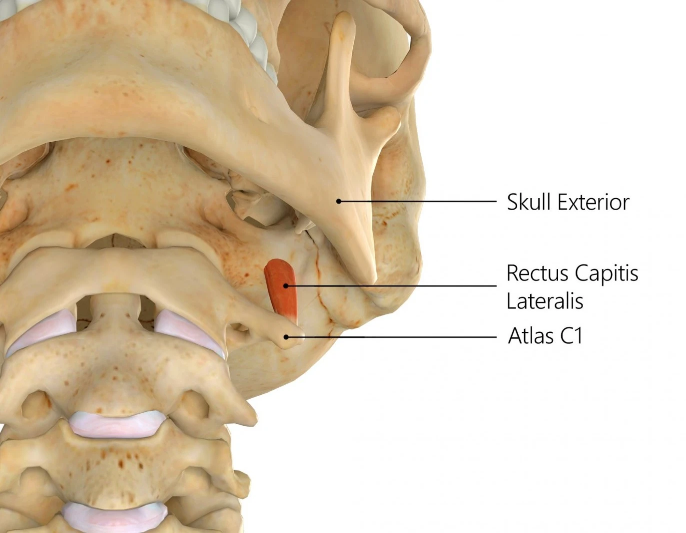
Rectus capitis lateralis muscle is a short and flat muscle. It originates from the superior surface of the transverse process of the atlas (C1) bone.
Insertion of Rectus capitis lateralis muscle
The vertically oriented fibers traverse superiorly to insert onto the inferior surface of the jugular process of the occipital bone.
Rectus capitis lateralis muscles have several muscular and also neurovascular relations. It is lateral to the rectus capitis anterior muscle and the cranial end of the longus capitis muscle. The levator scapulae muscle inserts on the opposing side of the C1 transverse process inferior to rectus capitis lateralis. Rectus capitis lateralis is also anterior to the obliquus capitis superior muscle, anterosuperior to the obliquus capitis inferior muscle, and superior to the anterior cervical intertransverse muscle.
The rectus capitis lateralis is posteriorly connected to the internal jugular vein. It is diverged from this structure by the carotid sheath and occasionally by the accessory nerve (CN XI). The posterior belly of the digastric muscles is also anterior to the muscle; with the occipital artery occasionally separating the two structures. The vertebral artery gives by the medial border of the rectus capitis lateralis muscles as it leaves the transverse foramen of the atlas. The glossopharyngeal, vagus, and accessory nerves (CN IX-XI) pass over the anterior surface of the rectus capitis lateralis muscle as they exit the jugular foramen.
Nerve supply
Motor innervation to rectus capitis lateralis muscle is provided by the division of the anterior rami of the first two cervical spinal nerves (C1-C2).
Blood supply
The muscle acquires blood from the ascending cervical artery, which is a small branch of the inferior thyroid artery from the thyrocervical trunk of the subclavian artery. It also acquires blood from the muscular branches of the vertebral artery. The muscle also acquires small muscular branches from the occipital artery as it passes its lateral part.
Function of Rectus capitis lateralis muscle
The primary function of the rectus capitis lateralis muscle is to stabilize the atlantooccipital joint during movement. Unilateral contraction prompts ipsilateral flexion of the neck. It helps weakly with lateral flexion for the head. The postural muscle assists in monitoring the position and motion of the head.
Clinical significance
The rectus capitis lateralis muscles can be used as a climacteric by neurosurgeons when approaching the jugular foramen. It permits the surgeons to determine important neurovascular entities early in the dissection. This becomes especially important if the natural anatomy has been distorted by pathological processes (e.g. tumors).
Some of the most common types of injuries to athletes are those of a critical strain or the sprain in the musculature of the neck, in complement to any soft-tissue contusions. Strains are injuries to the muscles that happen whenever the muscle is overfilled or stretched. Sprains are when the ligaments or the capsular structures that bind the vertebrae or the cervical facet joints become harmed. It may be quite challenging to separate the 2 injuries, which is why they generally occur at the same time. Symptoms may be explained as stiffness in the neck, muscle spasms in the contrived area, tenderness, or decreased range of motion of the head or neck.
Treatment of an injury to this muscle consists of icing the harmed place, isolating movement of the area, resting, & then massage therapy. Non-steroidal anti-inflammatory drugs (NSAIDs), such as ibuprofen, might be assumed to address pain or decrease inflammation. It has been said that rectus capitis lateralis muscles damage at a significantly higher level of load or strain than the other 3 pairs of muscles. The acquiescent stiffness of rectus capitis lateralis or the anterior muscles was more than the other muscle pairs.
Exercise of Rectus capitis lateralis muscle
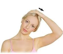
Side neck stretch: Sit up straight in a solid-based and also straight-backed chair. Relax the left hand into the lap while the right-hand starts to reach over the top of the head. Permit the right hand, with fingers closed, to apply on the left side of the head just above the left ear. Firmly presses the head to the right side as if to press the right ear to the right shoulder. Maintain this position for 10-15 seconds, then slowly release it back up to the starting position. Change hands and repeat to the other side. This may be accomplished two to three times a day.
FAQ
What is the origin and insertion of rectus capitis lateralis?
The rectus capitis lateralis, a short, flat muscle, originates from the upper surface of the transverse process of the atlas and is inserted into the undersurface of the jugular process of the occipital bone.
What is the blood supply of the rectus capitis lateralis muscle?
The rectus capitis lateralis muscle acquires arterial blood supply from the branches of the vertebral, occipital, and ascending pharyngeal arteries. The first artery is a division of the subclavian artery, while the occipital and ascending pharyngeal arteries are branches of the external carotid artery.
What is rectus capitis lateralis?
Rectus capitis lateralis muscle is a small, paired muscle that is seen within the neck, deep to the prevertebral portion of the deep cervical fascia. It is a portion of the prevertebral muscle group along with also splenius capitis, levator scapulae, and middle and posterior scalene muscles.
What are the symptoms of rectus capitis injury?
Symptoms of rectus capitis injury are muscle spasms with neck stiffness, Neck pain with tenderness, and reduced range of motion(ROM) neck.
Treatment is the use of the RICE Principle such as Rest, applying Ice, use of neck collar with Pain relieving medicine.
What does capitis mean?
A Latin term meaning “of the head,” is used in the names of some muscles that are connected to the head.