
Respiratory System
The Respiratory System is a complex network of organs and tissues that facilitates the exchange of gases, primarily oxygen and carbon dioxide, between the body and the external environment. This vital system is responsible for the intake of oxygen, essential for cellular functions, and the removal of carbon dioxide, a waste product produced by the body’s metabolic processes.
Key components of the respiratory system include the lungs, where gas exchange occurs, and a system of airways that transport air to and from the lungs. The process of respiration involves both inhalation, where fresh air is drawn into the lungs, and exhalation, where stale air and carbon dioxide are expelled.
In addition to the lungs, the respiratory system includes the nose, mouth, trachea (windpipe), bronchi, and bronchioles. These structures work together to filter, humidify, and warm the air as it travels into the lungs, optimizing conditions for gas exchange.
The respiratory system also plays a crucial role in maintaining the body’s acid-base balance by regulating the levels of carbon dioxide in the blood. Furthermore, it interacts with other bodily systems, such as the cardiovascular system, to ensure the delivery of oxygen-rich blood to various tissues.
Function
The respiratory system serves multiple functions. Apart from helping you to inhale (breathe in) and exhale (breathe out), it:
- Allows you to speak and smell.
- Warms the air to your body temperature and absorbs it to the humidity level your body requires.
- Provides oxygen to your body’s cells.
- When you breathe out, waste gases such as carbon dioxide are taken out of your body.
- Keeps harmful compounds and irritants out of your airways.
Anatomy
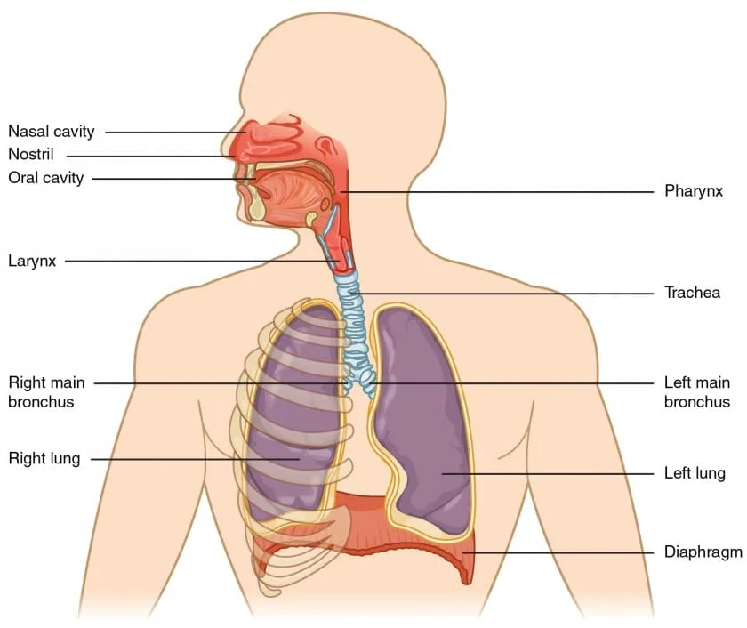
The respiratory system is made up of organs that interchange oxygen and carbon dioxide. The upper and lower respiratory tracts form the respiratory system.
The upper respiratory tract is made up of the following components:
- Nose
- The nasal cavity
- Mouth
- Sinuses
- Pharynx (throat)
- The larynx (voice box)
- The trachea (windpipe)
The lower respiratory tract is made up of the following components:
- Lungs
- Bronchi (large airways)
- Bronchioles (small airways)
- Alveoli (air sacs)
Upper Respiratory Tract
Nasal Cavity
The nasal cavity is the first step in the upper respiratory system. The nasal cavity opens anteriorly on the face via the two nares and posteriorly by the two choanae into the nasopharynx. The hard palate forms the floor of the nasal cavity, while the roof is made up of the cribriform plate of the ethmoid bone posteriorly and the frontal and nasal bones anteriorly. The nares and anterior nasal passages include sebaceous glands and hair follicles that serve to keep greater dangerous substances out of the nasal cavity.
The nasal cavity’s lateral walls have three bony extensions termed nasal conchae (superior, middle, and inferior), which increase the nasal cavity’s surface area. The nasal conchae also cause laminar disruption.
The olfactory epithelium, which is made up of specialized sensory receptors, is found on the roof of the nose cavity. These receptors detect airborne odorant molecules and convert them into action potentials that travel to the cerebral cortex via the olfactory nerve, allowing the brain to register them and create a sense of smell.
The mouth cavity is another entry point for air. Although it is not considered part of the upper respiratory tract, the oral cavity provides an alternative pathway in the event of nasal cavity obstruction. The oral cavity opens anteriorly on the face through the oral fissure, and posteriorly into the oropharynx via the oropharyngeal isthmus.
Paranasal Sinuses
Several bones that constitute the nasal cavity’s walls contain air-filled spaces known as paranasal sinuses, which are named after the bones with which they are associated: maxillary, frontal, sphenoidal, and ethmoidal sinuses.
The paranasal sinuses communicate with the nasal cavity through various apertures, receiving breathed air and contributing to its humidification and warmth. Furthermore, the mucous membrane and respiratory epithelium that line both the nasal cavity and the paranasal sinuses catch any potentially dangerous particles, dust, or germs.
Pharynx
Inhaled air exits the pharynx via the choanae after passing via the nasal cavity and paranasal sinuses. The pharynx is a muscular tube with three parts: the nasopharynx, oropharynx, and laryngopharynx.
The nasopharynx is the first and most superior section of the pharynx, located behind the nasal cavity. Because this region of the pharynx only serves as an airway, it is lined with respiratory epithelium. During swallowing, the uvula and soft palate move upwards to block off the nasopharynx and prevent food from entering the nasal cavity.
The laryngopharynx (hypopharynx) is the pharynx’s most inferior subsection. It is the connection of the digestive and respiratory systems. The laryngopharynx continues into the larynx anteriorly, and the esophagus posteriorly is located behind the oral cavity and interacts with it via the oropharyngeal isthmus. The oropharynx is a passageway for both air from the nasopharynx and food.
Larynx
The superior region of the larynx is the next and final segment of the upper respiratory system after the laryngopharynx. The larynx is a difficult hollow structure that is located anterior to the esophagus. It is supported by a cartilaginous skeleton that is linked by membranes, ligaments, and muscles. The larynx, like the laryngopharynx, is lined by stratified squamous epithelium above the auditory cords. This epithelium gives way to pseudostratified ciliated columnar epithelium with goblet cells (respiratory epithelium) below the vocal cords.
The larynx not only distributes air but also houses the vocal cords, which engage in voice production. During swallowing, the epiglottis closes the laryngeal inlet to prevent food or liquid from entering the lower respiratory tract.
Lower Respiratory Tract
The lower respiratory tract includes the elements of the respiratory system below the cricoid cartilage and voice cords, such as the inferior part of the larynx, the tracheobronchial tree, and the lungs.
Tracheobronchial Tree
The tracheobronchial tree is a branch of the respiratory system that connects the upper airways to the lung parenchyma. It is made up of the trachea as well as the intrapulmonary airways (bronchi and bronchioles). The trachea is the trunk of the tracheobronchial tree and is located in the upper mediastinum. The trachea divides into two major bronchi, one for each lung, at the level of the sternal angle (T5).
The left main bronchus enters the hilum of the left lung inferolateral. It travels inferior to the aortic arch and anterior to the esophagus and thoracic aorta.
The right main bronchus enters the right lung’s hilum inferolateral. The right main bronchus is bigger and shorter than the left, with a more vertical path. As a result, the right bronchus is more vulnerable to foreign body impaction.
The main bronchi branch out into increasingly smaller intrapulmonary bronchi as they reach the lungs. The left main bronchus separates into two secondary lobar bronchi, while the right main bronchus divides into three secondary lobar bronchi, each of which supplies the lobes of the left and right lung.
Each lobar bronchi divides further into tertiary segmental bronchi, which aerate the bronchopulmonary segments. The segmental bronchi give rise to many generations of intrasegmental (conducting) bronchioles, which gradually terminate as terminal bronchioles. Each terminal bronchiole produces multiple generations of respiratory bronchioles. Respiratory bronchioles develop into multiple alveolar ducts, which lead into alveolar sacs, which contain numerous grape-like outpocketings known as alveoli. Because they contain alveoli, these structures present the beginning of gas exchange.
Lungs
The lungs are two spongy organs that are located in the thoracic cavity. The right lung is larger than the left lung and is separated into three lobes (superior, middle, and inferior) by two fissures (oblique and horizontal). An oblique fissure divides the left lung into two lobes (superior and inferior).
The lungs have three different surfaces which are the apex, the center, and the base. The costal, mediastinal, and diaphragmatic surfaces of the lung are named after the surrounding anatomical tissue that they face. The hilum of the mediastinal surface connects the lung to the mediastinum. At the apex of the lung, the mediastinal and costal surfaces come together. It is the most superior part of the lung, that reaches into the root of the neck. The base of the lung is the lowest concave portion of the lung that lies on the diaphragm.
Each lung hilum contains the following:
- The main bronchus
- The pulmonary artery
- There are two pulmonary veins.
- Bronchial arteries
- Autonomic plexus of the lungs
- Nodes and vessels of the lymphatic system
Physiology
Ventilation is the movement of inspired gas into and exhaled gas out of the lung. Understanding lung volumes, lung compliance, ventilation-perfusion, and bronchomotor tone is vital for clinical respiratory physiology applications in anesthesia and critical care.
Lung-volumes
Typical tidal ventilation, which is roughly 4-8 ml/kg, may readily meet the body’s typical requirements. When extra ventilation is necessary (e.g., during exercise), the body maintains a system to provide it in the form of inspiratory reserve volume and expiratory reserve volume. After tidal expiration, a human takes a full inspiratory breath followed by an expiration to reserve volume, which is 4-5 L in an average 70 kg individual. The alveoli are always partially filled with air, which keeps them from collapsing. The volume left in the lungs following a vital capacity breath is referred to as residual volume.
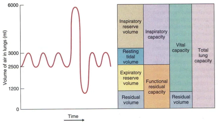
The sum of residual volume and expiratory reserve volume is the functional residual capacity (FRC). FRC is the quantity of air in the lungs following a typical expiration. The gases that linger in the lungs at the end of expiration not only prevent alveolar collapse but also continue to oxygenate the pulmonary blood streaming through the capillaries during this time. The reported FRC values vary, but in standing position, it is typically between 2.8 and 3.1 L. FRC changes with position, anesthesia, and body weight. FRC is the reserve that extends the period between non-hypoxic apneas.
Alveolar ventilation refers to the portion of minute ventilation that reaches the alveoli and participates in gas exchange. The normal amount of alveolar ventilation is about 5 L/min, which is comparable to the volume of blood moving through the lung (cardiac output 5 L/min). This results in an alveolar ventilation-to-perfusion ratio of about one.
Mechanism Of Respiration
The air pressure in and out of our lungs varies. So, as the air pressure drops, the alveolar gaps collapse and air enters the lungs (inspiration), and when the pressure of the alveoli within exceeds the atmospheric pressure, the air is blown out of the lungs (expiration). The size of the pressure difference determines the flow rate of air.
The breathing mechanism involves two processes:
- Inspiration
- Expiration
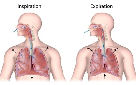
Inspiration
During inspiration, there is a contraction of muscles linked to the ribs on the outer side, which pulls the ribs out and causes the chest cavity to expand.
Later, the diaphragm brings downwards and widens the chest cavity, causing the abdominal muscles to contract.
The chest cavity expands, causing a partial vacuum to form, drawing air into the lungs and filling the expanded alveoli.
Inspiration Mechanism
- Inspiration is the process of getting atmospheric air. It is a dynamic process.
- Inspiration occurs when the volume of the thoracic cavity increases and the air pressure falls.
- The thoracic cavity is expanded when the external intercostal muscles contract.
- Contraction of the diaphragm enlarges the size of the thoracic activity even more. Simultaneously, the lungs expand.
- The air pressure inside the lungs reduces as the lungs expand.
- When the pressure in the lungs equalizes, atmospheric air rushes into the lungs.
Expiration
After the gaseous exchange occurs in the lungs and the air is released, the expiration process is regarded once. Expiration refers to the evacuation of air.
During this procedure, the rib muscles contract, and the diaphragm and abdominal muscles relax, resulting in a decrease in the volume of the chest cavity and an increase in the pressure of the lungs, allowing the air in the lungs to be forced out via the nose.
Expiration Mechanism
- Expiration is the process of exhaling carbon dioxide. It is a non-active process.
- It happens when the size of the thoracic activity reduces and the outside air pressure rises.
- The external intercostal muscles are now relaxed, whereas the internal intercostal muscles are contracted.
- As a result, the ribs are pressed inwards, and the thoracic cavity shrinks.
- The diaphragm relaxes and the lungs constrict.
- As a result, the pressure rises and the air is driven outside.
Respiratory System Disorders
What do Respiratory System Disorders consist of?
A disorder is described as an abnormal state of body functioning. Respiratory system disorders or respiratory illnesses are medical words used to describe the various forms of infections, allergies, and other diseases that affect the human respiratory system’s various organs, tissues, and specialized cells.
The upper respiratory tract, alveoli, bronchi, bronchioles, trachea, pleura, and pleural cavity are the main components of the respiratory system. A common cold is an example of a moderate respiratory condition, whereas other significant and potentially fatal respiratory disorders include pneumonia, lung cancer, asthma, influenza, tuberculosis, and others.
Factors Affecting Respiratory System Disorders
There are several factors related to respiratory system disorders. A few of these factors include:
- Genetics
- Allergies
- Smoking
- Air Pollution
- Bacterial and viral infections.
Disorders of the Respiratory System
Millions of individuals worldwide suffer from respiratory system problems. Respiratory illnesses are classified into three types:
Airway Disorder
It affects the bronchial tubes, which move oxygen and other gases into and out of the lungs. The path for air is decreased in airway disease, which is associated with either constriction or obstruction of the bronchial tubes.
Lung Tissue Illness
The pleura is a thin tissue surface that covers the human lungs. The structure of the lung tissues is damaged by some viral or bacterial infections, resulting in scarring or inflammation of the tissue that allows the lungs to expand correctly, making breathing difficult.
Disease of the Lung Circulation
This condition develops when the blood arteries in the lungs become coagulated, enlarged, or damaged. This impairs the lungs’ ability to accept oxygen and expel carbon dioxide. In severe circumstances, this condition might impair heart function.
Respiratory Disorders Causes
Respiratory ailments are caused by pollution, smoking, passively absorbing cigarette smoke, asbestos, radon, and other factors.
Diseases of the Respiratory System
The primary respiratory ailments or diseases are as follows:
Asthma is a chronic lung condition characterized by inflammation of the bronchi and bronchioles. It causes breathing difficulties and is accompanied by a severe cough, restlessness, cough, and a wheezing sound while breathing.
Asthma can be caused by the following factors:
- Cold air
- Allergens in the air
- Infections of the respiratory tract
- Pollutants in the air
- COPD (Chronic Obstructive Pulmonary Disease)
This comprises all respiratory disorders that build shortness of breath or difficulty in exhaling. It primarily affects persons who have been exposed to tobacco. It is a deadly condition that worsens even if you stop smoking.
Emphysema is a chronic condition that causes a reduction in the respiratory surface due to damage to the lung alveolar walls. Cigarette smoking is the primary culprit. Shortness of breath and cough are the most common symptoms of emphysema. Emphysema can cause the lungs to lose flexibility.
Emphysema can be caused by one or more of the following factors:
- Chemicals and Dust
- Pollution of the atmosphere
- Using tobacco
- Passive cigarette smoking exposure
Respiratory Disorders in the Workplace
Occupational respiratory disorders are defined as any condition caused by long-term inhalation of chemicals, proteins, or dust that impairs the respiratory system. Asbestosis, for example, is caused by inhaling asbestos parts:
- Metals emit fumes.
- Varnish, paint, acids, and pesticides are all sprayed.
- Cotton, silica, coal, medicine powders, and insecticides all produce dust.
- Industries’ gases. Ammonia, chlorine, and nitrogen oxides are a few examples.
Sinusitis
It is an inflammation of the nasal sinus mucous membranes. The mucus is produced by the mucous membranes and flows into the nasal passages. Mucous membrane inflammation is caused by bacterial or viral infections, as well as some airborne allergens.
Cancer of the Lungs
Lung cancer can manifest itself in any area of the lungs. It takes place in the main section of the lungs. Lung cancer treatment is determined by the kind, location, and extent of the disease.
Summary
The respiratory system controls bringing oxygen into the body and removing carbon dioxide. It also helps to filter the air we breathe and regulate our body temperature. The main parts of the respiratory system are the nose and mouth, throat (pharynx), larynx (voice box), trachea (windpipe), bronchi, and lungs. When we breathe in, air enters the nose or mouth and travels down the throat to the trachea. The trachea splits into the two bronchi, which lead to the lungs. In the lungs, the bronchi divide into smaller and smaller tubes, called bronchioles. The bronchioles close at tiny sacs called alveoli.
The alveoli are surrounded by blood vessels. When air enters the alveoli, oxygen diffuses from the air into the blood. Carbon dioxide spread out from the blood into the air. The oxygen-rich blood travels to the heart and is pumped to all parts of the body. The carbon dioxide-rich blood travels back to the lungs and is exhaled. The respiratory system also helps to filter the air we breathe. The nose and throat contain cilia, which are tiny hairs that trap dirt and dust particles. The lungs also contain mucus, which traps bacteria and viruses.
In addition to gas exchange and air filtration, the respiratory system also helps to regulate body temperature. When we are hot, we breathe more quickly. This increases the amount of air that passes through the lungs, which helps to cool the body. When we are cold, we breathe more slowly. This helps to conserve heat. The respiratory system is a vital organ system that is essential for life. Without a functioning respiratory system, we would not be able to get the oxygen we need to survive.
FAQs
What is the Function of the Respiratory System?
The respiratory system in humans is a complex network of organs and tissues that facilitate the process of breathing. Our airways, lungs, and blood arteries are all part of it. The muscles that power our lungs are part of the respiratory system. These components work together to transport oxygen and eliminate waste gases.
The respiratory system’s primary job is to exchange gases between the air and the blood. This process takes place in the lungs, where oxygen from the inhaled air diffuses into the bloodstream and carbon dioxide from the bloodstream diffuses into the exhaled air.
What are the primary components of the respiratory system?
The respiratory system can be subdivided into two main parts: the upper respiratory tract and the lower respiratory tract.
The upper respiratory tract includes the nose, mouth, throat, and larynx. The lower respiratory tract involves the trachea, bronchi, and lungs.
The upper respiratory tract is responsible for purifying and warming the air we breathe. It also contains the vocal cords, which allow us to speak.
The lower respiratory tract is responsible for gas interchange. This is the process of oxygen moving from the air into the blood and carbon dioxide moving from the blood into the air.
How can I maintain my Respiratory System?
Respiratory health must be able to release mucus from the lungs and airways.
To maintain the health of your respiratory system, you should do the following.
You should not smoke.
Drink plenty of water and eat a balanced diet rich in fruits and vegetables to stay hydrated.
Exercise daily to keep your lungs in good shape.
Wash your hands frequently and obtain a flu vaccine every year to avoid infection.
What is the role of the Respiratory System in cleaning the air?
Your respiratory system is designed to prevent hazardous substances in the air from entering your lungs.
The hairs on your nose filter out large particles. Tiny hairs called cilia move in a sweeping motion along your air passageways to retain them clean. However, if you breathe in toxic substances such as cigarette smoke, the cilia may cease operating. This can outcome in health issues such as bronchitis.
Cells in your trachea and bronchial tubes produce mucus, which keeps air passages moist and keeps allergens, bacteria, and viruses out of your lungs.
Mucus can drag things up from deeper within your lungs. Then you cough or swallow them.
References:
- Professional, C. C. M. (n.d.). Respiratory System. Cleveland Clinic. https://my.clevelandclinic.org/health/body/21205-respiratory-system#function
- Sendić, G. (2023, September 7). Respiratory system. Kenhub. https://www.kenhub.com/en/library/anatomy/the-respiratory-system
- Patwa, A., & Shah, A. (2015, January 1). Anatomy and physiology of respiratory system relevant to anesthesia. Indian Journal of Anaesthesia; Medknow. https://doi.org/10.4103/0019-5049.165849
- A. (2022, July 28). Respiratory System Disorders – Types and Causes of Respiratory Diseases. BYJUS. https://byjus.com/biology/respiratory-system-disorders/
- Respiratory System. (2002, February 11). WebMD. https://www.webmd.com/lung/how-we-breathe#1-6
- https://www.google.com/url?sa=i&url=https%3A%2F%2Fcourses.lumenlearning.com%2Fsuny-ap2%2Fchapter%2Forgans-and-structures-of-the-respiratory-system

4 Comments