
Stapedius Muscle
Introduction
The stapedius muscle is a muscle within the ear that support the stapes bone of the inner ear. It’s the very small skeletal muscle in the body. It plays an important role in hearing. It is Shaped like a sickle, the stapedius muscle is also described as the smallest skeletal muscle in the human body. However, some sources indicate its intermediate size may be larger than the often-cited 1-millimeter length.
The stapedius muscle is the smallest muscle of the human body, estimated at 6 millimeters in length. It is encountered in the tympanic cavity in the middle ear, connecting the pyramidal eminence of the petrous portion of the temporal bone to the posterior element of the neck of stapes.
The Stapedius muscles are innervated by the stapedial branch of the facial nerve ( CN VII). These autonomic fibers enable the muscle to be implicated in the auditory middle ear reflex, having a crucial role in covering the auditory system from damage.
Origin and insertion of Stapedius muscle
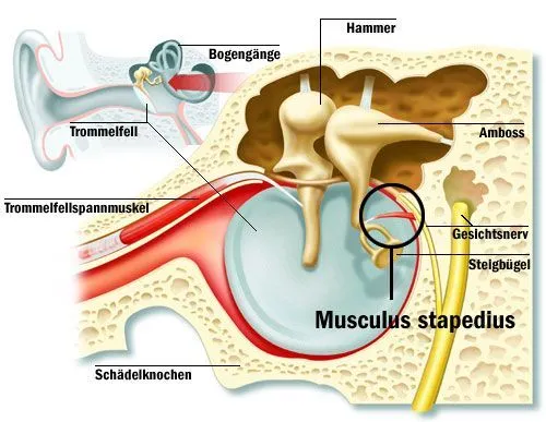
The stapedius emerges from a pinpoint foramen or opening in the apex of the pyramidal prominence (a hollow, cone-shaped eminence in the posterior wall of the tympanic cavity), and inserts into the neck of the stapes bone.
The stapedius muscle is located inside the ear cavity, particularly inside the tympanic cavity, in the middle portion of the ear. In a nourishing hearing canal, the tympanic cavity is filled with air. The muscle extends from the back part of the stapes bone, with the thinnest part of the muscle attaching to the neck of the stapes. It arises from a small opening of the pyramidal eminence, which is a hollow part of the tympanic cavity wall. The stapedial tendon attaches to the bottom portion of the stapedius muscle at the middle ear.
Blood supply
The blood supply is delivered by the stapedial branch of the posterior auricular artery, which branches give from the external carotid artery.
Nerve Supply
The stapedius muscle is innervated, or obtains nerve input from the brain, by the seventh cranial nerve. This can also be written as cranial nerve VII or as facial nerve VII.
The stapedius muscle is innervated by small branches that arise from the facial nerve (CN VII), also known as the nerve to the stapedius muscle. After branching off from the mastoid component of the facial nerve, the nerve gives posterior to the pyramidal process to innervate the stapedius muscle.
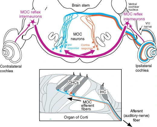
When the incoming sound is audible sufficiently to stimulate the receptor cells in the inner ear, the afferent signal arrives at the cochlear nucleus in the brainstem via the vestibulocochlear nerve (CN VIII).
From the brainstem, efferent signals are transmitted to the ipsilateral and contralateral middle ears triggering the contractions of the stapedius muscles. Their contractions result in posterior rotation and positioning of the base of the stapes into the oval window which closes it and attenuates further vibrations given to the cochlea. Thus the main function of the stapedius muscle is to cover the hearing apparatus when exposed to loud sounds.
Function of Stapedius muscle
The stapedius muscle damps the vibrations of the stapes by pulling on the neck of that bone. As one of the muscles affected in the acoustic reflex, it prevents extreme movement of the stapes, helping to maintain the amplitude of sound waves from the general external environment to the inner ear.
The small stapedius muscle recreates a crucial role in your ability to hear. Its main role is to support the stapes bone, the smallest bone in your body. The middle ear’s job is to stimulate the movement of sound waves from the outer ear to the inner ear.
This chain of circumstances is set in motion as you hear sounds. Sound waves generate inner ear receptors that transmit signals to your brain. Your brainstem then transmits an alert to your middle ear, telling your stapedius muscle to contract.
As sound waves pass through the middle ear’s three ossicles, the stapes bone strikes the oval window, the part of the ear that delineates the middle and inner ear. When the stapes bone activates the oval window, fluid inside the inner ear initiates to move, which starts the sound processing process.
By stabilizing the stapes, the stapedius damps sound vibrations that ultimately make their way to the inner ear. Think of the stapedius muscles as a kind of volume control mechanism. Without it, audible sounds could smoothly injure your inner ear. This reflex to save your hearing is part of the acoustic middle ear reflex, an involuntary reflex. Healthcare providers can test your auricular middle ear reflex using certain tools, and the results can provide valuable information about your hearing health. It is not a fail-proof means, though. You assumably know that you can experience hearing loss when hearing sudden loud sounds. The stapedius muscle and acoustic middle ear reflex have their limits. Loud noises can still generate inner ear damage.
The stapedius muscle is one-half of a muscular system that controls the middle ear in place. The other is called the tensor tympani, which works its way via the bones and muscles of the middle ear to hold the tympanic membrane, or eardrum, taut. If the tensor tympani evolved loose, the eardrum would vibrate too much and cause inaccurate auditory perceptions.
The stapedius muscle reflex, even known as the acoustic reflex, is the involuntary contraction of the stapedius muscle when the ear is presented with a high-volume sound. This contraction delivers pressure to the stapes bone, preventing it from vibrating too heavily. Even if the sound is presented only to one ear, or more instantly to one ear, both stapedius muscles will contract to protect the ear from loss of hearing or other damage. About 5% of the people that have been tested for acoustic reflexes do not have them present, which puts them at a more increased risk of hearing damage when exposed to loud noises.
Clinical significance
Paralysis of the stapedius permits more expansive oscillation of the stapes, resulting in a heightened reaction of the auditory ossicles to sound vibration. This condition, also known as hyperacusis, causes normal sounds to be perceived as extremely loud. Paralysis of the stapedius muscles may result when the nerve to the stapedius, a branch of the facial nerve, is damaged, or when the facial nerve itself is damaged before the nerve to the stapedius branch. In cases of Bell’s palsy, a unilateral paralysis of the facial nerve, the stapedius muscle is paralyzed and hyperacusis may occur.
Should the nerve supply of the stapedius muscle be impaired, or if the stapedius muscle is damaged in some other way, it can flutter. The involuntary contraction of the stapedius muscles can cause butterfly or birdwing sounds and sensations within the ear. This fluttering can also be voluntarily directed by some someones but is usually described as a symptom of neurodegenerative disorders like Bell’s palsy.
If irregularities occur in how the stapedial nerve reflex occurs, tinnitus can result. Tinnitus is a more common condition in which the patient hears continuous buzzing or ringing that isn’t caused by external auditory perception. Between 15% and 20% of older adults have tinnitus due to a variety of ear damage. It is evolving more prevalent in younger adults, as they are more likely to damage their hearing by hearing sounds through earbuds above recommended decibel levels.
The tensor tympani muscle is supplied by the fifth cranial nerve, also written as cranial nerve V or facial nerve V. It is important to note that the tensor tympani muscles and the stapedius muscle are innervated by two separate cranial nerves. This is well organized; because if one nerve becomes disabled for whatever reason, the other half of the hearing protection musculature may still be operating.
Anatomical Variations
In occasional cases, a person can be born without stapedius muscles or tendons. This may cause problems with hearing, such as listening loss.
Rehabilitation
While there’s no cure for hyperacusis, ministering the underlying issue may help. For instance, if you’re taking medicines that are causing ear damage, stopping the drug may prevent further damage.
Sound therapy may also help improve sound tolerance. In this therapy, you model a noise-generating device that produces barely audible white noise. It retrains the brain to receive sounds you hear throughout the day.
sound therapy to get you used to day-to-day sounds again, and may get affected by wearing earpieces that make white noise. cognitive behavior therapy (CBT) to alter the way you think regarding your hyperacusis and reduce anxiety.
FAQ
What is the role of the stapedius muscle?
The stapedius muscle stabilizes the stapes bone by contracting via the acoustic reflex. This holds the stapes bone in position when too-loud sounds enter the auditory system.
What occurs when the stapedius muscle contracts?
When the stapedius muscle contracts, it keeps the stapes bone in place to prevent it from damaging the tympanic membrane or other important elements of the middle ear.
What are the function of the tensor tympani muscle and stapedius muscles?
Both the tensor tympani muscles and stapedius muscles function jointly to prevent hearing loss and damage when too many decibels of sound come into the auditory system at once.
What is the stapedius muscle attached to?
The stapedius muscles attach the smallest bone in the body, the stapes, to the tympanic cavity. It’s the small skeletal muscle in the body.
What is the weakest muscle in the human body?
The Stapedius, the smallest skeletal muscle in the human body, which is approximately 1 mm in length, is regarded to be the weakest muscle. It originates from a prominence known as the pyramidal eminence at the posterior border of the tympanic cavity. It inserts into the stapes’ neck.


One Comment