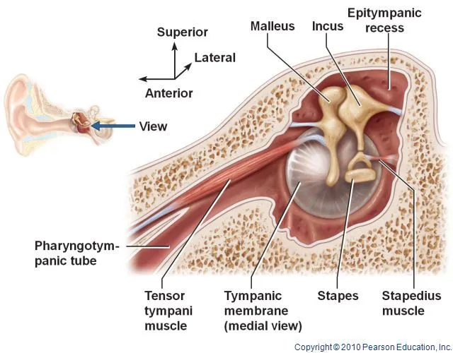
Tensor tympani muscle
Introduction
The tensor tympani is tiny, but the long paired muscles of the middle ear. Concurrently with the stapedius, it belongs to the group of intratympanic muscles. Tensor tympani regales a bony canal encountered superior to the osseous part of the auditory tube (pharyngotympanic tube; Eustachian tube).
Its attachment to the malleus, one of the three auditory ossicles, permits the tightening of the tympanic membrane, reducing its vibration amplitude and thus reducing the sound transmission into the inner ear. Thereby, it recreates an important function in the tympanic reflex – an evolutionary adaptation to protect the inner ear from excessively loud noises, which also aids speech coordination.
Tensor tympani transits in its canal together with the superior tympanic artery that supplies it. The bony canal for tensor tympani runs alongside the bony part of the auditory tube, divided from it by a thin bony septum.
Development
The tensor tympani muscle originates from mesodermal tissue in the 1st pharyngeal arch.
Origin
Tensor tympani is contained in a bony canal within the petrous portion of the temporal bone, known as the seminal for tensor tympani. This bony canal is one of the origin points of the muscles, along with the cartilaginous part of the auditory tube and the adjacent greater wing of the sphenoid bone. Tensor tympani travels posteriorly within the bony canal and goes into the tympanic cavity just above the opening of the auditory tube.
As it appears from the bony canal, the tensor tympani narrows into a long tendon that bends laterally as it passes over a pulley-like promontory of the bony canal called the processus cochleariformis.
Insertion
It inserts into the upper part of the medial element of the handle of the malleus, around its base.
Nerve supply
Tensor tympani is innervated by the nerve to the medial pterygoid, which arises from the mandibular branch of the trigeminal nerve (CNV3). The tensor tympani is innervated by a division of the fifth cranial nerve( CN V), whereas the stapedius muscle is innervated by a branch of the seventh cranial nerve(CN VII). The muscles obtain a dense innervation from motor neurons, it does not receive fibers from the trigeminal ganglion, which has sensory fibers only.

Blood supply
The tensor tympani muscle is vascularized by the superior tympanic branch of the middle meningeal artery.
Function
The tensor tympani muscle acts to dampen the noise created by chewing. When tensed, the muscles draw the malleus medially, tensing the tympanic membrane and damping vibration in the ear ossicles and thereby declining the perceived amplitude of sounds. It is not to be confused with the acoustic reflex but can be activated by the startle reflex.
Voluntary control
Contracting muscles produce vibration and sound. Slow twitch fibers of muscles produce 10 to 30 contractions per second (equivalent to 10 to 30 Hz sound frequency). Fast twitch fibers of muscles produce 30 to 70 contractions per second (equivalent to 30 to 70 Hz sound frequency). The vibration can be detected and felt by highly tensing one’s muscles, as when creating a firm fist. The sound can be heard by pushing highly tensed muscles against the ear, again a firm fist is a good example. The sound is also usually represented as a rumbling sound.
Some people can voluntarily create this rumbling sound by contracting the muscles. The rumbling sound can also be heard when the neck or/and jaw muscles are highly tensed as when yawning deeply. This phenomenon has been also known since (at most undersized) 1884.
Involuntary control (tympanic reflex)
The tympanic reflex assists prevent damage to the inner ear by muffling the transmission of low-frequency vibrations from the tympanic membrane to the oval window. The reflex has a response time of 40 milliseconds, not quick enough to cover the ear from sudden loud noises such as an explosion or gunshot.
Thus, the reflex multiple likely developed to protect early humans from thunderclaps which do not happen in a split second.
The reflex functions by contracting the muscles of the middle ear, the tensor tympani, and the stapedial muscle. However, the stapedial muscles are innervated by the facial nerve while the tensor tympani muscles are innervated by the trigeminal nerve. The tensor tympani draws the manubrium of the malleus inwards and tightens it while the stapedial muscles pull the stapes inward. This tightening dampens the sound vibration that is permitted to penetrate the cochlea. Withdrawal from medications such as benzodiazepines had been also known to cause tonic tensor tympani syndrome (TTTS) during withdrawal. The tympanic reflex will also be activated when loud vibrations are generated by the person. The tensor tympani can usually be observed vibrating while shouting at an improved volume, dampening the sound somewhat.
Clinical significance
In many individuals with hyperacusis, increased movement develops in the tensor tympani muscle in the middle ear as part of the startle response to some sounds. This lowered reflex threshold for tensor tympani contraction is activated by the perception/anticipation of loud sound and is also called tonic tensor tympani syndrome (TTTS).TTTS usually introduces symptoms such as fluttering of the eardrum, a sensation of heat, pain, blockage, fullness in the ear, tinnitus, hyperacusis, and dizziness.
In some individuals with hyperacusis, the tensor tympani muscle can contract just by thinking around a loud sound. Following exposure to unendurable sounds, this contraction of the tensor tympani muscle tightens the eardrum, which can lead to the symptoms of earache fluttering sensation of fullness in the ear (in the lack of any middle or inner ear pathology).
The mechanisms after the dysfunction of the tympanic tensor muscle and their significance are hypothesized. However, in a published study, researchers studied the case of an acoustic shock whose mechanisms present dysfunction of the tympanic tensor muscle. This study appears to be the first to provide an experimental asset suggesting that middle ear muscles (MEM) may behave abnormally after an acoustic shock. It is suggested that irregular contractions (e.g. tonic contractions) of the tympanic tensor muscles may trigger neurogenic inflammation. Indeed, fibers with substances P and CGRP were also found nearby. Placing a membrane on the eardrum allows the tensor tympani to relax so that some of the symptoms can be diminished or disappeared.
Treatment
Sound Therapy
Sound Therapy is a form of habituation therapy designed to help individuals who suffer from tinnitus or hyperacusis which is caused by acoustic trauma. It can contain intensified sounds from listening aids, environmental sounds, and music or “white noise”. Try Widex Zen Simulator to understand more about how music therapy can help relieve your symptoms.
Many individuals with tensor tympani syndrome had an anxiety disorder. High-quality magnesium complements reduce stress and also improves sleep.
Relaxation
Sit in a comfortable seat in a peaceful place
Do the exercises while hearing to relaxing music. Turn the music off if it diverts you.
Take off your shoes and wear loose and comfy clothing
Progressive muscle relaxation – While seating down, focus on the muscles in one place of your body – like your right foot. Inhale and tighten only the muscles you are focusing on for 8 seconds. Release them by abruptly letting go. Let the tightness and pain soak out of the muscles while you gradually exhale. Continue this progression systematically from your skull down to your feet.
Deep breathing – Reprise the following sequence 20 times: 1. Exhale completely through your mouth. 2. Inhale via your nose for 4 seconds. 3. Control your breath for 4 seconds. 4. Exhale via your mouth for 6-8 seconds
Guided imagery – Once you are comforted from your deep breathing exercise, close your eyes and restart to breathe deeply while you imagine yourself in the most relaxing environment possible. Imagine the scene with all your senses: The smell of the coast, the feel of the air, and the taste of your favorite beverage. Use relaxing background music to evolve even more relaxed.
Exercises for Stretching the Neck and Jaw Muscles to Acquire Rid of Tinnitus:
The cause of your tinnitus may be muscular tenseness in the area of the cervical spine. If you constantly press your upper and lower jaws together, you can build up high stress in the masticatory muscles and the surrounding fascial tissue. Thus, it is essential to reduce these tensions by stretching your jaw and neck muscles intensively to assemble the fasciae supple again. With the subsequent relaxation exercises, you can relax the tense muscles and fasciae in both areas.
A woman is seating straight and stretches her jaw muscles by opening her jaws widely
Sit up straight, hold your lower jaw with one hand, and open your mouth as wide as possible. Use your hand as a stabilizer to stretch the muscles in the jaw, cheek, and around your mouth as much as possible. Stay in this placement for two to three minutes. There is a prospect that the tinnitus initially alters its sound or becomes stronger before the symptoms subside considerably. Don’t let this discourage you! Just make sure that your pain perception is in an adequate range.
A woman is releasing her head and jaw muscle tension by using a mini ball, rolling the tense area on her cheek
Grab a massage ball or an equivalent instrument and roll off your jaw and chewing muscles on the cheek with a lot of pressure and spiraling movements. If you come along places with significantly noticeable tension, linger with the mini ball and circle around these spots.
Why is that so important? It’s really simple: Balling off your tissue assists you to dissolve fascial adhesions in tense areas. The precise pressure on your tissue provides that the shortened fasciae, which may be slagged with deposits, is properly loosened. As a consequence, the perceived pain might decrease.
A woman is sitting in a straight position and stretches her neck muscles by doing an exercise. Sit up straight and turn your left arm to control you are pulling your shoulder up during the stretching exercise. Reach over the head with your right hand to the left ear and carry the head into a stretching position. You will sense the stretch at the side of your neck. Stay in this arrangement for two to three minutes and breathe evenly. Change sides to relieve tension and muscle stiffness on the right side as well.
FAQ
What is the function of tensor tympani?
The tensor tympani is attached to the malleus; it is innervated by a division of the Vth cranial nerve. The motion of both muscles is to decrease sound transmission through the middle ear.
What causes tensor tympani syndrome?
Tonic tensor tympani syndrome (TTTS) has been described to cause TT spasms directing to tinnitus and hyperacusis. TTTS is believed to be an involuntary situation due to an overlying anxiety disorder, which induces a reduction in the threshold demanded to trigger the TT muscle reflex.
How does the tensor tympani muscle control damage to the internal ear?
The muscle reflects at the cochleariform process and then inserts into the upper end of the handle of the malleus. It serves to pull down the handle medially, which in turn draws the tympanic membrane and therefore reduces the amplitude of its oscillations. This controls damage to the inner ear from loud sounds
What is the role of the Stapedius and Tensor Tympani Muscles?
These muscles contract in reaction to loud sounds, thereby reducing the transmission of sound to the inner ear.
How does the tensor tympani protect hearing?
In reaction to loud sounds, the tensor tympani muscles tighten the eardrum and through the tendon between the hammer and anvil and shift the stirrup backward from the oval window of the inner ear. This movement of the ossicles lowers the transmitted force to the inner ear, protecting it.