Superior Tarsal Muscle
Introdution of Superior tarsal
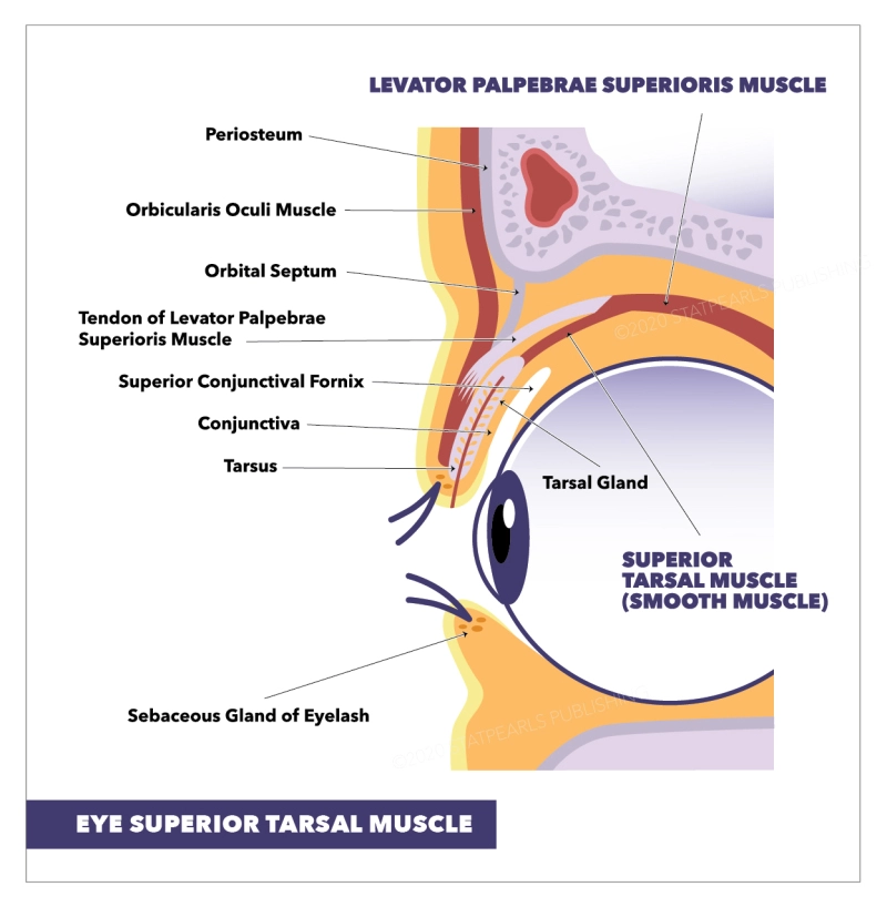
The superior tarsal muscle is a smooth muscle bordering the levator palpebrae superioris muscle that permits to raise of the upper eyelid. Not to be mistaken with the orbitalis muscle or the circular fibres of the ciliary muscle, both of which are also known as Müller’s muscle. The superior tarsal (Muller’s muscle) is a smallish muscle found within the superior eyelid. It is a smooth muscle, but it is viewed as a structural component of the more extensive skeletal muscle of the eyelid; levator palpebrae superioris. It rises from the deep aspect of the levator palpebrae superioris to the upper margin of the superior tarsal plate of the eyelid.
Existing a smooth muscle, the superior tarsal is controlled by the autonomic nervous system, specifically its sympathetic division. The main function of this muscle is to elevate the upper eyelid in a form of sympathetic predominance, such as excitement, fear, or amazement. Moreover, the fixed tone of the muscle also contributes to maintaining the eyes open and maintaining the size of the palpebral fissure constant in the state of awakeness.
Injury to this muscle, or the nerves which supply it, will result in ptosis of the affected eye as seen in Horner syndrome, a condition in which there is damage to the cervical sympathetic chain. Surgeries that involve repair of ptosis, or upper eyelid correction procedures, will usually face Muller’s muscle and should be very careful in adjustments when resecting portions of the upper eyelid.
What is the structure of Superior tarsal?
The superior tarsal muscle originates from the underside of the levator palpebrae superioris and inserts at the superior tarsal plate of the eyelid. The superior tarsal stands as a smooth muscle of the upper eyelid. It arises from the deep surface of the levator palpebrae superioris and inserts inferiorly, to the superior tarsal plate of the upper eyelid. The superior tarsal muscle is observable with the naked eye and is about 15 mm wide by 10 mm long.
The superior tarsal muscle obtains its innervation from the sympathetic nervous system. More particularly, the postganglionic sympathetic fibres stem from the superior cervical ganglion and form the internal carotid plexus around the cervical segment of the internal carotid artery. Following the petrous and cavernous segments of the internal carotid artery, these nerve fibres enter the skull and traverse the cavernous sinus. Finally, the nerve fibres for the superior tarsal muscle permit the orbit in a form of a tight nervous plexus bound around the ophthalmic artery, which is a branch of the internal carotid artery. Note that the ophthalmic artery supplies the blood supply for the superior tarsal muscle.
The main movement of the superior tarsal muscle is to elevate the upper eyelid in a state of sympathetic predominance. The function of this action is to allow a wider visual field in states of acute life-threatening situations (fight or flight response). In extra, the muscle has a static tone, which counts up to the tone of the levator palpebrae superioris muscle. The tones of these two muscles are in balance with the adversarial tone of orbicularis oculi, thereby keeping the eyes open and defining the size of the palpebral fissure.
What is the blood supply of the superior tarsal?
Oxygenated blood goes to the superior tarsal muscle through the lateral muscular branch of the ophthalmic artery. This branch also provides blood to the lateral rectus, superior rectus, superior oblique, and levator palpebrae muscles. The ophthalmic artery also holds a medial muscular branch supplying blood to the remaining extraocular muscles.
What are the lymphatics of the superior tarsal?
The superior tarsal muscle drains deoxygenated blood into the exact veins as the remaining extraocular muscles. These veins, known as vortex veins – arising from the posterior part of the eye, are where the blood exiting the superior tarsal muscle reaches first. This blood then empties into the superior and inferior orbital veins which eventually run to the cavernous sinus.
What is the nerve supply of the superior tarsal?
The superior tarsal muscle obtains its innervation from the sympathetic nervous system. Postganglionic sympathetic fibres derive in the superior cervical ganglion and transit via the internal carotid plexus, where small branches disseminate with the oculomotor nerve as it hands through the cavernous sinus. The sympathetic fibres continue to the superior branch of the oculomotor nerve, where they enter the superior tarsal muscle on its inferior part.
What is the Function of Superior tarsal?
Its function is not fully clear but may be an accessory muscle to raise the upper eyelid. The superior tarsal muscle has a remarkable function in that it helps the levator palpebrae superioris by sustaining the elevation of the upper eyelid after the levator palpebrae superioris has raised it. The superior tarsal muscle also functions to raise the upper eyelids a further 2 mm after the levator palpebrae superioris has already originally raised them as a sympathetic nervous system response.
The superior tarsal muscle was discovered to have a much more important role in levator action when compared to the levator palpebrae superioris than earlier thought. Research has resolved that the superior tarsal muscle shares a large amount of power with the levator palpebrae superioris when elevating the eyelid
What is the Clinical significance of Superior tarsal?
Harm to some elements of the sympathetic nervous system can inhibit this muscle, causing a drooping eyelid (partial ptosis). This is observed in Horner’s syndrome. The ptosis seen in Horner’s syndrome is of a smaller degree than is seen with oculomotor nerve palsy.
What is the History of Superior tarsal?
The muscle emanates its name from the Greek ταρσός ‘flat surface’, typically used for drying. The term Müller’s muscle is occasionally used as a synonym. Yet, the same term is also used for the circular fibres of the ciliary muscle, and also for the orbitalis muscle that protects the inferior orbital fissure. Given the possible confusion, the use of the term Müller’s muscle should be discouraged unless the context clears any mysteriousness.
What are the Clinical relations of Superior tarsal?
Horner’s syndrome
Horner’s syndrome happens when the sympathetic nerve supply to the face and eye is injured. This condition usually happens due to trauma of the neck and shoulder regions and consequential injury of the cervical sympathetic ganglia and/or nerves. Damages of this kind will result in loss of effective innervation of the smooth muscles and glands in the head. Horner’s syndrome is characterized by a trio of symptoms; partial ptosis (a drop of the upper eyelid), miosis (constricted pupil), and flop of hemifacial sweating (anhidrosis). Sometimes, it can also be tracked by the posterior displacement of the eyeball within the orbit (enophthalmos).
What is the Embryology of Superior tarsal?
The superior tarsal muscle materialises in a way, not unlike the other extraocular muscles. These subtypes of muscles are distinct in that; when compared to periocular tissues, these muscles came from paraxial mesoderm in the prechordal plate, which is unique in that connective tissue tendons arise from neural crest cells rather. Especially, around week five is when Muller’s muscle first appears in development.
What are the Muscle fibres of Superior tarsal?
The distinct muscle subtype of the superior tarsal muscle is different from the other extraocular muscles. The superior tarsal muscle is constructed of smooth muscles, especially thin fibres of smooth muscular tissue. This muscle originates at the bottom of the levator palpebrae superioris and attaches to the superior tarsal plate.
Precise differences which are seen in this muscle compared to other smooth muscles exist. One such distinction is the point that the superior tarsal muscle is made up not solely of smooth muscle but instead is mixed tissues. These tissues contain connective tissue, blood vessels, and fat. Also, one fundamental difference between the superior tarsal muscle and others is that the smooth muscle cells of this muscle are distributed sporadically and are not connected. Lastly, another difference indicates that the origin of the superior tarsal muscle may have fibres of striated muscle connected with fibres of the smooth muscle.
What are the physiologic Variants of Superior tarsal?
The superior tarsal muscle has typical physiologic variants when comparing different individuals, and these centre near the attachment of the muscle to the superior tarsal plate. Four variations live, and they are below:
- The superior tarsal muscle binding to the upper border of the superior tarsal plate
- 2M: The superior tarsal muscle binds to the medial aspect of the plate
- 2L: The superior tarsal muscle binding to the lateral aspect of the plate
- The superior tarsal muscle attaches along the whole area of the superior tarsal plate.
- The third variation was the most repeatedly observed.
FAQ :
What does the superior tarsal muscle do?
The superior tarsal muscle learned as Muller’s muscle is a structural muscle that functions to maintain the elevation of the upper eyelid. It obtains innervation from the sympathetic nervous system and is unique in that it consists of thin fibers of the smooth muscle.
What nerve innervates the superior tarsal muscle?
The superior tarsal muscle obtains its innervation from the sympathetic nervous system. More precisely, the postganglionic sympathetic fibers stem from the superior cervical ganglion and create the internal carotid plexus around the cervical component of the internal carotid artery.
Where is the right superior tarsal region?
The superior tarsal muscle is a smooth muscle connecting the levator palpebrae superioris muscle that allows it to raise the upper eyelid. The superior tarsal muscle initiates from the bottom of the levator palpebrae superioris and inserts on the superior tarsal plate of the eyelid.
What does the superior tarsal muscle do?
The superior tarsal muscle learned as Muller’s muscle is a structural muscle that functions to maintain the elevation of the upper eyelid. It obtains innervation from the sympathetic nervous system and is unique in that it consists of slim fibers of the smooth muscle.
What is the function of the tarsal plates?
The tarsal plates perform as the main structural component of the eyelids. They are constructed of dense connective tissue and have the Meibomian glands and eyelash follicles. There are around 30 meibomian glands in the upper eyelid and 20 meibomian glands in the lower eyelid.