
Cerebrospinal Fluid (CSF)
What is a Cerebrospinal Fluid?
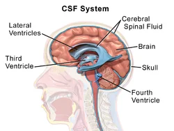
Cerebrospinal fluid (CSF) is a clear or transparent, colorless plasma-like fluid that bathes or cleans the central nervous system (CNS). The system of cavities is established inside the brain and spinal cord, including the ventricles, the subarachnoid cavity of the cerebral cortex, the spinal cord, and the central canal of the spinal cord. Most of the cerebrospinal fluid (CSF) is secreted or produced by the specialized tissue known as the choroid plexus, which is situated within the lateral, third ventricle, and fourth ventricles. The secretion or production of the cerebrospinal fluid (CSF) equals its removal, so there is about 150-270 ml of cerebrospinal fluid (CSF) within the central nervous system (CNS) at all times.
The cerebrospinal fluid’s (CSF) primary functions are to provide basic immunological defense for the brain and spinal cord and to cushion the brain and spinal cord when they are impacted by force from the outside. central nervous system (CNS), to eliminate metabolic waste, as well as to transport neuromodulators and neurotransmitters. cerebrospinal fluid (CSF) is also very convenient for assisting the clinical diagnosis, and its samples are normally obtained from the subarachnoid space (SAS) by lumbar puncture procedure.
Secretion
Choroid plexus
The choroid plexus, a specialized tissue, generates cerebrospinal fluid. The third and fourth ventricles’ rooftops, along with the lateral ventricles’ walls and roofs, contain choroid plexuses. A choroid plexus shows many villi, through which it produces cerebrospinal fluid (CSF). anatomically each villus comprises three components;
A layer of altered ependymal cells (choroid cells), faces the lumen of the ventricles and secretes the cerebrospinal fluid (CSF). The cells show numerous apical villous predictions and are tightly bound to each other through tight junctions.
A layer or coating of pia mater (tela choroidea)
A fenestrated or lancet was capillary directly within the pia mater.
Choroid cells take up different chemicals from the underlying blood vessel which they utilize to actively secrete or produce cerebrospinal fluid (CSF). Thus, cerebrospinal fluid (CSF) fluid differs from blood in terms of its electrolyte, glucose, and protein amount and is not merely an ultrafiltrate of blood The vascular source for the choroid plexuses varies between the lateral ventricle, third ventricle, and fourth ventricle.
The anterior region of the choroid plexuses in the lateral ventricle and third ventricle is innervated by the anterior choroidal artery (a branch of the internal carotid artery). While their posterior portion is vascularized by the posterior choroidal artery, lateral choroidal artery, and medial choroidal artery (branches of the posterior cerebral artery)
The choroid plexus in the fourth ventricle is innervated by the inferior cerebellar arteries.
from superficial to deep, the blood-brain barrier comprised of:
- Choroid ependymal cells and their tight junctions
- Pia mater
- Endothelial cells of capillaries
- The basal membrane of endothelial capillary cells
- The function of the blood-brain barrier is to regulate the motion of the water and solutes into the cerebrospinal fluid (CSF), as well as from the cerebrospinal fluid (CSF) into the neural tissue.
The BBB, or blood-brain barrier
The layers of the choroid plexus compose the barrier between the blood and the brain (also known as the barrier with selective permeability that also plays a role in the production of CSF.
For a quick review, the blood-brain barrier is made up of:
- ependymal cells found in the choroid and their tight connections
- Pia Mater
- endothelial capillary cells
- The endothelium capillaries cells’ basal membrane
- Regulating the flow of water and other solutes into the CSF and from the CSF into neural tissue is the responsibility of the blood-brain barrier.
Blood-CSF barrier (BCSFB)
The blood-CSF barrier is frequently confused for the blood-brain barrier (BBB), while in fact, it is only a portion of it. The blood-CSF (cerebrospinal fluid) barrier truly refers to the tight junctions in the middle of the choroid ependymal cells, which regulate or control the passage or pathways of molecules in the middle of the underlying capillaries and the cerebrospinal fluid (CSF).
If it weren’t for the tight junctions (i.e. blood-CSF barrier), the particles like electrolytes and blood cells that can convey through the lancets of the capillaries would enter the cerebrospinal fluid (CSF) and therefore interrupt the electrolytic and biochemical equilibrium in the blood. This barrier is functionally very important for the opposite direction as well, from the cerebrospinal fluid (CSF) to the capillaries; as it prevents or restricts large molecules (such as different types of drugs) from freely entering the brain blood vessels.
Circulation
There is 600-700 ml of newly created cerebrospinal fluid (CSF) every day when cerebrospinal fluid (CSF) is continually generated at a secretion rate of 0.2-0.7 ml/per minute. considering the total volume of the cerebrospinal fluid (CSF) ranges around 150-270 mL, this means that the whole volume of the cerebrospinal fluid (CSF) is replaced or changed around 4 times per day.
The cerebrospinal fluid (CSF) travels through a particular pathway:
Through the interventricular foramen, which is part of Monro, cerebrospinal fluid (CSF) flows via the lateral ventricle towards the third ventricle.
From the third ventricle, the cerebrospinal fluid (CSF) flows via the cerebral aqueduct of the Sylvius to the fourth ventricle.
From the fourth ventricle, some cerebrospinal fluid (CSF) flows or runs via a narrow passage or pathway called the obex and enters the central canal of the spinal cord.
However, the majority of cerebrospinal fluid (CSF) convey via the apertures of the fourth ventricle; the median aperture of the foramen of Magendie, and two lateral apertures of the foramen of Luschka.
Via these openings, the cerebrospinal fluid (CSF) enters the cisterna and cerebellopontine cisterns, respectively.
From there, the cerebrospinal fluid CSF runs through the subarachnoid space of the brain and spinal cord.
It is finally reabsorbed into the dural venous sinuses via the arachnoid granulations.
Absorption
There are three recognized pathway through which cerebrospinal fluid(CSF) leave the
subarachnoid space (SAS) to get into the cerebral venous system; arachnoid granulations, minute channels that convey through the cribriform plate of the ethmoid bone, and the glymphatic system.
Arachnoid granulations
Most of the cerebrospinal fluid (CSF) is absorbed into the venous system by the arachnoid granulations.
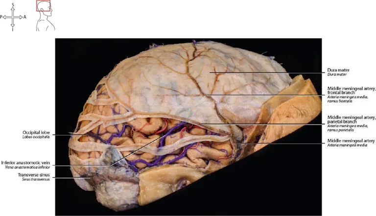
The arachnoid granulations are the protrusions or prominence of the arachnoid mater that penetrates the dura mater and protrudes or sticks out into the lumina of the dural venous sinuses.
The surface of each arachnoid granulation comprises smaller outpouchings called arachnoid villi that increase or rise its absorption surface.
The cerebrospinal fluid (CSF) leaves the subarachnoid space by diffusing via the walls of arachnoid granulations.
The valve mechanism for the flow of CSF, or cerebrospinal fluid, is offered by the arachnoid granulations, which enables or allows the flow of CSF into the bloodstream without enabling the backflow of blood into the cerebrospinal fluid (CSF).
Normally the pressure or the force of the cerebrospinal fluid (CSF) is greater or more than that of the venous system, so the cerebrospinal fluid (CSF) flows via the villi and granulations into the venous blood.
The greatest number of arachnoid granulations is present in the superior sagittal and transverse sinuses.
Thus, these are the regions where most of the cerebrospinal fluid (CSF) is get absorbed.
Minute channels in the cribriform plate
The second route comprises small channels that expand from the subarachnoid space via the openings in the cribriform plate of the ethmoid bone. These channels cross the cribriform plate with the fila olfactory of the olfactory nerve (CN I) and drain or strain into the lymphatic channels of the nasal mucosa.
Glymphatic system
The glymphatic system gives the third route. It is a newly discovered system of channels that are formed by the astroglial cells about the pial arteries. Its role is to give an entrance or opening route for the cerebrospinal fluid (CSF) interchange for the interstitial fluid of the brain and the spinal cord. This means that a small quantity of cerebrospinal fluid (CSF) enters the nervous tissue, whilst the same quantity of interstitial fluid leaves into the subarachnoid space in order to be eliminated or removed through the dural venous sinuses.
Spinal CcerebroSpinal Fluid (csf)
The subarachnoid space of the spinal cord is constant to that of the brain so that the cerebrospinal fluid CSF that is produced in the brain ventricles can easily reach or get to the spinal cord as well.
Functions
The cerebrospinal fluid (CSF) has many protective and metabolic functions.
The role of cerebrospinal fluid (CSF) is a shock absorber, giving a fluid buffer and thus protecting the brain from injury.
It gives neutral buoyancy that prevents or avoid the brain from compressing the blood vessels and cranial nerves against the internal surface of the bones of the skull.
It eliminates by-products of metabolism and plays a very important role in the homeostasis and metabolism of the central nervous system.
Homeostasis implies the regulation or control of the distribution of substances (e.g. electrolytes) between the neuroendocrine factors and cells of the brain. mild changes in the pH or the composition of the Cerebrospinal fluid (CSF) can disrupt temperature, blood pressure control, and hormonal exchange or interchange in subcortical structures.
clinical significance
Hydrocephalus
The most significant disease of the cerebrospinal fluid (CSF) flow is hydrocephalus. Hydrocephalus is caused by the accumulation of excessive amounts of cerebrospinal fluid (CSF), either caused by increased production of cerebrospinal fluid (hypersecretory hydrocephalus), an obstruction or blockage of its flow (non-communicating or obstructive hydrocephalus), or by impaired absorption via the arachnoid villi (malabsorptive hydrocephalus). Hydrocephalus is followed by an abnormal enlargement of the head in children due to an abnormal increase in the amount of cerebrospinal fluid (CSF). This creates increased pressure within the cranium causing degeneration of brain tissue.
Hydrocephalus is classified as obstructive (non-communicating) when there is when there is an obstacle or blockage limiting the movement of this compound from the ventricular system to the subarachnoid area, it may be interpreted as communicating. The interventricular foramen, the cerebral aqueduct, the median aperture, and the lateral apertures of the fourth and fourth ventricles constitute the tiny channels wherein obstructions are most likely to take place.
Meningitis and subarachnoid hemorrhage, both of which can cause or cause occlusive adhesions of the arachnoid granulations, are common causes of malabsorptive hydrocephalus. Traumatic brain injury and intraventricular hemorrhage are additional causes.
If the amount of pressure inside the ventricular system is constantly elevated, encephalopathy may also be active. Normal pressure hydrocephalus, in which cerebrospinal fluid (CSF) pressure is only periodically raised, is not the same as active hydrocephalus.
Syringomyelia
occurs due to blockage of CSF circulation.
Meningitis is a disorder in which the coverings of the brain become inflamed. There are two types of meningitis: aseptic infection and bacterial infection. Aseptic meningitis can be caused by fungus, drugs, and cancer metastases, however, the majority of cases are cancer-related. Although aseptic meningitis can be brought on by fungi, drugs, and cancer metastases, the majority of cases are brought on or triggered by viruses. viruses cause or influence events. Fever, photophobia nuchal rigidity are classic presenting symptoms. Diagnosis is made through an analysis of the cerebrospinal fluid (CSF) obtained through the Lumbar plexus.
Subarachnoid Hemorrhage (SAH) is the leakage of blood into the subarachnoid space where it mixes with the (cerebral spinal fluid) CSF. subarachnoid hemorrhage most commonly occurs by trauma with 80% of nontraumatic subarachnoid hemorrhage being caused by aneurysm rupture. Other nontraumatic causes of subarachnoid hemorrhage involve arteriovenous malformations and vasculitis.
Lumbar Puncture and cerebrospinal fluid Analysis – Lumbar puncture is a sterile procedure, done to obtain cerebrospinal fluid samples for diagnostic purposes. It includes passing a needle into the subarachnoid space at the region between the second lumbar and the fifth lumbar vertebrae. However, most commonly lumbar puncture is applied between L4 and L5. microbiologic and biochemical and cytologic studies are then carried out on the sample.
FAQ
Where is the cerebrospinal fluid found?
Cerebrospinal fluid (CSF) is a clear, colorless, watery fluid that travels in and around the brain and the spinal cord. the brain and the spinal cord are made up of the central nervous system (CNS). It regulates and coordinates everything we do, including the ability to move, breathe, vision, think, and much more.
What is a cerebral fluid made up of?
cerebrospinal fluid (CSF)is obtained from blood plasma and is most similar to it, except that cerebrospinal fluid (CSF) is almost protein-free compared with plasma proteins and has various electrolyte levels. Due to the way, it is generated cerebrospinal fluid (CSF) has a higher chloride level than plasma, and an equivalent sodium level.
Is cerebrospinal fluid water?
cerebrospinal fluid (CSF) is slightly alkaline and is around 99 percent water. There is around 100 to 150 ml of cerebrospinal fluid (CSF) in the normal adult human body. The exact method of the formation of cerebrospinal fluid (CSF) is uncertain.
What are the 3 main functions of CSF?
cerebrospinal fluid (CSF) assists the brain by offering protection, nourishment, and removal of waste.
How much CSF is produced per day?
around 500 ml/day
cerebrospinal fluid (CSF) is generated at an amount of around 500 ml/day; there are approximates that there is approximately 125 mL to 150 mL of cerebrospinal fluid (CSF) in the body at any given time. The cerebrospinal fluid (CSF) can be refilled or changed every 7.5 hours or 450 minutes of the day, depending on the amount for manufacturing and absorption (which varies differently).
Which cells produce CSF?
cerebrospinal fluid (CSF) is produced mainly by the choroid plexus epithelium and ependymal cells of the ventricles and travels into interconnecting chambers; termed the cisterns and the subarachnoid spaces.
What is brain fluid called?
Cerebrospinal fluid (CSF) is made by tissue that lines the ventricles (hollow spaces) in the brain. It travels in and around the brain and the spinal cord to assist in cushioning them from injury and give nutrients.
What absorbs CSF?
Most of the cerebrospinal fluid (CSF) is get absorbed into the venous system by the arachnoid granulations. The projections of the arachnoid layer through the dura mater (its outer layer) and into the lumina of the dural venous sinuses are referred to as arachnoid granulations.
What color is a CSF leak?
The classic presentation of cerebrospinal fluid (CSF) leaks or discharge is the expression or statement of clear, watery drainage from the nostrils. This takes place normally on one side; however if fluid gets drains into the back of the throat there may be a salty taste.
What is the clinical significance of cerebrospinal fluid?
Cerebrospinal fluid (CSF) acts like a cushion that assists protect the brain and spinal cord from sudden impact or injury. The fluid also eliminates waste products from the brain and assists the central nervous system work properly.
Which clinical disease is related to CSF infection?
meningoencephalitis, encephalitis, Viral meningitis
Infection of the meningeal structures or meninges with an endogenic inflammatory response results in meningitis. Clinically, the typical presenting symptoms are fever, headache, and stiff neck.
What diseases are caused by cerebrospinal fluid?
Hydrocephalus is caused by an imbalance between the range of cerebrospinal fluid produced and how much cerebrospinal fluid is absorbed into the bloodstream. Cerebrospinal fluid (CSF)is produced by tissues lining the ventricles of the brain. It flows via the ventricles by way of interconnecting channels.
What are the most common CSF infections?
Pneumococcal meningitis is the commonest form of recurrent or recurring meningitis in patients who have cerebrospinal fluid (CSF )leaks.
What is normal human CSF volume?
90 – 150 ml
The rate of cerebrospinal fluid (CSF) formation in humans is 0.3 – 0.4 ml per minute, and the total cerebrospinal fluid (CSF) volume is 90 – 150 ml in adults. It is also believed that Cerebrospinal fluid (CSF) circulates via the ventricles, the cisterns, and the subarachnoid space or gap ultimately getting absorbed into the venous blood at the region of the arachnoid villi.
What color is CSF in disease?
The color of the cerebrospinal fluid or CSF is most usefully or efficiently described as (
pink or orange, yellow, brown. These colors communicate with the main pigments or colors obtained from red cells, bilirubin, oxyhemoglobin, and methemoglobin. Oxyhemoglobin is red in color, but after dilution in the cerebrospinal fluid (CSF), it appears pink or orange in color.
What is the clinical importance of CSF in various diseases?
A CSF analysis can evaluate different substances in your cerebrospinal fluid. Tests to identify infectious disorders of the brain and spinal cord, such as meningitis and encephalitis, may be included. White blood cells, bacteria, and other elements in the cerebrospinal fluid are examined in CSF tests for infections.


13 Comments