
Sural Nerve
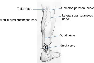
The sural nerve is a sensory nerve in the calf region (sura) of the leg. It is made up of branches of the tibial nerve and common fibular nerve, the medial cutaneous branch from the tibial nerve, and the lateral cutaneous branch from the common fibular nerve.
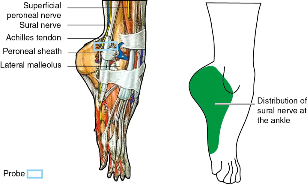
Once formed, the nerves run down the mid-calf to the ankle and along the skin from the mid-posterior popliteal fossa to just behind the lateral malleolus and then under the malleolus and forward along the lateral aspect of the foot.
Description:
The sural cutaneous nerve consists of the fusion of the medial sural cutaneous nerve (MSCN) which is a terminal branch of the tibial nerve and the lateral sural cutaneous nerve (LSCN) which is one of the terminal branches of the common fibular nerve.
These two branches, MSCN and LSCN, are connected by the sural communicating branch and form the sural nerve.
How the two branches fuse, the contribution of the fibular and tibial branches, the location of the connection, and differences between the two lower extremities contribute to the variability of this nerve.
From the mid-calf down to the ankle the nerve courses subcutaneously along a line drawn from the mid-posterior popliteal fossa to just posterior to the lateral malleolus and thence under the malleolus and forward along the lateral aspect of the foot.
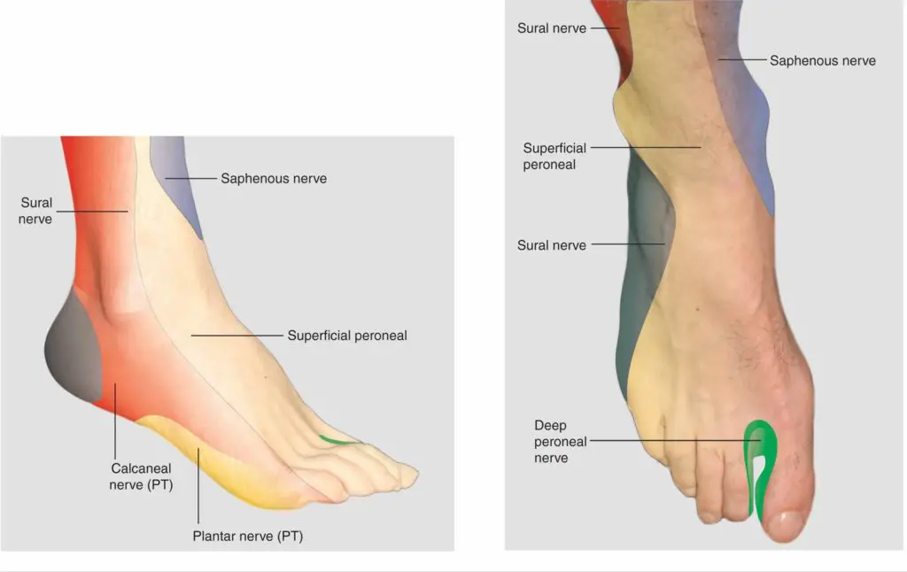
It supplies sensation to the skin of the lateral foot and lateral lower ankle.
Anatomy:
it receives innervation from the lumbosacral plexus (L5, S1). In the posterior thigh, the sciatic nerve divides into two main branches: the common peroneal nerve and the tibial nerve.
Each of these nerves gives off a branch, which together forms the sural nerve. This nerve arises below the knee and travels superficially in the posterior leg.
The nerve is located near the midline in the lower leg and travels behind the lateral malleolus to innervate the foot.
it supplies sensation to the posterolateral aspect of the calf and the lateral aspect of the foot.
Structure:
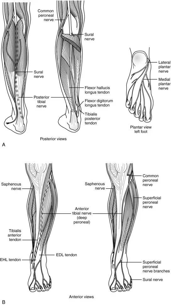
- The tibial nerve and the common fibular nerve arise as the sciatic nerve divides into two branches in the popliteal fossa.
- As the tibial nerve travels down the popliteal fossa, and before it goes beneath the gastrocnemius, it gives off a cutaneous branch which is the medial sural cutaneous nerve.
- This nerve courses laterally over the lateral head of the gastrocnemius.
- The common fibular nerve also gives off a small cutaneous branch which is the lateral sural cutaneous nerve.
- When the common fibular nerve is divided from the sciatic nerve, it travels parallel to the distal portion of the biceps femoris muscle and towards the fibular head.
- The small cutaneous branch arises as the common fibular nerve travels towards the fibular head.
- The nerve then continues down the leg on the posterior-lateral side, then posterior to the lateral malleolus where it runs deep to the fibularis tendon sheath and reaches the lateral tuberosity of the fifth toe, where it ramifies.
- From the mid-calf down to the ankle the nerve courses close to the skin along a line drawn from the mid-posterior popliteal fossa to just posterior to the lateral malleolus and then under the malleolus and forward along the lateral aspect of the foot.
Function:
- This nerve supplies sensation to the skin of the lateral foot and lateral lower ankle.
- The nerve transmits sensory signals from the posterior lateral corner of the leg and the lateral foot and 5th toe towards the spinal cord and brain.
Embryology
The neural tube and neural crest cells are created by the neural plate, which forms during the third week following fertilization. Peripheral nerves, such as the sural nerve, develop from neural crest cells. In order to improve the conduction of electrical signals, Schwann cells, which myelinate peripheral nerves in the nervous system, also originate from neural crest cells.
Blood Supply and Lymphatics
Peripheral nerves receive their vascular supply extrinsically from parallel-running arteries and internally from the vasa nervorum inside the nerve’s epineurium.
Anatomical Variations
The sural nerve is variables because of its position and the way in which the lateral sural cutaneous nerve and the lateral sural cutaneous nerve fuse, the fibular and tibial branch (since it can arise from only one of these), and variations in each lower extremity within an individual.
Clinical significance:
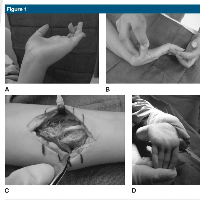
This nerve has a purely sensory function, and so its removal results in only a relatively minor deficit. For this reason, it is often used for nerve biopsy, as well as the donor nerve when a nerve graft is performed.
Sural nerve block:
- A sural nerve block can be used for quick anesthetization of the foot and lower leg. Because this technique requires few injections to reach adequate anesthesia, a smaller volume of anesthetic is needed.
- This nerve is rather superficial, which makes it more accessible to surgeons.
- Therefore, it is relatively easier than other procedures. Also, due to its superficial properties, this nerve is easily blocked at multiple levels at or above the ankle.
- In one study, regional anesthesia of the foot and ankle, when performed by surgeons, was completely successful 95% of the time.
- A sural nerve block is not advised if a patient is allergic to the anesthetic solution, has infected tissue at the injection site, has a severe bleeding disorder, or has preexisting neurological damage.
Disease:
- Sural mononeuropathy is uncommon. If affected, it can be due to a mass lesion such as a ganglion or trauma, which is the most common cause.
- Sometimes inflammatory or vasculitic diseases will selectively involve the sural nerve.
- In addition, the sural nerve will be involved in any kind of generalized peripheral sensory or sensorimotor neuropathy.
- Sensory changes from sural neuropathy are variable but usually occur in the postero-lateral aspect of the leg and the dorso-lateral foot.
- These can sometimes be painful with paresthesias and dysesthesias.
- Nerve conduction studies can be used to delineate sural nerve lesions.
- Treatment will depend on the cause of the neuropathy. Occasionally biopsy of the nerve is performed for diagnostic purposes.
- For example, ganglions are usually resected. Traumatic neuropathy is usually treated non-surgically. Sural nerve damage is usually part of a more generalized peripheral neuropathy.
Sural Nerve Biopsy
Finding the histological origin of peripheral neuropathy in patients without a clear underlying cause may be helped by sural nerve biopsy. Because of the development of less-invasive technologies like genetic and electrophysiological testing, it is not often used in modern medicine. In situations where these more advanced techniques provide no results or a confusing picture, a biopsy may still be helpful. Examples of these situations include vasculitic neuropathy, amyloid neuropathy, and multifocal leprosy.
Because it is entirely sensory, superficial, and easily located physically, the sural nerve is a suitable candidate for biopsy. Additionally, the distribution of persistent anesthetic over it is unlikely to cause long-term patient injury. Irritation, allodynia, and surgical wound infection are possible side effects. The degree of sensory loss and recovery of the sensory deficit are strongly correlated with the amount of nerve removed. Nonetheless, after 18 months, 91% of patients who had a nerve segment removed had recovered from their sensory loss, according to a research by Bevilacqua et al.
FAQs
What is your sural nerve?
A cutaneous nerve, or nerve that sends feeling to the skin, is the sural nerve. The sural nerve is situated under the skin at the rear of each leg’s calf. The back of your leg below your knee, as well as the outer portion of your foot, heel, and ankle, are all sensed by the sural nerve.
Is the sural nerve part of the sciatic?
The common peroneal nerve and the tibial nerve are the two primary branches of the sciatic nerve that split in the posterior thigh. The sural nerve is made up of the branches that each of these nerves produces.
What is the sural communicating nerve?
In contrast to the medial and lateral sural cutaneous nerves, the sural communicating nerve (SCN), also known as the peroneal communicating branch of the common fibular nerve, is a distinct and independent nerve that frequently emerges from the common fibular nerve’s trunk.
What is sural nerve a branch of?
In the distal part of the calf, the lateral sural cutaneous nerve, a terminal branch from the common fibular nerve, unites with the medial sural cutaneous nerve, a terminal branch from the tibial nerve, to produce the sural nerve.
What does the sural nerve branch into?
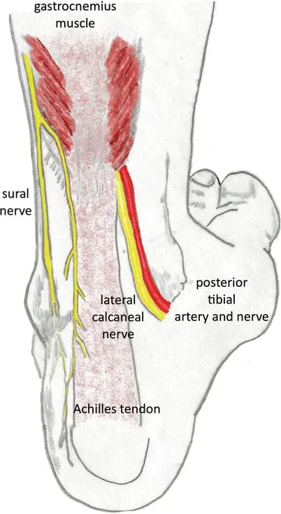
As the sural nerve enters the foot, it produces two terminal branches: The skin covering the lateral part of the heel is innervated by the sural nerve’s lateral calcaneal branch. The lateral side of the foot’s dorsum is supplied by the lateral dorsal cutaneous nerve.


4 Comments