
Tibialis posterior muscle
What is Tibialis posterior muscle?
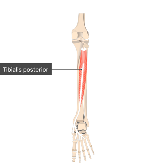
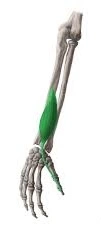
The muscle in the posterior aspect of the leg that is the most central and deepest is the Tibialis posterior. It is part of the deep group of muscles in the leg’s posterior compartment, along with the popliteus, flexor hallucis longus, and flexor digitorum longus.
The tibia, fibula, and interosseous membrane are all posterior to these muscles. Tibialis posterior is hidden from view by the huge, superficial muscles of the leg; gastrocnemius and soleus. This muscle inserts on the plantar surface of the foot after crossing the ankle joint. Thus, it assists with plantar flexion of the foot at the lower leg joint. It is likewise a synergist of the tibialis anterior in an inversion of the foot.
Origin of Tibialis posterior muscle
The muscle originates from:
A tibia’s postero-lateral proximal aspect.
A proximal posteromedial part of the fibula and the interosseous membrane
Mid part: located in the deep posterior compartment of the lower leg and extends proximally to the medial malleoli, where the flexor retinaculum holds it in place.
Insertion
The plantar slip attaches to the medial cuneiform bone, while the major insertion is on the navicular.
Relations
In the leg’s posterior compartment, the Tibialis posterior muscle is the deepest and most central muscle. It is found posterior to the tibia, fibula, and interosseous layer of the leg. The latter divides the anterior leg muscles from the tibialis posterior. The flexor hallucis longus and flexor digitorum longus muscles overlap the muscle’s belly. Additionally, the plantaris tendon, soleus, and gastrocnemius are all superficial to the tibialis posterior. As they move through the tarsal tunnel, the tendon of the flexor digitorum longus crosses the tendon of the tibialis posterior toward the ankle and lies medial to it.
There are also connections between the Tibialis posterior and important neurovascular structures. It is located, for instance, anterior to the posterior tibial artery, which generates the fibular artery branch. Between the tibialis posterior and the fibula, the fibular artery descends. The medial and lateral portions of the muscle are connected by the anterior tibial artery, which runs close to its point of origin. For the majority of its journey, the tibial nerve crosses the tibialis posterior.
Innervation
The tibial nerve, which originates from the L4 and L5 spinal nerves, innervates the tibialis posterior. The larger of the two sciatic nerve branches is the tibial nerve.
Blood supply
Blood supply to the tibialis posterior muscle is through parts of the posterior tibial artery, which comes from the popliteal vein. These branches incorporate the fibular and medial plantar supply routes. The medial malleolar blood vessel network additionally adds to the blood supply of the ligament. The Tibialis posterior is drained by the posterior tibial veins, which empty into the popliteal vein.
Function of Tibialis posterior muscle
The Tibialis posterior is engaged with motions at two unique joints, as follows:
The foot is in plantar flexion at the talocrural (ankle) joint.
Foot inversion at the subtalar joint.
The tibialis posterior assists the other, more powerful foot flexors in elevating the heel when the foot is planted on the ground by acting on the ankle joint. Walking, running, and various fitness exercises, like calf raises, are made easier by this. Additionally, the posterior contraction of the tibialis approx the tibia and fibula. This obtains the malleoli during plantar flexion, working on their grasp on the bone and supporting the lower leg. Inversion of the foot likewise has a few significant works. When standing on one leg, the tibialis posterior resists the body’s tendency to sway laterally, thereby facilitating balance.
By raising, tensing, and strengthening the foot’s medial longitudinal arch, this muscle also provides support. When the foot is planted on the ground, this action aids in the distribution of the body’s weight.
Clinical relevance
Tibialis Posterior Rupture
Tibialis posterior rupture is a condition in which the tendon that connects the tibialis posterior muscle to the bones of the foot becomes partially or completely torn.
Symptoms of a tibialis posterior rupture may include pain, swelling, and tenderness along the inside of the ankle and foot, as well as weakness or difficulty with walking or standing on the affected foot. In more severe cases, there may be a visible deformity or flattening of the arch of the foot.
Shin Splints
Shin splints (medial tibial stress syndrome) is a common overuse injury that affects the lower leg. It is characterized by pain and tenderness along the inner edge of the shin bone (tibia).
Shin splints typically occur in individuals who engage in repetitive physical activities that involve running, jumping, or other high-impact activities. These activities can place a significant amount of stress on the muscles, tendons, and bones of the lower leg, causing inflammation and pain.
Most cases of adult-acquired flatfoot are thought to be caused by Tibialis Posterior Tendon Dysfunction (TPTD).
In foot drop (High steppage gait) because of deep peroneal nerve paralysis, moving the posterior tibialis tendon to the dorsum of the foot is a surgical tendon transfer system to make up for the deficiency of dorsiflexion because of tibialis anterior loss of motion.
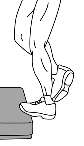
Tibialis posterior muscle stretching
Position of the beginning: Hold onto a banister while standing on one leg on a step. The front of the foot ought to be on the step, with the impact point hanging off the back. Bend your knee slightly and let the heel drop below the level of the step until you feel a stretch in your lower calf. Do this three times a day, on each side, for 30 to 50 seconds.
Tibialis posterior muscle strengthening
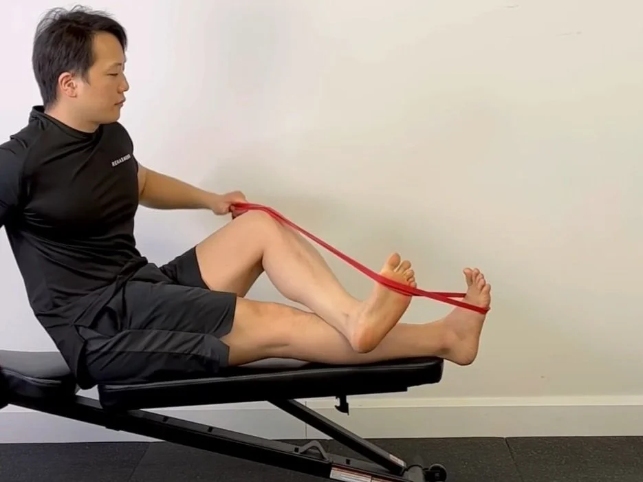
Ankle inversion with band
An ankle inverter strengthening exercise is what this is all about. Hold the target foot with the hand on the same side as you wrap a band around it. Use your foot to push the band to the side and secure it by crossing your other leg over top. By bringing the bottom of your foot in contact with your body’s midline, you can activate the ankle inverters. Hold for as long as 2 seconds and gradually release to finish a count.
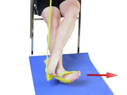
Banded ankle inversion
An ankle inverter strengthening exercise is what this is all about. Begin by tying a band around the foot you want to target. While crossing your other leg over top, grip the free end of the band with your hand and pull the band to the side with your foot. To stimulate the inner shin and foot muscles, pull the band inward with your target foot. Hold for as long as 2 seconds and gradually release to finish a count.
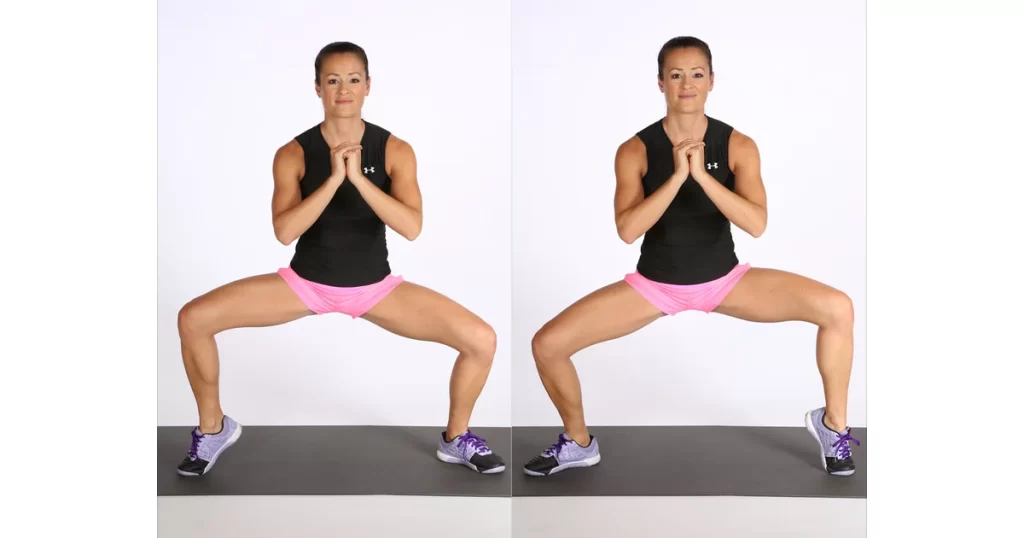
Alternating calf raise squat
This is a balance and calf-strengthening exercise. Put your hands on your knees to support your upper body as you take a wide-based squat stance. Lift one heel off of the bottom maximally and maintain for 3-5 seconds. Alternate sides by raising the opposite heel off the ground while simultaneously lowering the raised heel back to the ground. Repeat as necessary.
Band pull lunge sliders
The Tibialis Posterior and Intrinsic Foot muscles will benefit from this dynamic, full-body workout. On hard flooring, place a band between your foot and a slider disc or hand towel. The other end of the band should be anchored to the outside of the foot that is stepping on it. Slide the foot back behind you into a split squat stance while keeping the balls of your toes planted on the disc. To get back to standing, drive the foot into the floor and pull the disc backward.
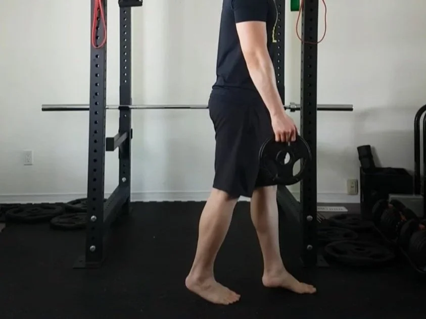
1-inch heel raise walks
The objective of this exercise is to strengthen your foot’s intrinsic muscles and medial longitudinal arch. Hold loads in the two arms and raise your heels only 1 inch off the ground. Keep in mind that raising them too high will result in the larger calf muscles taking over control of the foot and ankle. Before taking a break, move forward about 50-100 meters.
Short foot squat
You will learn how to engage and control the intrinsic foot muscles that stabilize the medial longitudinal arch (MLA) through this muscle activation exercise. It is essential to dissociate movement from the toes when contracting the foot’s intrinsic muscles. Pull the ball of your big toe toward your heel to raise the MLA while keeping the toes relaxed. To add dynamic resistance, maintain this muscular contraction after completing a squat.
Short foot side lean
You will learn how to engage and control the intrinsic foot muscles that stabilize the medial longitudinal arch (MLA) through this muscle activation exercise. It is essential to dissociate movement from the toes when contracting the foot’s intrinsic muscles. Pull the ball of your big toe toward your heel to raise the MLA while keeping the toes relaxed. Maintain this muscular contraction and apply additional resistance by leaning on one leg.
Short foot single leg stance
You will learn how to engage and control the intrinsic foot muscles that stabilize the medial longitudinal arch (MLA) through this muscle activation exercise. It is essential to dissociate movement from the toes when contracting the foot’s intrinsic muscles. Pull the ball of your big toe toward your heel to raise the MLA while keeping the toes relaxed. As you stand on one leg to provide additional resistance, maintain this muscular contraction.
Single leg pallof press (ipsilateral)
This is both an intrinsic foot muscle-strengthening exercise and a core anti-rotation stability exercise. Stand only with the leg that is closest to the band and anchor a band next to you so that it is about belly height. Hold the band with two hands and gradually broaden your elbows out before you. Maintain the outstretched position for up to five seconds without bending the side or rotating the body.
Ankle sways
Following an ankle sprain injury, this exercise aims to increase load tolerance and ankle capacity. By wrapping a strength band or exercise band around your foot and grabbing the ends with your hands, you can exercise. From here, slowly sway your foot from side to side and straighten your knee to tighten the band. Your foot should be pulled in either direction slowly and within a range of motion that is comfortable for you by the band.
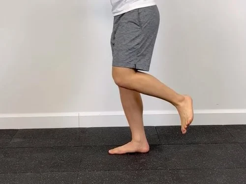
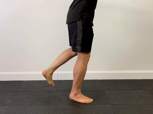
Unilateral one-inch heel raise
This is a boosting exercise for the intrinsic foot muscles that balance out your medial longitudinal arch. Start by standing on one leg and raising the heel just 2.5 centimeters, or 1 inch, off the ground. Maintain your balance in this position for 15 to 30 seconds, then rest for the same amount of time. Make certain to keep an even appropriation of your weight through the balls of your toes in general.
FAQ
Can posterior tibialis be healed?
Posterior tibial tendon dysfunction by and large requires 6 to 8 months to improve and early action on a recovery ligament can bring about a setback in healing. Patients can experience a lot of frustration as a result of non-compliance, which can increase recovery time by twice.
Can the tibialis posterior be massaged?
Regarding the massage of the posterior tibial tendon, it is liable for supporting the foot’s arch and assisting in the stabilization of the ankle during movement. The posterior tibial tendon can be made more flexible, strengthened, and less painful by massaging it.
How can I make my posterior tibialis stronger?
The most straightforward method for beginning to boost the tibialis posterior muscles is to perform heel raises. You could begin by performing these exercises while seated in a chair, and as your muscle strength improves, you could try doing them standing up.
What is a weakness in the posterior tibialis?
The term “posterior tibial tendon insufficiency,” also known as “adult-acquired flatfoot,” refers to the failure of the posterior tibial tendon. Nonetheless, this condition additionally includes the disappointment of related tendons and joints on the medial (internal) side of the foot and lower leg.


7 Comments