
Muscle Detail | Anatomy | Anatomy Of Muscle | Exercise Detail | Strengthening exercise | Stretching exercise
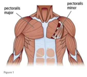
Pectoralis Major Muscle
What is the pectoralis major muscle?
- The pectoralis major muscle is a broader, fan-molded, or 3-sided focalized muscle, situated at the chest of the human body. It is made up of the majority of the chest muscles & lies beneath the breast. The pectoralis major is the pectoralis minor, a flimsy, 3-sided muscle. The pectoralis major primary functions are adduction of the arm, flexion & internal rotation of the humerus.
- It comes out from the foremost surface of the sternal portion of the clavicle from the expansiveness of the portion of the front surface of the sternum, a slow down as the joining of the ligament of the sixth or seventh rib; from the ligaments of the multitude of genuine ribs, with the exception, frequently, of the 1st or 7th, & from the aponeurosis of the stomach outer slanted muscle.
It consists of two laminae:
- The 1st lamina, which is larger, gets the clavicular & the most elevated sternal fibers. They are inserted in a comparative solicitation as that where they emerge: the most sidelong of the clavicular strands are embedded at the upper part of the front lamina; the highest sternal filaments pass downwards to the lower part of the lamina which reaches out as low as the ligament of the Deltoid & gets together with it.
- The posterior lamina of the tendon receives the joining of the greater part of the sternal portion & the deep fibers, i. e., these are from the costal cartilages.
- These profound filaments, & especially those from the lower costal ligaments, arise the humerus inclusion higher, turning in reverse progressively beside the shallow & upper ones, with the goal that the ligament seeing, by all accounts, to be curved. The beside lamina comes to higher on the humerus than the foremost 1, & from it, an extension is done which covers the intertubercular notch of the humeral and mixes with the case of the glenohumeral joint.
Location of Pectoralis Major Muscle
- Pectoralis major muscle is located in the upper part of the chest and ends at a ridge at the rear of the humerus (the bone of the upper arm).
- The muscle is the superior and largest muscle of the anterior chest wall. It is a thick, fan-shaped muscle that is located underneath the breast tissue and forms the anterior wall of the axilla.
Relation of Pectoralis Major Muscle
- The pectoralis major muscle is a broad superficial muscle located superficially in the anterior chest wall. In males, it is surfaced by the deep layer of fascia, subcutaneous tissue & the adjacent skin. In females, it is surfaced by the breast. The deep surface of the muscle covers the pectoralis minor & serratus anterior muscles & the anterior surface of the upper 6th ribs.
- The triangular depression between the pectoralis major muscle, deltoid muscle & clavicle is known as the infraclavicular fossa (Mohrenheim’s fossa) which gives us an important landmark in the surgical procedures on the subclavian artery.
Origin of Pectoralis Major Muscle
- The pectoralis major muscle consists of three parts:-
- The sternocostal part originates from the anterior surface of the sternum & the anterior aspects of the costal cartilages of ribs 1st & 6th.
- The smallest, abdominal part arises from the anterior layer of the rectus sheath.
- The clavicular part arises from the anterior surface of the medial half of the clavicle.
Insertion of Pectoralis Major Muscle
- The Pectoralis major muscle fibers from all three parts pass laterally, diverging towards the proximal humerus. They give off a large tendon that inserts along the crest of the greater tubercle of the humeral bone. The upper and lower fibers of the muscle are inserted at the crest of the greater tubercle of the humeral bone. Upper fibers are major anterior & inferior on the crest, while posterior fibers twist on themselves & are major posterior & superior to the upper fibers.
Nerve supply of Pectoralis Major Muscle
- The pectoralis major gets 2 engine innervation by the average pectoral nerve and the parallel pectoral nerve, otherwise known as the horizontal from the thoracic nerve.
- The sternal head gets nerve supply from the C7, C8 & T1 nerve roots, via the lower trunk of the brachial plexus & the average pectoral nerve.
- The clavicular head gets blood supply from the C5 & C6 nerve roots via the upper trunk & horizontal string of the brachial plexus, which elongates the sidelong pectoral nerve. The parallel pectoral nerve is rounding above the profound floor of the pectoralis major muscle.
Blood supply of Pectoralis Major Muscle
- The blood supply of the pectoralis major muscle is from the pectoral branches of the thoracic acromial artery & the perforating branches of the internal thoracic artery.
The action of the Pectoralis Major Muscle
- Pectoralis major muscle primarily does the adduction of shoulder joint or depression of the arm & rotation of the arm forward about the axis of the body (medial/internal rotation).
- It also helps in inspiratory & expiratory breathing patterns.
Pectoralis major muscle Function
- At the point when the arm is in the physical position, the pectoralis significantly moves about as a solid adductor & internal rotation of the shoulder joint. Pretending autonomously, the clavicular portion of the muscle flexes the humerus up to 90 degrees in a flat plane. The sternocostal part of the muscle will be delivered the adversarial development & stretch outward the humerus back into the physical position.
- Working together with the latissimus dorsi muscle, the pectoralis major muscle pulls the trunk forwards or upwards when its humeral stabilization is fixed. This motion is important in activities such as climbing. When acting from the humeral attachment, the pectoralis major muscle also helps out the act of inspiration. This is significantly important during forced breathing in physical distress.
The antagonist for pectoralis major muscle :
- Deltoid muscle
- Trapezius muscle
- The Deltoid muscle is a larger, triangular in shape glenohumeral muscle. The deltoid muscle (acromial part) is the primary abductor of the arm at the shoulder joint (Glenohumeral joint).
- The trapezius muscle is a large paired surface muscle that extends longitudinally from the occipital bone to the lower thoracic vertebrae of the spine & laterally to the spine of the scapula. The primary function of the trapezius is stabilizing the scapula in its anatomical place, as well as controlling it during movements of the shoulder & upper limb.
Clinical Significance of Pectoralis Major Muscle
- Poland syndrome is an embryonic malformation of a thoracic wall, which occurs by a defect of the pectoralis major muscle in combining with different malformations of the upper limbs (e.g. malformations of the fingers). The muscle can be partially or completely missing. Depending on the degree of severity, the adduction & internal rotation of the shoulder joint are more or minimum difficult to perform (for example, the crossing of the raised arms). In addition, infected patients have visibly distorted breasts because of the aplasia of the pectoralis major muscle, which is why plastic-surgical rearrangement can be used therapeutically.
Other diseases
- Pectoralis major muscle on rare conditions may develop intramuscular lipomas. Such rare tumors might cause malignant breast tumors as they will look like enlargements of the breasts. They are well-encapsulated radiolucent tumors of fat tissue density. Their location can be accurately identified through computed tomography(CT SCAN) & magnetic resonance imaging (MRI). The treatment in these cases required complete surgical removal because of the risk of liposarcoma as they post especially large intramuscular lipomas. Partial excision is risky because chances of recurrence may occur.
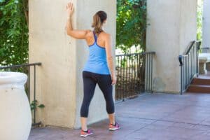
Pectoralis Major Stretching Exercise
Doorway Pectoral Stretch:
- Standing aside from the door frame or corner of a wall.
- Maintain the back extended & the inner core contract, bring the arm raised up against the wall with the elbow and shoulder flexed at 90 degrees.
- With the arms placed on the wall, draw the opp side shoulder back followed by the torso in a parallel line. Keep the back extended & core contracted.
- Hold this for 30 seconds & repeat three times on each side two times per day.
Camel Pose Stretch:
- Take a kneeling position on the floor with knees hip-width apart and hands on the waist. Tuck the toes or put them flat against the floor.
- Gently reach behind & place 1 hand on each heel.
- Keep the chest up, shoulders back and down, engage the core, and gently push the hips forward.
- Hold for 15-20 secs and repeat three times.
Hand Behind the Back Stretch:
- Standing tall with the feet shoulder-width apart. Interlace the fingers beside the back & extended the arms.
- Maintain the chest up and pull the shoulder blades downward.
- Hold for 15-20 seconds & repeat three times.
Floor Angels:
- In supine position with feet hip-width apart and flat on the surface.
- Position both arms to the behind at a 90-degree angle with palms facing up toward the roof.
- Maintaining in contact with the surface at all times, gently bring the arms raised over the head until they are fully straightened.
- Then gently bring both arms beside to the 90-degree initial position.
- Repeat 10 times for three sets.
- Remember to keep the back flat against the surface and ribs tucked at all times.
Above-the-head Chest Stretch:
- The patient is in a sitting /standing position, interlock the fingers, then flex the elbow joint and lift the arms above the head.
- Then slowly squeeze the shoulder joint blades combine and move the elbow joint and hands behind.
- Change the height of the hands to focus on the shoulder joint and chest means hands are beside the head, hands are on top of the head, and the hand is also a few inches raised to the head.
Bent-arm Wall Stretch:
- The patient assumes a split stance, the left leg is in the front and the right leg is behind, in a doorway, and at the end of a wall.
- Then Bring the right arm up to shoulder level and position the palm and inside of the arm on the wall surface or doorway.
- The patient arm seems like a goal post.
- Then slowly press the chest via the open space to feel the stretch.
- Movement of the arm higher and lower which is allowed to you stretch to the various sections of the chest.
- Then Repeat this exercise on the opposite side.
Extended Child’s Pose on Fingertips stretch:
- The patient is Kneeling on the surface.
- Try to Touch the big toes combine and the patient is sitting on the heels.
- Next, differentiate the knee joint about as wide as the hip joint.
- Then flex the forward from the hip joint and walk the hands outwards as far in front of them as possible.
- With the arms straightened palms are facing down then come up into the fingertips as if the ball beneath the palms melts the chest toward the floor.
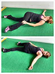
Side-Lying Parallel Arm Chest Stretch:
- The patient is lying down prone on the stomach then collides both arms out to the sides then palms facing down to create the letter, T.
- Then begins to roll into the left side by pushing with the right hand.
- Raise the right, flex the knee joint and place the right foot beside you on the floor for stability.
- Then Rest the left temple on the floor.
- Must Keep the right hand on the floor for balance.
- For the feel of the extra stretch, raise the right hand toward the ceiling.
- Then Repeat this stretching on the opposite side.
Pectoralis Major Strengthening Exercise
Push-Ups:
- Begins this Exercise in the push-up position as demonstrated.
- Maintaining the back and neck straight, gently straighten the elbows until they are extended tightening the pectoral muscles, then back to the starting position.
- Perform three sets of 7 to10 reps, if the exercise is pain-free.
Incline push-up:
- This exercise begins with the hands-on wall & a little higher the height surface.
- Then Walk the feet behind so that the body makes roughly a 45-degree angle with the surface.
- Keep the body straight & spine neutral & lower the chest to the floor leaning against it.
- Pause for a moment, & after that return to the initial position.
- This resistance must be little enough & do complete up to 20 reps.
- For a simple exercise, a step nearer to the hands
- To make the harder exercise, step farther away.
Flat bench press:
- Lie down on the back on the bench with the knees flex and foot flat on the floor.
- Hold the barbell, with the thumb, wrapped surround the barbell, and palms facing toward the feet.
- Press the arms extended towards the ceiling which raises weight off the rack.
- Then movement the weight over chest level.
- Flexing the elbow joint down at a 45-degree angle gently lowers the weight of the chest.
- Must maintain the bar approximately in line with the nipples.
- Then stop for a moment after that press the weight back into the initial position.
- Do the Complete three sets of the 8–12 reps.
Incline bench press:
- The patient is lying down on the back on the incline bench with the knees flexed and the foot flat on the floor.
- The patient is holding the barbell, with the thumb which is wrapped surrounding the barbell, and palms facing toward the feet.
- Compress the arms extended toward the ceiling to raise the weight off the rack.
- Placing the weight above the collarbone.
- Then gently lower the weight downwards to the chest, approx. in line with the mid-chest to just above the nipples.
- Then stop for a moment after that press the weight back into the initial position.
- Do the Complete three sets of the 8–12 reps.
Decline bench press:
- The patient is lying down on the back on the decline bench, with the knees joint bent and ankles which is secured beside the ankle rests.
- The patient is grabbing a barbell, with the thumb wrapped to surround the barbell and palms facing toward the feet.
- Compress the arms extended to raise the weight off the rack.
- Place the weight above the lower chest to the upper abdomen region.
- Then gently flex the elbow joint to lower the weight downwards to the chest, approx. in line with the nipples.
- Then stop for a moment after that press the weight back into the initial position.
- Do the Complete three sets of the 8–12 reps.
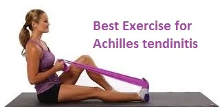
Resistance Band Adduction:
- Start this exercise by standing or kneeling with the back extended and holding a resistance band as illustrated.
- Maintain the back and elbows extended, and gently pull the resistance band to the hip as seems, tightening the pectoral muscles.
- Perform three sets of 10 reps provided the exercise is pain-free.
Pectoral Dumbbell Pull-on Swiss Ball:
- Gently raise arms overhead and then gently back to the initial position tightening the pectoral muscles.
- Perform three sets of 10 reps when the exercise is pain-free.
Cable crossover:
- This exercise begins with the standing position far from a set of high pulley cable machines and a resistance band anchored overhead.
- Select light to a moderate weight which is adding to the challenge but gives success.
- Hold the hands (or the ends of the band) as they step ahead with one foot.
- Maintain enough tension and control on the handles to sustain them in front of the chest.
- Contract the chest muscles and bring the handles downwards extended across the body at randomly belly button level.
- The hands are crossing to increase the importance of the serratus anterior muscles.
- Hold for a moment and then gently return to the initial position.
- Then repeat to the opposite side.
- Do three sets of 8–12 reps.
Chest dip:
- The patient is in the Standing position which is seeing the 2 parallel bars & grasps them, palms facing in.
- Then straight the elbow joint and press into the hands, lifting the body so that it is in parallel with the hands.
- Then, flex the elbow joint and lower the chest toward the hands.
- Pause, then press back into the initial position.
- Repeat Do the three sets of the 8–12 reps.
Shoulder joint internal rotation isometric exercise:
- To do shoulder internal rotation you are standing seeing a door frame & outside the wall corner.
- The bad shoulder joint is closer to the door corner.
- Then flex the elbow joint up to 90 degrees and make a fist.
- Try to slowly compress the towel into the corner wall and door jamb like as you rotate the arm inward towards the belly.
- Hold this pressing for 5 to 10 secs & gently release.
- Do the 10 to 15 reps.
Shoulder Extension isometric exercise:
- To do shoulder extension you are standing to six inches away from the wall and the back is seeing the wall.
- The elbow joint is extended so the hand stays just near the buttocks and makes a fist.
- Try to smoothly compress the towel into the wall beside you.
- Hold this pressing for 5 to 10 secs and gently release.
- Complete the 10 to 20 reps.
Shoulder adduction isometric exercise:
This exercise mostly targets the rotator cuff muscles.
- To do shoulder adduction you stand 6 inches away from the wall.
- Then turn the body perpendicular to the wall and close to the wall at the side which is affected by the shoulder joint side.
- Make a fist on that side and compress the towel into it.
- You are also using a wrapped towel for more support.
- You are only gently compressed into the wall and try to elevate the arm outside.
- Hold this pressure for 7 to 10 seconds and gently release pressure from the wall.
- Do the 10 to 15 reps.
- Repeat three sets at a time.
Chin-Ups:
- Grab the bar, hands shoulder-width apart palms with an underhand grip
- Start with arms completely extended and pull the chest up to the bar as high as you can
- Hold briefly at the top then back to the starting position
- Complete the 5 to 10 reps.
- Repeat three sets at a time.
- Every exercise does the two to three times per day.
Lateral raises:
- First, gran a pair of light dumbbells for this exercise.
- You are standing with the foot slightly wider than the hip joint width apart.
- Try to elevate the weights to the sides till are at shoulder level.
- Must be remembered to contract the core muscles and gently lower the weights down to the sides.
- Repeat this exercise for two sets, in every set consisting of 12 to15 reps and three to four times per week.
Bent-Over Horizontal Abduction:
- You are lying on the stomach means in a prone position on a table and bed with the injured arm hanging over the side.
- Must keep the arm straight and gently raise it to eye level.
- Then gently lower it back to the initial position and repeat this exercise.
- Do this exercise three to five times per day.
Finger ladder Exercises:
- The patient is in a standing position and facing a ladder that is hanging over a wall.
- Ask them to patient place the bad hands on the ladder at a low level.
- Then gently start an upward climb on the finger ladder till it reached the top and gently down back to the beginning.
- This exercise is performed ten times in one session and three sessions per day.
Breathing Exercises
Diaphragmatic Breathing Exercise :
In supine position
- Lying on the back on a surface or in bed, together with the knees flexed & the head supported to the bottom. you’ll use a pillow beside the knees to support the legs.
- Place 1 hand on his higher chest & therefore the alternative is slightly below the skeletal structure. as you are feeling the diaphragm move as you breathe.
- Breathe in gently exploiting the nose so the abdomen moves away and it’ll permit the hand to rise. The hand on the chest ought to persist as still as connected.
- Tight the abdominal muscles & allow them to fall within as you expire exploitation of the lips. The hand on the belly ought to move down to its normal position
- In a sitting position in a chair, with the knees flexed & the shoulders, head, & neck neutral position. you can perform for seven to ten minutes, several five to six times a day. Diaphragmatic exercise is also called a relation exercise.
In sitting position
- Sitting in a chair straight position & lengthen the distance between the umbilicus & sternum.
- The shoulders & neck should be relaxed.
- The pelvis is in the normal position.
- The hand’s position should be at the side of the lower ribs.
- Breath in slow using the nose. As you inspirited feel the ribs should be outwards & upwards. During inspiration expansion of the trunk in 3 areas forward, sides, & back. expiration using the nose. As you expire the lower ribs move inward direction.
Segmental Breathing Exercise :
Lateral costal expansion
- it is also known as lateral basal expansion. it was usually done unilaterally or bilaterally. it can also do in a sitting or lying position place the hand lateral aspect of the rib cage & ask the patient to breathe out & the rib cage should be downward & inward as the patient expired therapist should.
- Apply pressure on the rib cage with the palms of the hand before inspiration, you need to downward & inward stretch to the chest to facilitate the external intercostal contraction.
- Apply normal force on the lower rib cage to get stimulated & patient to get inspirited deeply & the chest will expand when the patient expiration therapist will squeeze the rib cage downward & inward the patient may perform manually & ask to apply force with his hand or with the towel.
Posterior basal expansion
- This form of deeper breathing exercise accents post-surgical maneuvers who have been bedridden & will not sit up for a prolonged-time period. because of that time secretion is moved to the posterior segment of the downward side of the rib.
- sit & lean onward on a pillow, slightly flex the hips
- place the therapist’s hand over a posterior segment of the rib
- place the hand lateral aspect of the rib cage
- ask the patient to breathe out & the rib cage should be downward & inward
- as the patient exhale therapist should apply pressure on the rib cage with the palms of their hand
- before inspiration, you need to downward & inward stretch to the chest to enhance the external intercostal contraction
- apply normal force on the lower rib cage to get stimulate & the patient to get inspirited deeper & the chest will expand
- when the patient exhales therapist will squeeze the rib cage downward & and inward
- the patient may do this indirectly ask to apply force with his hand or with a towel
Right middle lobe expansion(lingual expansion)
- the patient is in a sitting position therapist’s hand should be left or right side at the below axilla level
- ask the patient to breathe out & the rib cage should be downward & inward
- as the patient exhale therapist should apply pressure on the rib cage with the palms of their hand
- before inspiration, you need to downward & inward stretch to the chest to enhance the external intercostal contraction
- apply normal force on the lower rib cage to get enhance & patient to get inspirited deeper & the chest will expand
- when the patient expiration therapist will contract the rib cage downward & the inward
- the patient may do this manually & ask to apply force with his hand or with a towel
Apical expansion
- The patient is in a sitting position on a chair/bed therapist’s hand should be under the clavicle
- As the patient exhales out apply pressure with the palm or fingertip just below the clavicle
Pursed lip breathing exercise
- Sitting with using the back straight or lying down. Relax each shoulder & neck the maximum amount as potential.
- inspiration using the nose for 2 to 3 secs, the air is getting into the abdomen. fill the abdomen with air instead of the lungs.
- Purse the lips like you are processing on hot food then expire gently, taking double as long to exhale as you took to respire.
- you can repeat several times. you’ll rise the inspiration & expiration counts from a pair of seconds to 4 secs, & so on pursed lip breath effective.

16 Comments