Calcific tendinitis of the longus colli
What is Calcific tendinitis of the longus colli?
Calcific tendinitis of the longus colli muscles is an inflammatory or granulomatous reaction to the deposition of calcium hydroxyapatite crystals in the tendons of the longus colli muscle. It is occasionally more generically called calcific prevertebral tendinitis or, less accurately, retropharyngeal calcific tendinitis.
The disease is rare and self-limiting but can mimic several severe conditions. The longus colli is a prevertebral muscle that bends and turns the neck. The deposition tends to happen predominantly in the superior fibers expanding from the transverse process at C3 – C5 to the anterior tubercle of C1 (atlas)
Epidemiology
Like different forms of calcific tendinitis, this disease generally happens in grown-ups in middle age (20-50 years old). There is no consistent gender preference across patient series.
In a retrospective study of the grown-up neck and cervical spine CTs in one American health system, the frequency of acute longus colli tendinitis was 1 in 1000 studies.
What are the Causes of Calcific tendinitis of the longus colli?
The causes of the disease stays unclear. One hypothesis is that degeneration, trauma, or ischemia of the tendon predisposes to the deposition of crystals in an try to compensate for decreased tendon rate. Rupture and release of the crystals cause a strong inflammatory foreign body reaction in the surrounding soft tissue. The crystals are resorbed throughout one to two weeks, and symptoms generally resolve after a few days. One analysis saw a mean symptom time of 4.6 days
Symptoms of Calcific tendinitis of the longus colli
A person can present with debilitating symptoms that are irrelevant to the degree of calcification noticed on CT. Symptoms form acutely and contain fever, neck pain, odynophagia, dysphagia, and less neck range of motion. White blood cell calculation and erythrocyte sedimentation rate may be increased.
Differential diagnosis
retropharyngeal abscess
- prominent surrounding enhancement
- diffusion restricting fluid within the collection
- cervical lymphadenopathy
trauma
- prevertebral bone fragments from acute fractures may be misunderstood for calcification in the longus colli tendons
tumor
- soft tissue mass
- contrast enhancement
- cervical lymphadenopathy
Diagnosis
Plain radiograph and CT
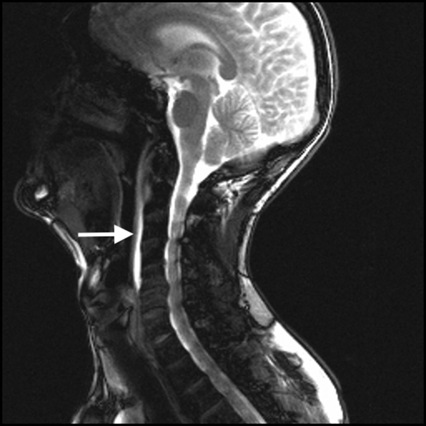
Calcifications may be noticed on radiographs, but the desired imaging modality is contrast-enhanced CT. On CT, unstructured calcifications are generally noticed in the superior fibers of the longus colli muscle tendons (at the C1-C2 level). Sometimes the inferior fibers may be involved, as low down as T3. The longus colli muscles may also seem hypo-attenuating due to edema.
Little retropharyngeal effusions and edema of the contiguous prevertebral soft tissues are also generally seen.
Significantly, enhancement near the effusion is generally minimal and if present, should shift the diagnosis toward a retropharyngeal abscess. Adenopathy and bone destruction, too, should indicate alternative diagnoses.
MRI
MRI will easily show edema, yet, a high level of suspicion is required as the calcifications are much more difficult to visualize.
As is the issue with CT, the presence of peripheral enhancement and/or suppurative lymphadenopathy should indicate infection as the underlying reason.
At least one issue of localized marrow signal inflammation has been noted.
Treatment of Calcific tendinitis of the longus colli
Conservative management with NSAIDs is normally all that is needed. Symptoms tend to settle within a few weeks.
In some patients, along with a solution of the fluid, the mineralization can be reabsorbed and will not be clear on follow-up imaging.
Pain-free Isometric neck exercise is also helps to maintain strength of Neck muscles.
FAQ
Is calcific tendonitis serious?
Frequently, calcific tendonitis doesn’t cause issues. But if the calcium deposits get larger or evolve inflamed, they can generate extreme pain. This disease most frequently involves the shoulder. The calcium deposits generally form in the rotator cuff – a group of muscles and tendons that cover the shoulder joint.
Is exercise good for calcific tendonitis?
In extra to rehabilitating your shoulder joint itself, at Humpal Physical Therapy & Sports Medicine Centers, we highly suggest keeping the rest of your body’s fitness with regular activity even while you heal from the calcific tendonitis in your shoulder.
Will calcific tendonitis go away?
Although calcific tendonitis can be painful for some patients, a fast resolution is probable. Most patients can be managed in a doctor’s office, and only 10 percent of patients need some form of surgery. Calcific tendonitis does finally disappear on its own, but it can lead to difficulties if left untreated
Is massage good for calcific tendonitis?
If calcific tendinitis is doubted, the massage practitioner should direct the customer to a physician. Yet, massage could be used for general pain ease in associated tissues and broad relaxation, unless it creates pain
How long does calcific tendonitis last?
Calcium generally disappears spontaneously with a period. A complete solution of symptoms can bring 12 to 18 months. If symptoms are extreme or resolution slow, then surgery is an option.