Cardiac plexus
The cardiac plexus is a network of autonomic nerves and ganglia located at the base of the heart. It is formed by cardiac branches originating from both the sympathetic and parasympathetic nervous systems.
Gross anatomy of the cardiac plexus
- Parasympathetic cardiac nerves reach the heart from the vagus nerve (cranial nerve X) through several cardiac nerves. An upper cardiac nerve get arises below the inferior cervical ganglion and a lower branch get arises in the root of the neck. The nerves descend anterior to the brachiocephalic artery on the right and over the aortic arch on the left side.
- Sympathetic cardiac nerves are originated from the T1 to T4 segments and partly from the T5 segment of the spinal cord.
- Both sympathetic and parasympathetic nerves end in the cardiac plexus which has two interlinked connected components, which in turn contribute to the left and right coronary plexuses:
- a superficial portion inferior to the aortic arch
- a deep portion posterior to the aortic arch
- Postganglionic fibers from both portions pass into the heart and are most deeply distributed to the sinoatrial node and atrioventricular node. Some fibers pass directly into the atria and ventricular myocardium. The coronary arteries and cardiac veins also collect adrenergic fibers.
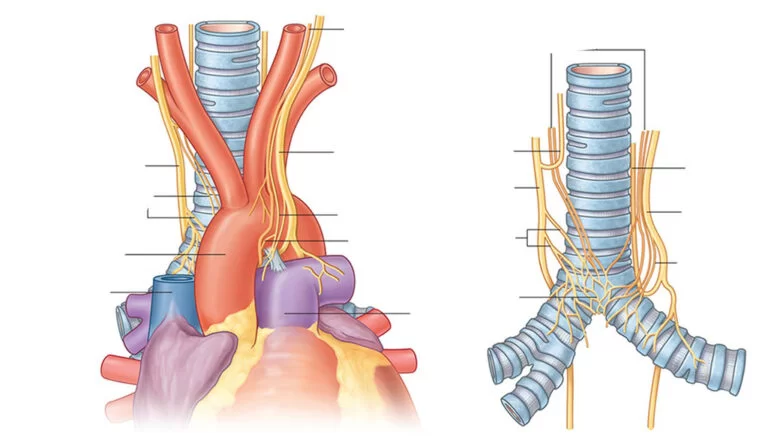
The superficial part of the cardiac plexus
The superficial part of the cardiac plexus is placed beneath the aortic arch and anterior to the right pulmonary artery. It is composed of the superior cervical cardiac branch of the left sympathetic trunk and the lower superior cervical cardiac branch of the left vagus trunk. A small ganglion, the cardiac ganglion of Wrisberg, is sometimes found that forms a junction for these nerves.
The superficial cardiac plexus gives branches to the anterior coronary plexus, left anterior pulmonary plexus, and deep cardiac plexus.
The deep part of the cardiac plexus
The deep part of the cardiac plexus is placed posterior to the aortic arch and anterior of the tracheal divarication (carina angle) above the point of division of the pulmonary trunk. It is constructed by the cardiac nerves get originate from the superior, middle, and inferior cervical ganglia of the sympathetic trunk and the cardiac branches of the vagus and repetitive laryngeal nerves except those that supply the superficial cardiac plexus.
The deep cardiac plexus diverges into halves:
the right half lies of the cardiac plexus along with the brachiocephalic trunk
A branch passes anterior to the right pulmonary artery supplying the right pulmonary plexus and continues to provide to the anterior coronary plexus
A branch passing posterior to the right pulmonary artery supplies the right atrium and continues to provide to the posterior coronary plexus
the left half of the cardiac plexus lies along with the aortic arch
- connects with the superficial cardiac plexus
- allow small branches to the left atrium and to the anterior pulmonary plexus
- continues to provide the posterior coronary plexus
Coronary plexuses
Both superficial and deep cardiac plexuses expand laterally becoming coronary plexuses. The left coronary plexus travels along with the left coronary artery and supplies the left atrium and the left ventricle. The right coronary plexus travels along the right coronary artery and supplies the right atrium and right ventricle.
FAQ
What contributes to the cardiac plexus?
The structure of nerves supplying the heart is called the cardiac plexus. It receives an allowance from the right and left vagus nerves, as well as an offering from the sympathetic trunk. These are responsible for affecting heart rate, cardiac output, and contraction forces of the heart.
Where does the cardiac plexus come from
The deep cardiac plexus is present at the anterior aspect of the tracheal bifurcation behind the aortic arch and is formed by cardiac nerves which get arising from the cervical ganglia of the sympathetic trunk and cardiac branches of the vagus & reoccurring laryngeal nerves.
How many cardiac plexuses are there?
Branches of these nerves construct the cardiac plexuses on the epicardium. The cardiac plexuses could be divided into six plexuses, which include;
the right and left coronary plexuses,
pericardiac transverse sinus plexus,
caudal cardiac plexus,
right and left superior cardiac plexuses.
What does the cardiac plexus innervate?
The cardiac plexus is a plexus of nerves present at the base of the heart that supplies the heart.