Carpal Bones
Introduction
Together with the distal extremities of the radius and ulna, the carpal bones are a collection of small bones in the human hand that constitute the wrist.
They are therefore also referred to as wrist bones. Combined, they are referred to as the carpus, and they individually articulate with the metacarpals, the long bones of the wrist, and the ulna and radius of the lower arm.
The highest density of bones is seen in the human upper extremities. This skeletal segment ranges in complexity from simple to sophisticated.
The hand’s diverse range of motions is made possible by its numerous articulations and structural variations. The wrist is one of the more intricate anatomical and functional components of the upper extremity.
The wrist bone also referred to as the carpal bone in biology, connects the arm’s dangling and fixed parts.
What is Carpal bone?
The eight little bones that make up the wrist (carpus), which joins the hand and forearm, are known as the carpal bones. The Latin carpus and the Greek word καρπός (karpós), which means “wrist,” are the sources of the terms “carpus” and “carpal.”
The primary functions of the carpal bones in human anatomy are to form a highly mobile condyloid joint by articulating with the radial and ulnar heads, to supply attachments for the thenar and hypothenar muscles, and to comprise a portion of the rigid carpal tunnel, which facilitates the transmission of the median nerve and the tendons of the anterior forearm muscles to the hand and fingers.
The carpus is the only group of bones in the wrist of tetrapods, situated between the metacarpus, ulna, and radius.
In contrast to those of the metacarpus, the carpus’s bones do not belong to specific fingers or, in the case of quadrupeds, toes. The wrist can rotate and move vertically thanks to the carpal bones.
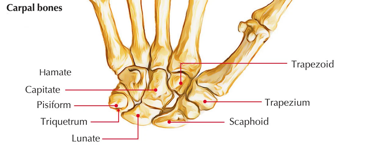
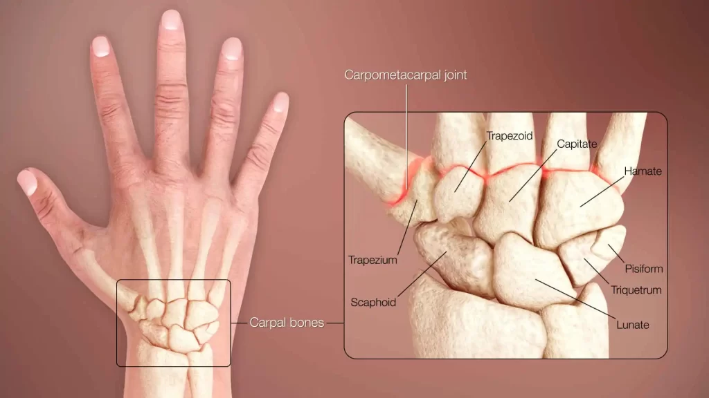
There are eight irregularly formed bones in the wrist region that are called the carpal bones (carpus). These bones link the proximal portions of the metacarpal bones to the distal aspects of the radius and ulna, the long bones of the forearm.
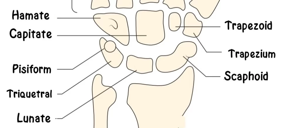
The proximal and distal carpal bones are arranged in two rows. The scaphoid, lunate, triquetrum, and pisiform bones are part of the proximal row of carpal bones, which runs from radial to ulnar. The hamate, capitate, trapezium, and trapezoid bones are in the distal row.
| Proximal row (From the Radial side to the Ulnar side) | Distal row (From the Radial side to the Ulnar side) |
| Scaphoid | Hamate |
| Lunate | Capitate |
| Triquetrum | Trapezium |
| Pisiform | Trapezoid |
Every carpal bone is different from the others and has several facets, which allow it to articulate with the surrounding bones, muscles, and ligaments in the hand and forearm.
In this manner, the carpal bones give the hand’s soft tissues flexibility and a range of motions. Additionally, they supply the majority of the wrist’s skeletal framework, which creates a pathway for the various neurovascular systems in the hand.
How Many Carpal Bones are in the Hand?
The human wrist consists of eight bones, each of which is called based on its shape:
- Scaphoid (form of a boat)
- Lunate (moon-shaped crescent)
- Triquetrum (pyramid)
- Pisiform (pea-shaped)
- Trapezium (formatively irregular trapezium)
- Trapezoid (formed wedge)
- Capitate (like a head)
- Hamate (a bony protrusion that resembles a wedge or “hook”)
Of all the carpal bones, the capitate is the largest.
The wrist gets its distinct shape from the transverse rows. The proximal surface of the wrist bone exhibits convexity, while the distal section of the bone is concave.
It is also possible to organise the eight bones into longitudinal columns. There are three of them, specifically:
- The trapezium, trapezoid, and scaphoid make up the radial scaphoid column.
- The lunate column is made up of capitates and lunates.
- The triquetrum and hamate make up the ulnar triquetral column.
Where are the Carpal Bones Located?
The human skeleton’s upper extremities contain the carpal bone. The bones serve as a connecting link between the metacarpals, radius, and ulna. On the proximal side, the radius and wrist bone articulate. On the distal surface of the carpal bones, the metacarpals are articulated.
The eight bones are located in the following locations:
- The scaphoid is oriented towards the thumb on the lateral side and is situated near the radial border.
- The lunate is situated where the radius and ulna articulate. The middle bone in the proximal carpal row is this one.
- Without any articulation with the ulna, the triquetral is situated on the ulnar side. Located on the medial side of the proximal row is this pyramidal bone.
- Marking the ulnar boundary, the pisiform is the final structure in the proximal row.
- The radial border of the distal transverse row is formed by the trapezium, which is located above the scaphoid.
- The second distal row bone is the trapezoid, which is nearly pierced between the capitate carpal bones and the trapezium.
- The wrist is commonly referred to as the capitates, which is the wrist bone situated in the centre. It is placed on the distal surface of the ulna and the radius.
- The distal transverse row’s ulnar boundary is indicated by the hamate.
Structure of Carpal Bones
The eight carpal bones have distinct structures, with varying numbers of flat surfaces on which other bones can articulate.
Two transverse rows or three longitudinal columns are the two possible conceptual arrangements for the eight carpal bones.
Each row forms an arch that is convex proximally and concave distally when viewed as paired rows. The flexor retinaculum covers the concave carpus on the palmar side, which produces the carpal tunnel.
Because the proximal row articulates with the distal carpal row and radius surfaces, it is always adjusting to these moving surfaces. Every carpal bone in the proximal row can move somewhat independently of one another.
The scaphoid, for instance, articulates distally with the trapezium and trapezoid, so contributing to midcarpal stability. The distal row, on the other hand, moves with the metacarpals, making it more stiff.
Three longitudinal columns provide a better way to conceptualise the carpal bones from a biomechanical and clinical standpoint:
- Radial scaphoid column: scaphoid, trapezium, and trapezoid.
- Capitate and lunate in a lunate column.
- Triquetrum and hamate make up the Ulnar triquetral column.
In this context, the pisiform is seen as a sesamoid bone inserted in the tendon of the flexor carpi ulnaris.
The radial or scaphoid and central or capitate columns are the only ones that articulate with the radius since the ulnar column leaves a space between the ulna and the triquetrum.
The strength of several capsules and ligaments, rather than the interlocking elements of the skeleton, is what makes the wrist more stable in flexion than in extension.
With the exception of the pisiform, almost all carpals have six surfaces. For ligamentous attachment, the palmar, or anterior, and dorsal, or posterior, surfaces are rough; the dorsal surfaces are wider, with the exception of the lunate.
Where they come into contact with adjacent bones, the medial and lateral surfaces are also articular; otherwise, they are rough and tuberculated.
The superior, proximal, and inferior, or distal, surfaces are articular, with the superior typically convex and the inferior concave.
Proximal row of bones
Scaphoid bone
The largest carpal bone in the proximal row is the scaphoid bone. The scaphoid is a boat-shaped bone that is located just below the anatomical snuffbox. The word “scaphos” in Greek means “boat,” hence the name.
It connects proximally to the radius, and distally with the trapezium and trapezoid. Additionally, the scaphoid articulates to the lunate and capitate bones.
The scaphoid tubercle, a bony protrusion on the palmar surface of the scaphoid, is easily perceptible on the palm of the hand due to its subcutaneous location.
The scaphoid is the most often fractured bone of the wrist. Most cases of scaphoid fractures happen when someone falls onto an outstretched hand.
- The largest bone is in the anterior row.
- It articulates distally with the hamate bone and laterally with the lunate bone.
- The scaphoid articulates with the radius and defines convexity on the proximal side.
- The semi-lunar, distolateral facet of the scaphoid is formed by the articulated trapezium and trapezoid.
- The concave facet of the scaphoid’s distomedial surface is where the head of the capitate articulates.
- The scaphoid bone’s flat surface articulates with the lunate.
The dorsally slender scaphoid bone has a groove where ligaments can be attached.
Lunate bone
The proximal row’s next bone, the lunate, is situated between the scaphoid and triquetrum bones.
The lunate articulates distally with the capitate bone and proximally with the head of the radius (carpal articular surface) and the articular disc of the distal radioulnar joint. Its look resembles the moon, hence its name.
- The crescent moon is represented by the lunate.
- Its proximal side contacts the radius.
- It articulates into the scaphoid’s flat surface on the lateral side with a facet that resembles a crescent.
- The lunate bone’s medial face has a quadrilateral facet that articulates with the triquetral.
- It is articulated distally using capitate.
- On both its distal and medial surfaces, the lunate exhibits articulation with the hamate.
- The dorsal side of the bone is spherical, whereas the distal surface is concave.
Triquetrum bone
Positioned on the medial aspect of the carpus, the triquetrum is a pyramid-shaped bone, meaning “three-cornered bone” in its name.
It articulates distally with the hamate bone and laterally with the lunate bone. Furthermore, on its distal palmar surface, the triquetrum has a single, oval-shaped facet for articulation with the pisiform bone.
- Having three sides, the bone has a pyramidal shape.
- One of the carpal bones that makes up the carpal arch is the triquetrum.
- Its oval-shaped facet articulates with the pisiform bone.
- The bone is largely unseen since it is located beneath the pisiform.
Pisiform bone
The distal palmar surface of the triquetrum bone contains a small, pea-shaped bone called the pisiform. In order to articulate with the triquetrum, it possesses a dorsal articular facet.
Since the pisiform bone is totally immersed in a tendon—more precisely, the tendon of the flexor carpi ulnaris muscle it is classified as a sesamoid bone. This bone can also be easily felt and is located superficially in the palm.
- The pisiform is situated at the wrist’s junction with the ulna at the ulnar border.
- Pisiform has a single point of articulation, which is with the triquetral bone.
- The pisiform bone’s dorsal surface has an oval shape.
- The bone’s palmer side is rounded.
Distal row
Trapezium bone
The first and most lateral carpal bone in the distal row is the trapezium. It articulates with the first and second metacarpals, the trapezoid, and the scaphoid. The rough palmar surface of the trapezium bone features a tubercle and groove.
Tendons and ligaments can either adhere to or pass through these bony structures. The radial artery and the dorsal surface of the bone are tightly connected.
- It is encircled by trapezoid and scaphoid bones on the ulnar side.
- It articulates with the trapezoid bone at the intercarpal joint.
Trapezoid bone
Located in the distal row of the carpal bones, the trapezoid is a wedge-shaped bone. From a palmar view, it could appear to be relatively little in relation to the other bones, yet its dorsal side is significantly wider.
It articulates proximally with the scaphoid bone, laterally with the trapezium, and medially with the capitate, and it can articulate with the second metacarpal bone thanks to its distal facet.
- There are four surfaces on it.
- The scaphoid and superior surface articulate.
- The second metacarpal bone articulates with the inferior surface.
- Both surfaces offer space for the ligaments to attach, with the dorsal surface being larger than the ventral one.
- The surface is articulated with the trapezium and is convex laterally.
- Medially, the capitate bone articulates into the trapezoid.
Capitate bone
Out of all the carpal bones, the capitate bone is the largest. It primarily articulates with the third metacarpal bone in a distal manner. It also articulates with the trapezoid, scaphoid, lunate, and hamate, the surrounding carpal bones.
- the largest of the eight carpal bones.
- The trapezoid is situated on the lateral aspect of the hamate bone, which is articulated on the ulnar side.
- The lunate and scaphoid of the proximal transverse row encircle it on the proximal end.
- There are two ridges separating the three facets on the distal, or inferior, surface of the bone. Number II, III, and IV metacarpal bones articulate these three aspects.
- The ligaments’ connection is supported by the palmar or ventral surface.
Hamate bone
The most medial bone in the distal row is the wedge-shaped hamate. The hamate creates articulation distally with the 4th and 5th metacarpals, with the capitate laterally and triquetrum proximally.
The hamulus, an extended bony structure that protrudes from the distal aspect of the palmar surface of the hamate bone, is the primary anatomical feature of the bone. The lateral wall of the ulnar canal (also known as Guyon’s canal) and the medial wall of the carpal tunnel are formed in part by the hamulus.
Additionally, the flexor retinaculum and several other hand and forearm muscles and ligaments join to the hamulus.
- The metacarpals IV and V are attached to the hamate bone.
- The Lunate is used to articulate the wedged bone’s superior, thin surface.
- Two concave facets on the distal surface allow the metacarpals to articulate.
- The hamate has a feature called the hamulus that resembles a hook on its palmar surface.
- The lateral side of the capitate is articulated.
The function of Carpal bones
There are two main aspects to the carpal bones’ functions:
Attachment of Ligaments
- Ligaments aid to connect the eight bones to the other bones as well as to each other. Three groups comprise these ligaments:
- the wrist ligaments that join the ulna and radius to the carpal bones
- the inter-carpal articulation ligaments that connect the inter-carpal bones
- The carpometacarpal ligaments connect the carpals to the metacarpals.
Movements
When the third finger of the hand is parallel to the forearm and crosses the capitate bone, the hand is said to be in a straight position.
This should not be confused with the midposition of the hand which correlates to an ulnar deviation of 12 degrees.
When the arm is in strict supination and slightly greater in strict pronation, two pairs of hand movements are possible from the straight position: abduction (movement towards the radius, also known as radial deviation or abduction) of 15 degrees and adduction (movement towards the ulna, also known as ulnar deviation or adduction) of 40 degrees.
There is a 170-degree range for flexion (also known as palmar flexion, or tilting towards the palm) and extension.
- Both ulnar adduction and radial abduction
The scaphoid is inclined towards the palmar side during radial abduction, allowing the trapezium and trapezoid to get closer to the radius.
Radial abduction effectively drags this combined structure towards the radius because the flexor and extensor carpi radialis are likewise linked to the second metacarpal bone, to which the trapezoid is permanently attached.
During radial abduction, the pisiform crosses the greatest path of all carpal bones.
The muscles that induce radial abduction are the extensor carpi radialis longus, flexor carpi radialis, abductor pollicis longus, extensor pollicis longus, and flexor pollicis longus, in that order of significance.
The dorsal displacement or tilting of the proximal row of carpal bones is brought on by ulnar adduction.
It is generated by the following muscles: extensor digitorum, extensor digiti minimi, flexor carpi ulnaris, and extensor carpi ulnaris.
Around a dorsopalmar axis that passes through the head of the capitate bone, radial abduction and ulnar adduction take place.
- Dorsiflexion and palmar flexion
During palmar flexion, the proximal carpal bones are shifted towards the dorsal side and towards the palmar side during dorsiflexion.
While dorsiflexion mostly happens in the midcarpal joint and palmar flexion primarily occurs in the radiocarpal joint, flexion and extension are motions around two transverse axes that travel through the capitate bone for the distal row and the lunate bone for the proximal row.
Dorsiflexion is performed by (in order of importance) extensor digitorum, extensor carpi radialis longus, extensor carpi radialis brevis, extensor indicis, extensor pollicis longus, and extensor digiti minimi.
Flexor digitorum superficialis, flexor digitorum profundus, flexor carpi ulnaris, flexor pollicis longus, flexor carpi radialis, and abductor pollicis longus are the muscles that cause palmar flexion, in that order of importance.
- Combined movements
Intermediate or combined wrist movements, when joined with those of the elbow and shoulder joints, resemble those of a ball-and-socket joint with the necessary limitations (maximal palmar flexion obstructing abduction, for example).
- Accessory movements
Anteroposterior gliding movements between neighbouring carpal bones or along the midcarpal joint can be done by stabilising specific bones while moving another (i.e. holding the bone between the thumb and index finger).
Carpal Bone Joints
All the joints involving the carpal bones are synovial joints, where the articulation surface contains a flexible cartilage layer, combined with a fluid liner to allow for more range of movement.
The proximal carpal bones, with the exception of the pisiform, are known as the radiocarpal joints.
Intercarpal Joints: Gliding joints, a kind of synovial joint, are exemplified by the articulations that separate the hand’s carpal bones. When moving, the bones must pass one another in various directions as they meet and articulate on a roughly flat surface.
Carpometacarpal Joints: These join the distal carpal bones to the metacarpals. Because of its distinctive saddle-shaped carpometacarpal joint, the thumb has far greater flexibility than the other fingers.
| Name | Proximal/Radial Articulations | Lateral/Medial Articulations | Distal/Metacarpal |
| Scaphoid | Radius | Capitate, Lunate | Trapezium, Trapezoid |
| Lunate | Radius, Articular disk | Scaphoid, Triquetral | Capitate, Hamate |
| Triquetrum | Articular disk Lunate | Lunate, Pisiform | Hamate |
| Pisiform | Triquetral | ||
| Trapezium | Scaphoid | Trapezoid | First and Second Metacarpal |
| Trapezoid | Scaphoid | Trapezium, Capitate | Second Metacarpal |
| Capitate | Scaphoid, Lunate | Trapezoid, Hamate | Third, Partly Second and Fourth Metacarpal |
| Hamate | Triquetral, Lunate | Capitate | Fourth and Fifth |
Development of Carpal bones
The ossific centres of the carpal bones do not become visible until after birth because they are ossified endochondrally, or from within the cartilage.
During the first year of life, these centres begin to emerge in the capitate and hamate and essentially follow a chronological spiral pattern.
| Bone | Average | Variation | Variation |
|---|---|---|---|
| Capitate | 2.5 months | 1–6 months | 1–5 months |
| Hamate | 4–5.5 months | 1–7 months | 1–12 months |
| Triquetrum | 2 years | 5 months to 3 years | 9 months to 4 years and 2 months |
| Lunate | 5 years | 2–5.5 years | 18 months to 4 years and 3 months |
| Trapezium | 6 years | 4–8 years | |
| Trapezoid | 6 years | 4–8 years | |
| Scaphoid | 6 years | 4–7 years | |
| Pisiform | 12 years | 8–12 years |
At birth, all of the carpal bones are cartilaginous; nevertheless, by a month or two of age, they begin to ossify one by one.
By the time a child is six or seven years old, seven of these eight bones have fully grown. The order of ossification is the capitate first, then the hamate, triquetrum, lunate, scaphoid, trapezium, and trapezoid.
The pisiform develops last, taking 12 years to fully ossify.
The sesamoid pisiform develops in the tendon of the flexor carpi ulnaris tendon after more than ten years, and the ulnar bones ossify before the radial bones.
Every bone undergoes ossification throughout time, just like other bones. This helps in forensic age determination.
The Carpal Tunnel
The medial nerve and nine tendons that run from the wrist into the hand and fingers pass via the carpal tunnel.
It is situated on the palmar side of the wrist, and the flexor retinaculum—a fibrous band that curves over the carpal bones on the palmar side—and the carpal bones define its bounds.
Blood Supply
The deep palmar arch and the radial, ulnar, and anterior interosseous arteries supply the carpal bones with their principal blood supply.
Ligament
Depending on which bones are involved in their connection, the ligaments in this region can be divided into distinct categories.
The radius is joined to the different carpal bones by the radioscaphocapitate and the long and short radiolunate ligaments.
In a similar vein, the ulna is connected to the lunate and capitate bones, respectively, via the ulnolunate and ulnocapitate ligaments.
Furthermore, the carpal bones are held in place by a number of ligaments that connect them to one another.
These ligaments are the lunotriquetral, capitotrapezoid, scaphocapitate, scaphotrapezial, scaphotrapezoidal, and triquetrohamate ligaments.
The ligaments in the wrist region belong to four groups:
- The ulnar and radial collateral ligaments, the palmar and dorsal radiocarpal ligaments, and the palmar ulnocarpal ligament are the wrist ligaments that connect the ulna and radius with the carpus.
- The radiating carpal ligament, the dorsal, palmar, and interosseous intercarpal ligaments, as well as the pisohamate ligament, are the ligaments of the intercarpal articulations that connect the carpal bones to one another.
- The pisometacarpal ligament, as well as the dorsal and palmar carpometacarpal ligaments, are the ligaments of the carpometacarpal articulations that connect the carpal and metacarpal bones.
- The dorsal, interosseous, and palmar metacarpal ligaments are the ligaments of the intermetacarpal articulations that connect the metacarpal bones.
Carpal Bones Nerves
The posterior interosseous nerves innervate the radiocarpal joint.
The mid-carpal and intercarpal joints are innervated by the ulnar, median, and anterior interosseous nerves, which are located in the anterior part of the carpal bones.
The carpometacarpal joint is innervated by the ulnar nerve and its deep-seated branch.
Clinical conditions
Fracture and Dislocation:
Of all the joints in the human body, injuries to the wrist occur most frequently. Owing to their location in the hand, carpal bones are prone to fractures and dislocations from sports injuries, particularly those sustained when participating in sports like tennis and hockey, as well as accidents like falling on an outstretched hand.
One distinguishing indication of a broken or dislocated carpal bone is that the pain gets greater with movement.
The most frequent shattered carpal bone in this region is the scaphoid, and the lunate is the most common type of dislocation.
Carpal Tunnel Syndrome:
The compression of the medial nerve during its journey through the wrist results in carpal tunnel syndrome, another frequent wrist ailment.
It typically results in the typical tingling, discomfort, and numbness in the fingers (which may not be as noticeable in the little finger).
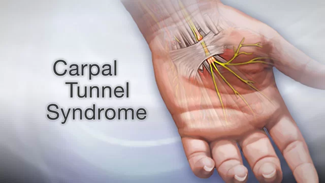
The most prevalent type of compression-related neuropathy is known as median nerve compression, and the infamous term for this ailment is carpal tunnel syndrome.
It is mainly brought on by occupational habits, including frequent wrist flexion and extension, which misuse the anatomical structures involved in these actions directly.
Consequently, the median nerve experiences compression from an increase in content mass and pressure within the carpal tunnel, causing sensory and motor abnormalities within its innervation.
Prolonged compression can cause the thenar muscles to atrophy and result in permanent nerve injury, which can cause weakness in the thumb and index finger. Resting the wrist and not using it for a while is part of the treatment.
In severe situations, anti-inflammatory medications and painkillers may be recommended in addition to bandages and splints to assist in stabilising the affected area.
Carpal Avascular Necrosis (Kienbock’s Disease):
A disorder in which the carpal bone cells sustain significant damage before dying as a result of a lack of blood flow. This degenerating condition most commonly affects the lunate and scaphoid.
Fracture of the scaphoid bone:
The scaphoid bone fracture is the most frequent type of carpal fracture. Usually, it happens when someone trips over an outstretched hand while attempting to stop the incident. The anatomical snuffbox is the area where discomfort and oedema are present.
The palmar carpal branch of the radial artery supplies insufficient blood to the affected area, which leads to complications.
Avascular osteonecrosis may result from this inadequate blood supply and the accompanying nutrient shortage.
Infections in the joints, overuse injuries, arthritis, and torn ligaments are some disorders that can affect the wrist.
Keep in mind
The following is a helpful mnemonic to help you recall the position of the carpal bones in the proximal row and subsequently the distal row, going from lateral to medial:
She looks Too Pretty Try To Catch Her:
- Scaphoid
- Lunate
- Triquetral
- Pisiform
- Trapezium
- Trapezoid
- Capitate
- Hamate
FAQs
What are carpal bones?
Together with the distal extremities of the radius and ulna, the carpal bones are a set of small bones in the human hand that constitute the wrist. They are therefore also referred to as wrist bones. Combined, they are referred to as the carpus, and they individually articulate with the metacarpals, the long bones of the wrist, and the ulna and radius of the lower arm.
How many bones are in the human wrist?
The human wrist consists of eight bones, each of which is called based on its shape: Out of all the carpal bones, the capitate is the largest. Every carpal bone plays a crucial role in the formation of the carpus, or wrist joint, which is essential for hand movement and enables us to perform a wide range of tasks, including eating, writing, and holding objects in our hands.
How many digits does a carpus have?
Even among tetrapods that maintain their entire set of five digits, there are significant differences in the structure of the carpus between other taxa. The carpus of primitive fossil frogs, like Eryops, is made up of three rows of bones: a distal row with five bones, a second row with four bones, and a proximal row with three carpals.
Which is the largest carpal bone?
Out of all the carpal bones, the capitate bone is the largest. It primarily articulates with the third metacarpal bone in a distal manner. It also articulates with the trapezoid, scaphoid, lunate, and hamate, the surrounding carpal bones.
Which is the smallest carpal bone?
Trapezoid bone
In tetrapods-including the trapezoid bone, also known as the smaller multangular bone, is a carpal bone. The distal row of carpal bones that give the hand’s palm its structure, is the smallest bone.
What is the main function of carpal bones?
Four carpals in each row make up a total of eight carpals. The wrist can rotate and move up and down thanks to the carpal bones. The carpal bones are necessary for wrist mobility. They allow the wrist to move up and down vertically as well as rotate.
Reference
- Carpal Bones (Wrist Bones): Definition, Names, Anatomy, Diagram. (2022, August 24). TheSkeletalSystem.net. https://www.theskeletalsystem.net/carpal-bones
- Agarwal, A. (2018, April 6). Carpal Bones (Wrist Bones) – Anatomy, Structure and FAQs. Human Anatomy. https://www.knowyourbody.net/carpal-bones.html
- Carpal bones. (2023, September 20). Wikipedia. https://en.wikipedia.org/wiki/Carpal_bones
- Grujičić, R. (2023, October 30). Carpal bones. Kenhub. https://www.kenhub.com/en/library/anatomy/carpal-bones