
Posterior Interosseous Nerve (PIN)
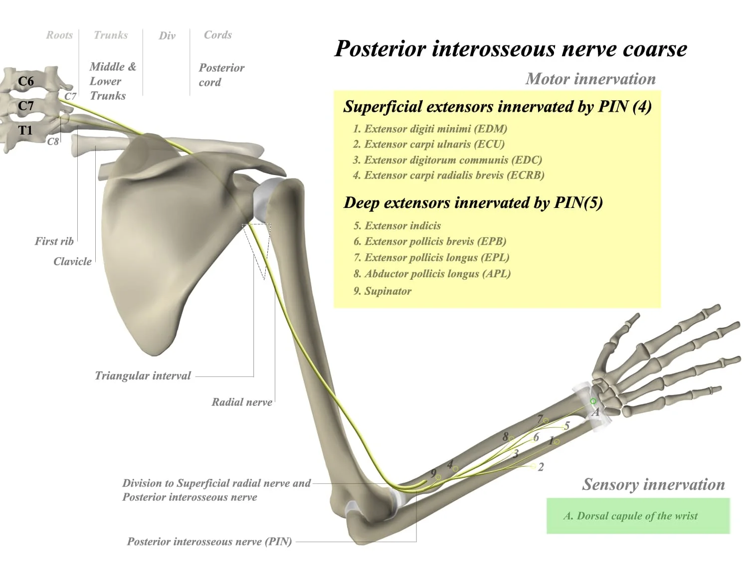
The posterior interosseous nerve (PIN) is a branch of the radial nerve, a major nerve in the upper limb. It is an important nerve that supplies several muscles in the forearm, playing a crucial role in hand and wrist movements.
After crossing the supinator muscle, it is the deep branch of the radial nerve that continues. When compared to the deep branch of the radial nerve, it is significantly smaller. The spinal column’s cervical segments C7 and C8 are where the nerve fibres start.
Structure
Course
The deep branch of the radial nerve enters the supinator muscle and innervates it before spiraling laterally around the radial head to become the posterior interosseous nerve. The arcade of Frosche (supinator arch), a fibrous arch at the superior portion of the supinator muscle, is where the posterior interosseous nerve is said to have its formal start.
The nerve finally reaches the interosseous membrane after traveling in a fascial plane between the superficial and deep extensor muscles. All of the muscles in the forearm’s posterior compartment receive motor innervation from the posterior interosseous branch along the way, and it also sends sensory fibres to the dorsal portion of the carpus.
Nerve Supply to Muscles
All of the muscles in the posterior compartment of the forearm, with the exception of the anconeus, brachioradialis, and extensor carpi radialis longus muscles, are supplied by the posterior interosseous nerve.
The primary function of the posterior interosseous nerve is to provide motor innervation to the deep muscles of the forearm. These muscles include:
Superficial
- extensor carpi radialis brevis (ECRB)
- extensor digiti minimi (EDM)
- extensor carpi ulnaris (ECU)
- extensor digitorum
Deep
- Supinator: deep branch of radial nerve
- extensor indicis
- abductor pollicis longus (APL)
- extensor pollicis longus (EPL)
- extensor pollicis brevis (EPB)
The distal radioulnar articulation’s joint capsule receives proprioception from the posterior interosseous nerve but not the sensation of pain.
Relations
The posterior interosseous artery, which similarly travels along the posterior side of the interosseous membrane, is located near to the posterior interosseous nerve. The main artery supplying the middle third of the forearm’s posterior compartment is called the posterior interosseous artery. The anterior interosseous artery, which pierces the interosseous membrane at the proximal aspect of the pronator quadratus muscle, eventually replaces the posterior interosseous artery.
Variation in Anatomy
The posterior interosseous nerve can occasionally leave the supinator muscle in two places: under its distal border and distal to the articular surface of the radial head. In the posterior compartment, the nerve’s two pathways merge and travel through the superficial and deep muscle layers.
Clinical significance
The arcade of Frohse, a component of the supinator muscle, may entrap the posterior interosseous nerve. The proximal head of the radius bone dislocating in a Monteggia fracture might damage this nerve.
The only motor syndrome posterior interosseous neuropathy causes finger drop because the IP joints do not extend and radial wrist deviation during extension.
Summary
In summary, the posterior interosseous nerve is a branch of the radial nerve that passes through the forearm along with the posterior interosseous artery. It provides motor innervation to the deep muscles of the posterior compartment of the forearm, facilitating various hand and wrist movements. Any damage or injury to this nerve can lead to weakness in the affected muscles and impairments in specific hand and wrist functions.

3 Comments