Coxa Magna
What is Coxa Magna?
Coxa magna refers to a medical condition characterized by an abnormal enlargement of the femoral head, which is the ball-shaped top portion of the thigh bone (femur) that fits into the hip socket (acetabulum). This condition can result from various underlying factors, including developmental abnormalities, genetic predisposition, trauma, or certain medical conditions.
Coxa magna can lead to hip pain, limited range of motion, and other complications, necessitating medical evaluation and potentially surgical intervention to manage the condition and alleviate symptoms. Treatment options vary based on the severity of the enlargement and its underlying cause, to improve joint function and minimize discomfort.
Although there are many different definitions in the literature, a suitable cut-off for diagnosis is enlargement with asymmetry >10% in size. Due to a variety of causes, it could be present from birth or only manifest later in life.
This especially impacts overweight males between the ages of 8 and 15 alone.
The term “coxa magna” is used in medicine to denote an enlargement or hypertrophy of the neck and femoral head of the hip joint. The Latin word “Magna” implies huge or great, while “Coxa” alludes to the hip.
Coxa magna can develop later in life as a result of some events or it can arise as a congenital disorder, meaning it is present at birth. Various anatomical or developmental disorders of the hip joint, such as hip dysplasia or slipping capital femoral epiphysis (SCFE), are frequently linked to it.
Mechanical problems with the hip joint may result from coxa magna patients’ bigger femoral head and neck. This may lead to hip instability, hip pain, and reduced range of motion. Degenerative joint illnesses like osteoarthritis may thus become more probable as people age.
The condition known as coxa magna occurs when the head is larger than it is on the often unaffected side. It is a classic Legg-Calve-Perthes sequela and represents the configuration of a repair process that is somewhat hyperactive but inadequately managed. Coxa Magna may only occur as a result of an excessive amount of repair because the cartilage model develops larger than anticipated.
Through the diffusion of nutrients from the synovial fluid, the articular and surrounding epiphyseal cartilage in the superior weight-bearing side of the skull continues to expand. The synovial fluid does not appear to be reduced by the pathoanatomic process; rather, it appears that the normal synovial diffusion nutritional source is continued and supplemented by stimulation from the larger mass of tissue produced than would have otherwise been the case from the extra blood supply that was delivered into the hip area during the repair process.
Coxa magna can come in two different forms, one with no long-term implications and the other with more serious long-term effects. When the femoral head and acetabulum connection is stable and does not lead to long-term issues, the development of the acetabulum may coincide with that of the femoral head. The coxa magna should connect with a still spherical head. Long-term issues arise when the femoral head’s growth interferes with the acetabulum’s growth, causing the head to be larger than the acetabulum and malformed so that it improperly joins to the acetabulum. The size and shape of the cartilage model of the femoral head can be measured by arthrography / magnetic resonance imaging (MRI).
Cause of Coxa Magna
coxa magna is caused by a reduced blood supply to the femoral head but the reason for this is unknown. Insufficient blood flow prevents the bone from receiving adequate oxygen and nutrients, which causes the bone cells to degenerate. This causes the femoral head to become flattened rather than rounded, which may cause it to fit improperly inside the hip socket and impair hip motion another related diagnosis are:
- Legg-Calve-Perthes disease
- transient synovitis of the hip,
- developmental dysplasia of the hip,
- trauma,
- Septic arthritis/osteomyelitis
- Juvenile rheumatoid arthritis
- chronic slipped capital femoral epiphysis.
Symptoms:
The following list of symptoms could be present and usually appear gradually:
- Limping that could progressively get worse over a few weeks.
- discomfort around the hip and groin.
- The knee or thigh may also be involved.
- Stiffness and a decreased range of motion in the hip that is affected.
- Due to pain and inactivity, the muscles in the affected area may weaken over time.
- Eventually, the damaged leg could grow shorter than the other leg.
The symptoms include hip discomfort, a constrained range of motion, instability, and a pronounced limp. Hip pain Individuals with coxa magna may experience persistent/ intermittent pain in the hip joint. The pain could be dull /sharp and may worsen with activity.
Limited range of motion: The normal range of motion of the hip can be restricted by an enlarged femoral head and neck, making movements like walking, jogging, or bending challenging.
Hip instability: Coxa magna can result in hip instability, which causes a “giving way” or unsteady-feeling hip joint when moving.
Limping: People with coxa magna may start to walk or run with a pronounced limp as a result of their discomfort and limited mobility.
Diagnosis:
Diagnosis involves physical examination and imaging tests.
Physical examination: A healthcare provider will search for symptoms of enlargement, restricted range of motion, instability, and any accompanying pain or discomfort in the hip joint.
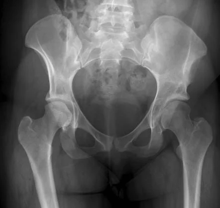
Imaging tests: X-rays, MRIs, or computed tomography (CT) scans can produce fine-grained pictures of the hip joint, indicating the degree of femoral head and neck enlargement as well as, as well as any associated structural abnormalities.
Treatment of Coxa Magna
Non-surgical options include using aids like crutches or canes, drug-assisted pain management, activity modification, physical therapy hip strengthening, and flexibility exercises…
Physiotherapy treatment
Physiotherapy Treatment for coxa magna will promote healing and ensure that the femoral head is positioned well in the hip socket as it heals and regrows. To retain the femoral head in a proper position, some youngsters receive treatment with plaster casts or specialized braces that must be worn for several months. In some circumstances, surgery may be an additional choice that can enhance the hip joint’s form and functionality and offer stability for the femoral head in the hip socket.
Conventional physiotherapy for coxa Magna
Most children with coxa magna disease are treated with physiotherapy, exercises, and hydrotherapy. specialist rehabilitation programs for children with coxa magna( Perthes disease) to enhance the movement of the joint, increase muscle strength, and keep the femoral head in an optimal position in the hip socket. Physiotherapy treatment will be tailored to your child and will aim to:
Improve range of movement in the hip
Increase muscle strength in the muscles in the leg and around the hip
Reduce pain
Facilitate mobility and independence with everyday tasks
To evaluate your child’s progress and make sure that their treatment objectives are satisfied. Exercises to strengthen the quadriceps, hamstrings, and glutes are among the physiotherapy treatments available at Stretching exercises to increase the flexibility of the hip muscles
Education of a normal (walking) gait pattern with gait training
Activities to improve balance and coordination
How to add low-impact exercises like walking and stationary cycling into your child’s daily routine.
Hydrotherapy helps maximize mobility by improving the range of movement and muscle strength.
Surgical Treatment
Surgery may be necessary in patients with severe symptoms or those with lingering symptoms. Hip osteotomy, which includes reshaping or realigning the hip joint, as well as complete hip replacement, which involves replacing the damaged joint with artificial components, are also possible procedures.
It’s crucial to remember that each patient’s unique circumstances, including age, general health, the underlying cause of coxa magna, and the severity of symptoms, will determine the exact treatment strategy.
Physiotherapy following surgery for coxa Magna
Following hip surgery, physical therapy to increase muscular strength and flexibility and assist your child in quickly regaining ambulation (walking) and function. Physiotherapy can speed up your child’s recovery and help them get back to moving around and being independent right away.
Your child’s range of motion, muscle strength, and capability with routine functional tasks will all be evaluated during the initial visit with your physiotherapist.
Following this evaluation, your child’s rehabilitation goals will be created and may include the following:
- reducing discomfort and swelling
- Restoring and maintaining a full range of movement at the hip joint
- Increasing muscle strength
- Improving mobility
- Increasing fitness and endurance
- Promoting independence
- Improving quality of life
A comprehensive treatment program will be developed to ensure that your child recovers as much as possible and reaches their full potential. Both the use of ice and ultrasonography are options in this program. This could be used in the first few weeks after your child’s procedure to lessen any discomfort and swelling and to encourage healing.
Teach your child how to mobilize safely and effectively using assistive devices such as crutches.
Promoting independence with transfers such as getting in and out of bed, and getting up and down the stairs.
Improve muscular strength and regain hip range of motion with a graduated workout program combining active and passive movement.
Activities to enhance your child’s coordination and balance
Increased mobility, pain relief, and muscular relaxation are all benefits of hydrotherapy.
Your child will receive physiotherapy care that is customized to their needs and conditions. Our mobile physiotherapy clinic’s, pediatric physiotherapist specialists have experience treating kids with hip issues.
Treatment is conservative measures or surgical intervention. Exercise, is suited to each person’s needs, and may include low-impact aerobic workouts, strengthening exercises, and range-of-motion exercises. For individualized advice, speaking with a healthcare expert is necessary.
FAQs
Why does Coxa Magna occur?
Between the ages of two and eight, the capital femur epiphysis experienced relative hyperemia, which is thought to be the cause of coxa magna. Reactive hyperemia brought on by synovitis causes the metaphysis and epiphysis peripheral cartilage to grow.
What causes hip coxa magna deformity?
The asymmetrical growth or distortion of the femoral head and neck is known as coxa magna. Although there are many different definitions in the literature, a suitable cut-off for diagnosis is enlargement with asymmetry >10% in size.
What distinguishes the Magna from the Coxa Plana?
Coxa plana, or the vertical femoral head (osteochondrosis), is an inflamed or inflamed femoral head, respectively, of the capitular epiphysis of the femur.
What is the largest part of the coxa?
The ilium, which forms the “wing” that is the most noticeable element of the coxal bone, is the most superior portion in a fully fused coxal bone. The iliac fossa refers to the side of this “wing” that faces inward. The sacrum joins each coxal bone at the ileum, completing the pelvic bowl.
What differentiates Coxa Magna from Coxa Breva?
Coxa breva: An early closure of the epiphysis that results in a short femoral neck and a small femoral head.expanded femoral head: coxa magna. Coxa plana, commonly known as Legg-Perthes, is a flattened femoral head (osteochondrosis) of the capitular epiphysis of the femur.
.