Dorsal interossei muscles of the foot
What are the Dorsal interossei muscles of the foot?
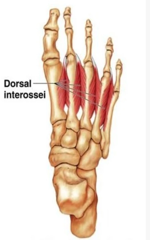
The four feather-like muscles in the center of the sole are called dorsal interossei. Dorsal interossei can be grouped because the plantar foot muscles can be isolated into layers (superficial to deep) or groups (medial to lateral).
The central plantar muscles, alongside the flexor digitorum brevis, quadratus plantae, lumbricals, and plantar interossei
The fourth (and deepest) layer of the plantar foot muscles, along with the plantar and dorsal interossei of the foot
Dorsal interossei are counted from one to four in the transverse plane, with the first being the most medial and the fourth being lateral. These muscles are known as bipennate muscles, and that implies that they comprise two muscle bellies that mix toward the centrally situated tendon. Toe flexion and abduction at the metatarsophalangeal joints and toe extension at the interphalangeal joints are the works of the dorsal interossei.
Origin
The origins of the dorsal interossei muscles of the foot vary slightly depending on the muscle. Here are the specific origins for each muscle:
- Dorsal Interosseous I: This muscle originates from the medial side of the first metatarsal bone.
- Dorsal Interosseous II: This muscle originates from the medial side of the second metatarsal bone.
- Dorsal Interosseous III: This muscle originates from the lateral side of the second metatarsal bone.
- Dorsal Interosseous IV: This muscle originates from the lateral side of the third metatarsal bone.
Insertion
The insertions of the dorsal interossei muscles of the foot are as follows:
- Dorsal Interosseous I: This muscle inserts into the medial side of the proximal phalanx of the first toe (hallux).
- Dorsal Interosseous II: This muscle inserts into the lateral side of the proximal phalanx of the second toe.
- Dorsal Interosseous III: This muscle inserts into the medial side of the proximal phalanx of the third toe.
- Dorsal Interosseous IV: This muscle inserts into the medial side of the proximal phalanx of the fourth toe.
In summary, the dorsal interossei muscles of the foot insert onto the medial or lateral sides of the proximal phalanges of the respective toes they correspond to.
Relations
The angular spaces in the proximal portions of intermetatarsal intervals are bound by the heads of the dorsal interossei, which converge in an anteromedial fashion toward one another. These openings between the tops of the second to fourth muscles act as paths for posterior perforating arteries en route to the sole, while the comparing space between the heads of the first dorsal interosseus muscle sends the dorsalis pedis artery.
Innervation
The lateral plantar nerve (S2-S3), a tibial nerve branch, innervates the foot’s dorsal interossei muscles.
Blood supply
Numerous tiny arteries in the foot supply the dorsal interossei muscles with their vascularization.
Anterior tibial artery, through dorsalis pedis and dorsal metatarsal arteries
Posterior tibial artery, through lateral plantar and plantar metatarsal arteries
Functions
Even though they are small, the strong muscles in the dorsal interossei work together with the plantar interossei to flex the three lateral toes at the metatarsophalangeal joints. Because it positions the toes so that the flexor digitorum longus and brevis can perform their activity while jumping and running, this action is crucial.
Dorsal interossei assist the extensor digitorum longus and extensor digitorum brevis muscles in toe extension at the interphalangeal joints of the second, third, and fourth toes in addition to being involved in flexion. Even though it is of less significance, it is worth focusing on that these muscles likewise abduct similar three toes at metatarsophalangeal joints. By supporting the anterior metatarsal as well as the medial and lateral arches of the foot when running, walking, and jumping, the dorsal interossei muscles also contribute to the stability of the foot.
Clinical relevance
Even though the dorsal interossei are a small set of muscles, they are essential for maintaining balance while standing and walking. On account of walking or running barefoot for an unusually significant stretch, these muscles can support harm due to over-burdening. When standing, walking, or exercising in footwear that does not adequately support the foot’s arches, this may also occur. Metatarsalgia, or sharp ache while coming down on the foot (for example in standing up or walking), situated on the distal instep in the middle of between the metatarsal bones can be a marker for at least one stressed dorsal interossei muscles.
The M. interosseus dorsalis pedis IV is an effective open muscle and is probably going to be utilized for the routine electromyographic conclusion of neurological sicknesses: Unusual [spontaneous activity] in [fourth dorsal interosseus pedis] corresponds well with the generally neurologic condition, and it could be a valuable muscle to remember for routine electrodiagnostic assessment.
Assessment
An Oxford scale muscle strength evaluation should be possible as follows for the scales of 0/1 and 2/3. Resistance tests are usually skipped because the interossei dorsales pedis can rarely be activated voluntarily.
0/1: supine position, the knee is in a supported bend. The patient is instructed to spread his or her toes while the therapist holds the foot in a neutral position.
2/3: the knee is supported in flexion when supine. The therapist instructs the patient to try to spread her or his toes while holding the foot in a neutral position and observing for minute toe movements.
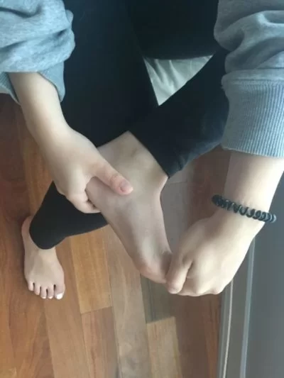
Dorsal interossei muscle stretching
Toe scrunch
Pull or curl your toes under your foot while seated. Repeat this three times after holding this position for twenty seconds.
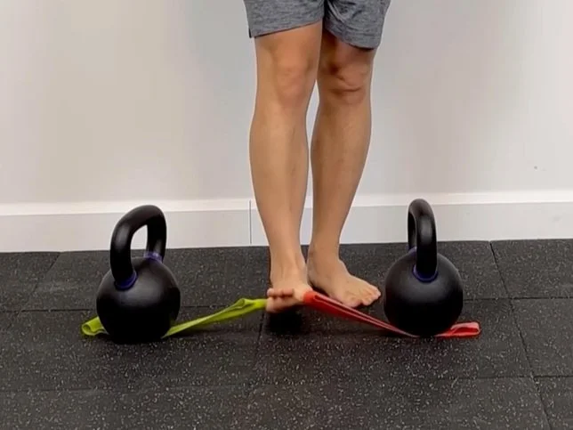
Dorsal interossei muscle strengthening
Toe Extension
Put a band of rubber around your toes. Try to separate each toe as you stretch your toes and hold for five seconds. For each foot, release and repeat five times.
Assisted toe spread
A band is used in this exercise to help stretch the adductors of the big and pinky toes. To get started, attach a band to a stable object inside the foot by looping it between the big and second toes. Circle one more band around the pinky toe and fourth toe and anchor down to a steady item beyond your foot. Place the foot on the floor after allowing the band to pull the big and pinky toes into the abduction. To stretch from a variety of angles, slowly lift the forefoot off the ground and back down.
FAQ
What is the primary movement of the dorsal interossei?
A group of paired intrinsic hand muscles that are arranged between the metacarpals are the dorsal interosseous muscles. The fingers are held in place by four muscles in the dorsal area. The dorsal interossei moreover aid the flexion of the metacarpophalangeal joints and extension of the interphalangeal joints.
What is the function of the dorsal interossei of the foot?
Description. Four bicephalic muscles in the shape of feathers make up the dorsal interossei muscles, which occupy the space in the foot between the metatarsal bones. Their primary functions are to spread the toes apart and flex the metatarsophalangeal joints of the second to the fifth toe.
What is the function of palmar interossei vs dorsal interossei?
Function. The palmar interosseous muscles adduct the fingers toward the middle finger. The dorsal interossei, on the other hand, adducts the fingers ( adduction ) away from the middle finger.
What is the first dorsal interossei muscle?
Dorsal Interossei
The first dorsal interosseous muscle enters the lateral base of the second phalanx and the extensor hood of the second digit from the surfaces of the first and second metacarpal bones that are adjacent to it.
What Innervates the dorsal interosseous of the foot?
The lateral plantar nerve innervates all dorsal interossei (S2–3). The superficial branch innervates those in the fourth interosseous space, while the deep branch innervates the other. The lateral branch of the deep fibular nerve also supplies innervation to the first and second dorsal interossei muscles.