
Masseter muscle
Introduction
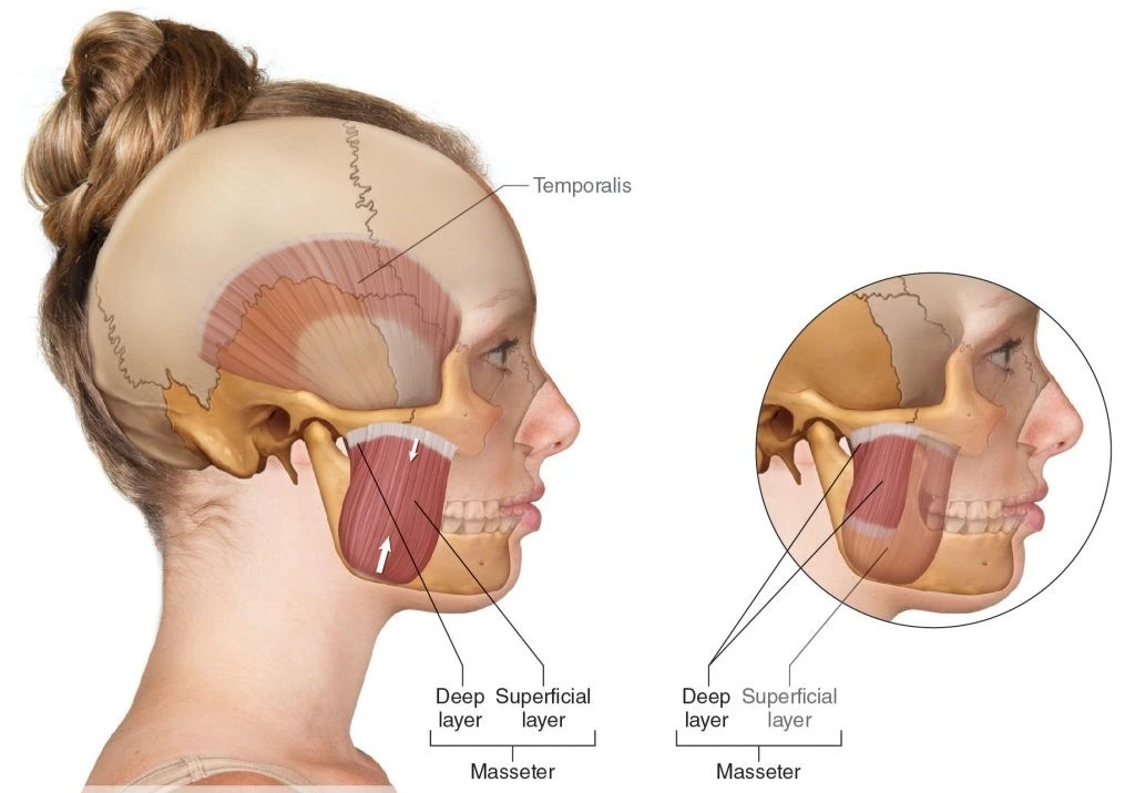
In human anatomy, the masseter muscle is one of the muscles of mastication. Found exclusively in mammals, it is particularly powerful in herbivores to encourage the chewing of plant matter. The most obvious muscle of mastication is the masseter muscle since it is the most superficial and one of the most powerful.
Origin of Masseter muscle
The masseter is a thick, relatively quadrilateral muscle, consisting of three heads, superficial, deep, and coronoid. The fibers of superficial and deep heads are continued at their insertion.
Superficial head
The superficial head, the larger, arises by a thick and tendinous aponeurosis from the temporal process of the zygomatic bone, and the anterior two-thirds of the inferior boundary of the zygomatic arch. Its fibers run inferior and posterior, to be inserted into the angle of the mandible and the inferior half of the lateral exterior of the ramus of the mandible.
Deep head
The deep head is extensively smaller and more muscular in texture. It originates from the posterior third of the lower border and the total of the medial surface of the zygomatic arch. Its fibers pass downward and forward, to be inserted into the upper half of the ramus as high as the coronoid process of the mandible bone. The deep head of the muscle is partially concealed, anteriorly, by the superficial portion. Posteriorly, it is also covered by the parotid gland.
Coronoid head
The coronoid head of the masseter’s tendon and its muscle fibers run posterolaterally from the coronoid process of the mandible towards the posterior third of the zygomatic arch. Its function is supposed to be the retraction of the mandible and the stabilization of the mandibular coronoid process.
Insertion of Masseter muscle
The superficial fibers run downwards and backward at an angle of 45 degrees. The superficial fibers are inserted into the lower portion of the lateral surface of the ramus of the mandible. The middle and also deep fibers pass vertically downwards. the middle fibers are inserted into the middle part of the ramus, and the deep fibers into the upper portion of the ramus and at the coronoid process.
Relation
The whole superficial (lateral) aspect of the muscle is covered with thin but very strong masseteric fascia.
Also on the superficial side, below the zygomatic process, the duct of the parotid gland gives across the surface of the muscle. This duct routes mostly over the anterior part of the lateral aspect, i.e. it is found anterolaterally.
The superficial portion of the gland itself is correspondingly located superficially to the masseter, but a bit additional posteriorly, i.e. posterolaterally to the muscle. Along with the parotid gland, a few more additional structures can be located over the lateral side of the muscle:
Terminal branches of the facial nerve, Facial vein, Facial artery, Risorius muscle, and Zygomaticus major muscle. The medial, or inner, surface of the muscle is mostly related ramus of the mandible almost entirely covering its superficial surface.
The medial characteristic of the masseter muscle forms the lateral wall of the facial area called the sub-masseteric space. It is a paired conceivable space between the lateral aspect of the mandible and the medial aspect of the masseter muscle.
Located deep to the posterior part of the inner aspect of the muscle, or simply posteromedially, the temporal muscle is seen.
Anterior to the muscles, the buccinator muscle is located. The overhead mentioned duct of the parotid gland penetrates the fibers of the buccinator muscle to find its way to the interior of the oral cavity and finally open onto the inner side of the cheek.
The posterior element of the muscle is located anteriorly to the deep part of the parotid gland. This practically signifies that the parotid gland surrounds the entire posterior and most of the superficial aspects of the muscle.
Nerve supply
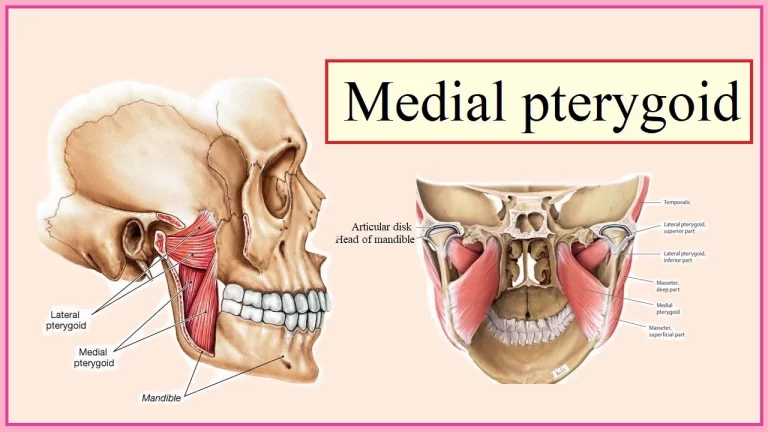
Along with the additional three muscles of mastication (temporalis, medial pterygoid, and lateral pterygoid), the masseter is innervated by the anterior division of the mandibular branch (V3) of the trigeminal nerve. The innervation course is gyrus precentralis > genu Capsula interna > nucleus motorius nervi trigeminal> nervus trigeminus > nervus mandibular> musculus masseter.
Blood supply
The masseter muscle is supplied with blood by the masseteric artery, it is a branch of the maxillary artery.
Function of Masseter muscle
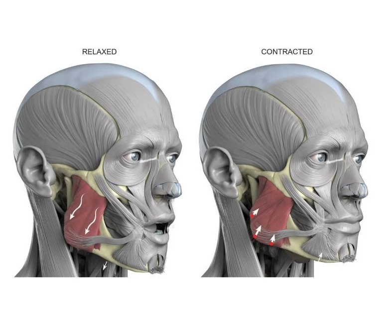
The action of the muscles during bilateral contraction of the entire muscle is to elevate the mandible, raising the lower jaw. Elevation of the mandible appears during the closing of the jaws. The masseter parallels the medial pterygoid muscles, but it is stronger and superficial fibers can induce protrusion.
Clinical significance
Examination
To perform an extraoral assessment, stand around the patient and visually examine and bilaterally palpate the muscle. Place the fingers of a separate hand over the muscle and ask the patient to clench his or her teeth for several duration.
Pathology
The masseter muscle can evolve enlarged in patients who habitually clench or grind (with bruxism) their teeth and exactly in those who continually chew gum. This masseteric hypertrophy is asymptomatic and soft; it is usually bilateral but can be also unilateral. Even if the hypertrophy is bilateral, asymmetry of the face may still happen due to unequal enlargement of the muscles. This extraoral enlargement may be mistaken for parotid salivary gland disease, dental infections, and maxillofacial neoplasms. However, no other signs are present excluding those involved in changes in occlusion intraorally such as pain, and the enlargement corresponds with the overview of the muscle. Most patients desire medical attention because of statements about facial appearance, and this problem may be associated with further pathology of the temporomandibular joint.
Finally, the muscle experiences spasm with malignant hyperthermia as do other skeletal muscles, but this one is easily noted since it occurs on the face.
Singers usually experience various kinds of masseter tension, which is often treated with transdermal massages or stretches as a vocal warm-up.
Masseter Muscle Rigidity
Masseter muscle rigidity (MMR), also understood as “jaws of steel,” happens following a dose of succinylcholine and is defined as limb muscle flaccidity with jaw muscle tightness. There is a varying presentation from entire trismus and severe spasticity to a tight jaw response, which becomes concerning as the patient is incapable of being adequately intubated, leading to an increased chance of malignant hyperthermia in the patient.
Masseter Reflex
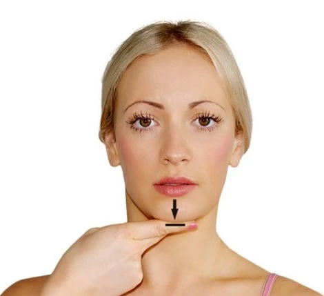
The masseter reflex, also known as the mandibular reflex or jaw-jerk reflex, implicates opening the mouth and placing a finger on the chin of the patient, and striking the finger with a reflex hammer. The downward force can provoke a reflex action in the masseter muscles resulting in an elevation of the mandible. The normal response is very small to no reflex activity, but with a brainstem lesion affecting an upper motor neuron lesion, hyperreflexia of the masseteric reflex is possible.[8]
Tetanus Toxin
Tetanus toxin arrives from the gram-positive spore-forming bacteria Clostridium tetani. Tetanus toxin penetrates the body, binds to peripheral neurons, and is transported retrogradely to the spinal cord or brainstem. Once in the central nervous system tetanus toxin hampers the vesicular release of inhibitory neurotransmitters, glycine. This disinhibition of lower motor neurons leads to sustained contractures and muscle rigidity including opisthotonus, respiratory failure, trismus, and also dysphagia. Trismus is muscle rigidity and spasms primarily of the masseter and temporalis muscle leading to an incapability to open the mouth and a smile characteristically called a risus sardonicus.
Treatment
Masseter Muscle Exercise
Some of these strategies can contain masseter muscle exercises such as stretching and massage.
Some of these activities may be performed by a physical therapist or given to you to do at home. Kaiser Permanente suggests some simple exercises to do on your own. Below are three to try for an at-home masseter muscle exercise:
Move 1: Tongue Rest Position
With your jaw relaxed and teeth negligibly apart, place your tongue on the roof of your mouth in a resting position.”Cluck” your tongue while maintaining your teeth slightly apart.
Replicate six times, up to six sessions per day.
Move 2: Controlled Opening
With your tongue in a resting position at the roof of your mouth, place the index fingers of separate hands over your temporomandibular joints. Without allowing your tongue to leave the roof of your mouth, open your jaw as far as you can. Inspect in the mirror for your jaw deviating to one side or the other. Try this while chewing, yawning, and also biting. Replicate six times, up to six sessions per day.
Move 3: Head Flexion and Head Extension
Intertwine the fingers of your hands behind your neck. Keeping your head in an upright position, shake your head forward while tucking in your chin. Replicate six times, up to six sessions per day.
Open and close: Begin this by resting the chin on the tip of the thumb. Open mouth while using light consistent pressure with the thumb. Persist holding the mouth open while applying pressure for ten seconds and then closing the mouth. Replicate this exercise five times every day.
Side-to-side Exercise: Place a pencil or pen in the mouth and maintain it between your teeth. Gradually move the jaw from one side to the other. Replicate this exercise ten times a day.

Stretch: To stretch the Masseter, look straight ahead and open your mouth. Place your index and middle finger against the interior of the teeth in the front of the lower jaw. Utilize your fingers to gently apply pressure downwards. Next, gradually tilt the head back to look up just slightly as you hold the downward pressure. Hold this position for 30 sections to stretch the Masseter muscles.
FAQ
Where are the masseter muscle located?
The masseter muscle courses from your cheekbone to your jaw and is the main muscle of chewing. It works with additional muscles to move and stabilize your jaw and temporomandibular joint.
What is the masseter used for?
The masseter muscle is a facial muscle that connects your lower jaw to your cheek. It is one of several muscles reliable for chewing.
Why masseter is called the strongest muscle?
The strongest muscles based on their weight are the masseter muscles. With all muscles of the jaw working concurrently, it can complete the teeth with a force as great as 55 pounds (25 kilograms) on the incisors or 200 pounds (90.7 kilograms) on the molars.
Is a masseter a head muscle?
The masseter is a thick, partially quadrilateral muscle, consisting of three heads, superficial, deep, and coronoid. The fibers of superficial and deep heads are continued at their insertion.
Does exercise reduce masseter muscle?
It naturally grows stronger when you use it, but there are a few easy exercises you can do for additional training. Regular exercise can help you gain a tighter jawline with a stronger masseter muscle.


2 Comments