Palatine Bone
Overview
Two L-shaped bones around the back of the nasal and mouth cavities are called the palatine bones. These bones are categorized as irregular and play a role in the development of the nasal cavities, viscerocranium, and bony hard palate.
The os palatinum, often known as the palatine bone, is an uneven, flat, paired facial bone. It is a component of the oral, nasal, and orbital cavities. Either the left or right maxilla bone’s processes and the single sphenoid bone are positioned between the two plates that make up each bone. There are apertures (foramina) on paired palatine bones that lead to the greater and minor palatine canals. Nerves and blood vessels can be accommodated by these canals.
Location
Running the tongue toward the rear of the throat will reveal part of the position of the palatine bones; this is the palatine surface of the horizontal plate, which forms the back portion of the hard palate (palate – palatine). Stopping just before the hard and soft tissue meet.
The placement of the palatine bone base is also indicated by the tops of the palatine tonsils, which are lymphoid tissue masses on either side of the back of the throat.
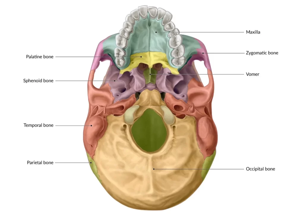
Between the maxillae and the pterygoid process of the sphenoid bone is the paired palatine bone. It takes a role in the development of the nasal cavity, orbits, and oral cavity—the three cavities that make up the skull. Five bones—the maxilla, sphenoid, ethmoid, inferior nasal concha, and vomer—articulate to do this.
An L-shape is formed by each palatine bone in the face. The horizontal lines (the horizontal plates) of the two Ls meet at the point where one bone is a mirror copy of the other. Each horizontal plate’s front segment articulates with a portion of the maxilla. The pharyngeal cartilage meets the posterior edge, or back, of the horizontal plates.
The vertical line of the L is the palatine bone’s perpendicular plate. The nasal conchae is behind this plate. The inferior nasal concha and sphenoid bone are the points at which the two plates articulate as they rise from the horizontal plate.
Anatomy
Two bones join to form the palatine bone. They form a L shape, mirroring each other, and come together at the intersection of their horizontal segments. This is at the median palatine suture, which runs the length of the upper mouth.
The palatine aids in forming the orbital process’s basis on the top border. The boundaries and articulations of the palatine bone, which serve as its points of connection with other structures, provide the clearest picture of its position.
The structure of palatine bones can be broadly divided into two subcategories: the horizontal plate and the perpendicular plate. It is important to note that they are paired bones, left and right.
The horizontal and perpendicular plates that make up the palatine bone are joined to produce the distinctive L-shaped bone. Three processes are present in the bone: sphenoidal, orbital, and pyramidal.
Horizontal Plate
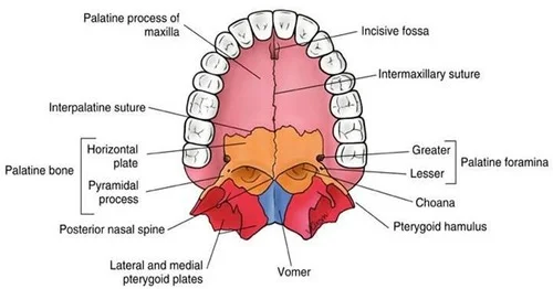
The transverse plane contains the palatine bone’s horizontal plate. It consists of a portion of the nasal cavity’s floor and the bone core of the hard palate’s posterior quarter. The plate has four borders: medial, lateral, anterior, and posterior. Its shape is quadrangular. It displays two surfaces that face the nasal and oral cavities, respectively: the palatine and nasal.
- The horizontal plate of the contralateral palatine bone articulates with the medial border of the horizontal plate. The posterior portion of the nasal crest, which articulates with the vomer, is formed by the articulating line that separates the two plates from the nasal side.
- The perpendicular plate and the horizontal plate’s lateral border are continuous. The greater palatine nerve and arteries pass through the greater palatine foramen, which is a feature of it.
- The maxilla’s palatine process and anterior border articulate. The maxilla and the palatine bone’s horizontal plate together make up the hard palate.
- The pharynx’s rear wall is faced by the posterior border. The posterior surfaces of both horizontal plates come meet at their medial ends to produce the median posterior nasal spine, a bony projection in the midline. This spine is where the uvular muscle attaches itself.
Perpendicular Plate
From the lateral edge of the horizontal plate, the palatine bone continues as a perpendicular plate. It gets its name from the fact that it makes a 90° angle with the horizontal plate, which gives the palatine bone its recognizable L-shape. The nasal and maxillary surfaces, as well as the anterior, posterior, superior, and inferior borders, make form the perpendicular plate.
Surfaces
Part of the lateral wall of the nasal cavity is formed by the perpendicular plate’s nasal surface, which faces the nasal cavity. A concavity that contributes to the inferior nasal meatus characterizes the inferior portion of this surface. The conchal crest, which articulates with the inferior nasal concha, is located above this. Part of the middle nasal meatus is located in a shallow concavity above this crest. The middle nasal concha attaches to the ethmoidal crest, which is located above this concavity.
The nasal surface of the maxilla articulates with the maxillary surface of the perpendicular place. Except for its anterior and posterosuperior regions, it is primarily uneven and rough. The anterior smooth area comprises the posterior part of the maxillary sinus’s medial wall, whereas the posterosuperior smooth area forms the pterygopalatine fossa’s medial wall.
The greater palatine groove is an oblique groove that also designates the maxillary surface. The structures that go via the larger palatine foramen are transmitted by it. The larger palatine canal, which carries the greater palatine nerve, artery, and vein, is created when the surrounding surface of the maxilla closes this groove.
Borders
The articulations between the perpendicular plate and its surrounding bones occur along its boundaries.
- At the level of the conchal crest, the anterior border exhibits a laminar projection that articulates with the inferior nasal concha and forms a portion of the maxillary sinus’s medial wall.
- The medial pterygoid plate of the sphenoid bone articulates with the serrated posterior border. The posterior border’s superior part is where the sphenoidal process continues, whereas the inferior part is where the pyramidal process continues.
- The sphenoid bone’s body and superior border articulate. It is distinguished by the sphenopalatine notch, which becomes the sphenopalatine foramen after being closed superiorly by the sphenoid body. The anterior portion of the superior border extends into the palatine bone’s orbital process, creating a foramen that connects the pterygopalatine fossa and superior nasal meatus and transmits the posterior superior nasal nerves and sphenopalatine arteries between them.
- The lateral border of the horizontal plate and the inferior border of the perpendicular plate are continuous.
Processes Of Palatine Bone
Pyramidal process
The intersection of the perpendicular and horizontal plates gives rise to the palatine bone’s pyramidal process. It passes between the lateral and medial pterygoid plates of the sphenoid bone and is orientated posterolaterally. The inferior portion of the pterygoid fossa is formed by the posterior aspect of this process articulating with the pterygoid plates.
The inferior surface has the lesser palatine foramina, which allows the lesser palatine nerves and arteries to pass through, while the lateral surface serves as the attachment point for the deep head of the medial pterygoid muscle.
Orbital process
The front portion of the perpendicular plate’s orbital edge is where the orbital process begins. It flows toward the orbit’s floor in a superior and slightly anterior direction. A thin neck, three articular surfaces, and two non-articular surfaces are shown in this procedure.
- The anterior (maxillary) surface articulates with the maxilla and is oriented laterally and anteriorly.
- The air sinus that is housed within this process and connects to the sphenoidal sinus via this hole marks the posterior (sphenoidal) surface, which faces both superiorly and posteromedially.
- Both anteriorly and medially is the path of the medial (ethmoidal) surface. It articulates with the ethmoid bone labyrinth. Occasionally, this surface—rather than the posterior surface—is the one with the air sinus opening. Here, the sphenoidal sinus is not the point of contact between the bone and the posterior ethmoidal air cells.
The superior (orbital) and lateral surfaces of the orbital process are its two non-articular surfaces. Whereas the lateral surface is part of the pterygopalatine fossa, the superior (orbital) surface makes up the floor of the bony orbit. A notch that forms the medial portion of the inferior border of the inferior orbital fissure divides these two surfaces.
Sphenoidal process
The superior portion of the posterior border of the perpendicular plate is where the sphenoidal process travels superomedially.
- Its superior surface articulates with the medial pterygoid plate root and the sphenoidal concha. Additionally, a groove that is a component of the palatovaginal canal marks this surface.
- The nasal cavity’s floor and lateral wall are partially formed by the concave anteromedial surface.
- A portion of the pterygopalatine fossa’s medial wall is contributed by the lateral surface.
There are three borders to the sphenoidal process. The medial border articulates with the vomer’s all, while the posterior border articulates with the medial pterygoid plate. The sphenopalatine notch’s posterior margin is formed by the anterior border.
Anatomical Variations
The larger palatine foramen, an aperture toward the back that permits the greater and descending palatine nerves to pass through, is the most often observed anatomical variation in the palatine bone.
According to one study, this aperture was situated across from the third upper molar teeth in about 73% of the cases. Additionally, it observed placement in between the second and third molars approximately 16% of the time, and opposite the second molar approximately 7% of the time.
Palatine Process
A section of the maxilla bone: It is a horizontal projection extending medially from the maxilla, not a distinct bone.
Forms the hard palate’s anterior 3/4: To complete the hard palate, it unites posteriorly with the palatine bone.
Comparatively easy structure: Its nasal (upper) and palatine (lower) flat surfaces are divided by a ridge.
Articulates with: Other sections of the maxilla laterally, the incisive bone anteriorly, and the palatine bone on the midline.
Palatine Bone
Independent bone: In the skull, it is a distinct pair of bones.
Forms the hard palate’s posterior 1/4: It connects anteriorly to the maxillary palatine processes.
A more intricate structure: It contributes to the walls of several cavities, including the nasal cavity, orbit, and pterygopalatine fossa, with its two horizontal and perpendicular plates and three processes (pyramidal, orbital, and sphenoidal).
Articulates with: Vomer bones, ethmoid, sphenoid, inferior nasal concha, and maxilla.
| POINTS | Palatine Process | Palatine Bone |
| Articulations | Articulates that have fewer bones, primarily the palatine and maxilla | Articulates with multiple cranial bones. |
| Location in palate | Covers the anterior 3/4 | covers the posterior 1/4 |
| Structure | More simple | More complicated with many plates and processes. |
| Relationship to skull | Part of the maxilla bone | Independent |
Function
The palatine bone primarily has structural significance; its form defines the lower wall of the cranium and aids in the carving out of significant structures within the head.
This bone aids in the formation of the roof of the mouth, the nasal and oral cavities, and the lower part of the eye sockets (orbits).
They also have holes through which the palatine nerves can flow, as was already mentioned. In this way, the palatine bones aid in housing the mouth’s and teeth’s main pain-signaling channels.
The horizontal plates of the palatine bones and the palatine process of the maxilla combine to generate the hard palate. It is the nasal cavity’s floor and the oral cavity’s roof.
The orbit bones comprise the palatine and seven other facial bones. The maxillary, palatine, and zygomatic bones make up portions of the orbit’s floor, or bottom. Examine the eye sockets beneath; the numerous tiny fissures show the locations where these distinct bones meet.
The nasal cavity collects pollutants and possible germs while also warming and humidifying the air we breathe. Although we only have one nose, the nasal canal is divided into two nasal cavities by the cartilaginous septum, which is the somewhat deflated structure seen in the accompanying photo. The nasal cavity floor is made up of the palatine process of the maxilla bone and the palatine bone’s horizontal plate.
Additionally, face muscles link to the os palatinum. Attached to the palatine bone are four muscles:
- The medial side of the pterygoid
- Uvula muscle
- Tensor Palitan
- muscle of the superior pharynx constrictor
Lastly, there are three more foramina in the facial palatine bone that permit blood vessels and nerves to pass through. The nasal cavity, hard and soft palate, and motor and sensory pathways are supplied by these nerves.
There are three foramina in a set of bones:
- Between the pterygopalatine fossa and the oral cavity, the greater palatine foramen serves as the entrance for the greater palatine nerve and blood vessels.
- Lesser palatine foramen: the opening through which the lesser palatine canal and the oral cavity are connected by the lesser palatine nerve and blood vessels.
- The sphenopalatine foramen serves as the entrance to the nasal nerves, nasopalatine nerve, and sphenopalatine artery and vein, which are located between the pterygopalatine fossa and nasal cavity. Between the pterygopalatine fossa and the oral cavity, the greater palatine foramen serves as the entrance for the greater palatine nerve and blood vessels.
- Lesser palatine foramen: the opening through which the lesser palatine canal and the oral cavity are connected by the lesser palatine nerve and blood vessels.
Situated between the pterygopalatine fossa and nasal cavity, the sphenopalatine foramen is the entrance of the nasal nerves, nasopalatine nerve, and sphenopalatine artery and vein.
Associated Conditions
The palatine bone is associated with multiple disorders. It is possible to sustain fractures and nerve damage, as well as to have bone outgrowths and congenital deformities.
Nerve Damage
Because the larger and lesser palatine nerves are known to be incredibly sensitive, the palatine bone is most frequently taken into consideration in dentistry. These nerves need to be anesthetized (numbed) for dentists to extract the upper molars and premolars.
The injection sites, which are normally located approximately 1 centimeter (cm) from the gingival edge, or the “height” of the gums, must be closely watched because there is a chance that the syringe may pierce the greater palatine foramen.
Fractures
Accidents or falls can break palatine bones. The majority of these palatal fractures, which are rather uncommon, affect adult men. Their position in the face poses a challenging issue for healthcare providers.
Depending on where the bone break occurs, there are six main forms of palatine fractures:
- Sagittal fractures
- Para sagittal fractures
- Anterior and posterior alveolar fractures
- Transverse fracture fractures
- Complex fractures
- Para alveolar fractures
Not only can these problems cause discomfort and swelling in the surrounding structures, but they can also cause the teeth and bite to become misaligned.
Cleft Palate
A congenital malformation called a cleft palate is usually identified at birth. It occurs when the palatine bone’s two halves fail to fuse during fetal development. Roughly one in 500 to 700 babies are born with a cleft palate or cleft lip.
Individuals who are born with a cleft palate may experience speech, hearing, feeding, and drinking difficulties in addition to dental issues. They may also have ear infections and colds frequently.
Torus Palatinus
The term “torus palatinus” refers to the development of palatine bone outgrowths that are often benign and painless. They can appear on one or both sides of the palate and usually start in the mid-plate.
Even though most occurrences are asymptomatic and people frequently never detect them, some do result in:
- Pain
- Oral ulcers
- Abnormal chewing
- Distorted speech
The illness known as torus palatinus typically affects people in the
Conclusion
In addition to linking to other bones, like the sphenoid, the paired palatine bone is an essential component of the orbitals, or eye sockets, nose, and skull. It allows vital blood arteries and nerves to pass through and be protected.
For some tooth extraction procedures, numbing the palatine nerves is necessary. Surgery may also be necessary to treat palatine bone fractures. One of the most prevalent palatine diseases is cleft palate, which is usually successfully repaired if detected at or before delivery.
The primary purpose of the palatine bone is structural. Speak with your healthcare practitioner if you have any unsettling symptoms or think you may have been injured so you can receive a proper diagnosis.
FAQs
What is another name for the palatine bone?
A section of the palate and nasal cavity is made up of the paired, L-shaped palatine bones (os palatinum). It is located between the sphenoid bone, whose wings aid in forming the base of the skull and the base of the eye sockets, and the maxilla bone, which is the fixed top bone of the jaw.
What is the palatine part of the maxilla?
The thick, horizontal palatine process of the maxilla is known as the palatal process in human mouth anatomy. It makes up the anterior three-quarters of the hard palate, with the palatine bone’s horizontal plate making up the remaining portion. Maxilla inferior surface.
Is palatine a facial bone?
Located above the uvula in the neck, the palatine bones (/ˈpaelətaɪn/) are two irregular bones of the face skeleton found in many animal species. They make up the hard palate along with the maxillae. (The word palate comes from the Latin palatum.)
Is the palatine bone part of the nose?
Between the sphenoid and maxillae in the rear of the nasal cavity are the palatine bones. Every bone is made up of an L-shaped plate that is perpendicular to the horizontal. The pyramidal, orbital, and sphenoidal processes are the three.
Reference
- Knapp, S. (2020, August 4). Palatine Bone – The Definitive Guide | Biology Dictionary. Biology Dictionary. https://biologydictionary.net/palatine-bone/
- Palatine Bone | Complete Anatomy. (n.d.). www.elsevier.com. https://www.elsevier.com/resources/anatomy/skeletal-system/axial-skeleton/palatine-bone/17739
- What Is the Palatine Bone? (2023, March 22). Verywell Health. https://www.verywellhealth.com/palatine-bone-anatomy-location-and-function-4707217
- Palatine bone. (n.d.). Palatine Bone: Plates, Borders, Processes, Articulations | Kenhub. https://www.kenhub.com/en/library/anatomy/the-palatine-bone
- Yahoo is part of the Yahoo family of brands. (n.d.). https://www.yahoo.com/lifestyle/palatine-bone-045142340.html?