
Wrist Joint
Introduction
The wrist is a compound joint structure that bridges the hand to the forearm. It is a group of many bones and joints. The distal portions of the radius and ulna, the carpal bones, and the five metacarpal bones make up the bones that collectively make up the wrist. The radiocarpal joint is another name for the wrist joint. It serves as a transition point between the forearm and the hand and is a condyloid synovial joint of the distal upper extremities. Condyloid joints are a type of modification of ball and socket joints that give permission for flexion, extension, abduction, and adduction motions.
The wrist joint is made up of three major joints. This gives the wrist more stability than having only one joint. It also permits your wrist and hands a wider range of motion, giving permission for so many different motions and positions.
The wrist joints let your wrist move your hand upward and downward, such as when you raise your hand for a wave. These joints give permission for rotation of the hand and bend the wrist forward and backward also side to side.
Radiocarpal joint: The radiocarpal joint is formed with the radius which is a thicker forearm bone, and connects with the baseline of the wrist bone (scaphoid, lunate, and triquetrum bones). This joint is mainly formed at the thumb side of the wrist.
Ulnocarpal joint: This joint is made up of the ulna which is the thin forearm bone, the lunate, and the triquetrum. This joint is formed at the side of the little finger of the wrist.
Distal radioulnar joint: This joint is formed in the wrist but does not involve the wrist bones. It joins the base ends of the radius and ulna.
Structure
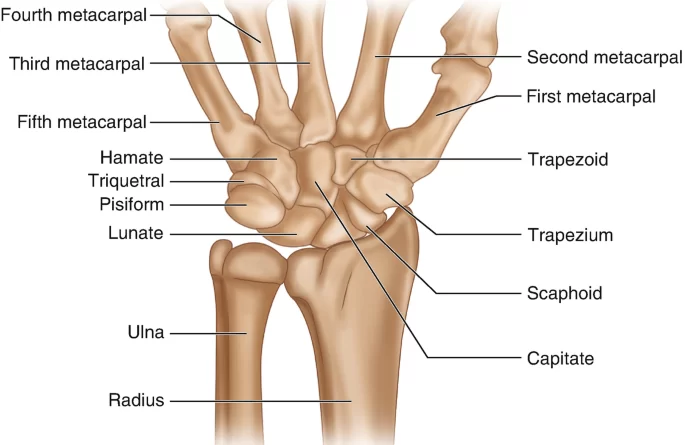
The structure of the wrist joint includes several bones, ligaments, and muscles. The distal end of the radius bone forms the primary articulation with the carpal bones. The ulna bone also contributes to the joint by forming a small articulation with the triangular fibrocartilage complex (TFCC) at the distal end of the radius. The carpal bones are divided into two rows: the distal row is made up of the trapezium, trapezoid, capitate, and hamate bone, and the proximal row is made up of scaphoid, lunate pisiform, and triquetrum bone.
Around the wrist joint, there is a double-layered joint capsule. It is very common in all synovial joints. The outer side layer is fibrous and connected to the radius, ulna, and carpal bones. The inside layer makes a synovial membrane which produces synovial fluid and lubrication of the joint.
Blood supply
The blood supply of the wrist joint is provided by several arteries, including the radial artery, ulnar artery, and interosseous arteries.
The radial artery is the main source of blood supply to the wrist joint. It enters the wrist through the anatomical snuffbox, which is a depression on the lateral aspect of the wrist. The radial artery then gives off several branches that supply the wrist joint and surrounding structures.
The ulnar artery also contributes to the blood supply of the wrist joint. It enters the wrist through Guyon’s canal, which is a tunnel formed by the pisiform bone and the hook of the hamate bone. The ulnar artery gives off several branches that supply the wrist joint and surrounding structures.
The interosseous arteries are small arteries that run between the radius and ulna bones. They provide additional blood supply to the wrist joint.
The blood supply to the wrist joint is important for maintaining the health and function of the joint.
Injuries or conditions that affect the blood supply can lead to decreased healing ability and increased risk of complications.
Movements occurring in the wrist joint

The wrist joint is a complex joint that allows for a variety of movements. The movements of the wrist joint are as mentioned below
- Flexion: This movement involves bending the wrist towards the palm of the hand. It is primarily controlled by the flexor muscles, which are located on the front side of the forearm. The flexor muscles contract, causing the wrist joint to move downwards towards the palm of the hand.
- Extension: This movement involves bending the wrist backward, away from the palm of the hand. It is primarily controlled by the extensor muscles, which are located on the back side of the forearm. The extensor muscles contract, causing the wrist joint to move upwards away from the palm of the hand.
- Abduction: This movement involves moving the wrist away from the midline of the body, towards the thumb side. It is primarily controlled by the abductor muscles, which are located on the side of the forearm closest to the thumb. The abductor’s muscles contract, causing the wrist joint to move away from the midline of the body.
- Adduction: This movement involves moving the wrist towards the midline of the body, towards the little finger side. It is primarily controlled by the adductor muscles, which are located on the side of the forearm closest to the little finger. The adductor muscles contract, causing the wrist joint to move toward the midline of the body.
- Pronation: This movement involves turning the wrist and forearm so that the palm faces downwards. It is primarily controlled by the pronator muscles, which are located on the front side of the forearm. The pronator muscles contract, causing the wrist and forearm to rotate inwards towards the body.
- Supination: This movement involves turning the wrist and forearm so that the palm faces upwards. It is primarily controlled by the supinator muscles, which are located on the back side of the forearm. The supinator muscles contract, causing the wrist and forearm to rotate outwards away from the body.
- Circumduction: This movement involves making a circular motion with the wrist joint, combining all of the above movements. It is controlled by a combination of all the muscles involved in the other movements. The wrist joint moves in a circular motion, allowing for a wide range of motion and flexibility.
Ligaments
Radial collateral ligament: This ligament runs from the styloid process of the radius to the scaphoid and trapezium bones. It provides stability to the lateral side of the wrist joint.
Ulnar collateral ligament: This ligament runs from the styloid process of the ulna to the triquetrum and pisiform bones. It provides stability to the medial side of the wrist joint.
Dorsal radiocarpal ligament: This ligament runs from the radius to the scaphoid, lunate, and triquetrum bones on the dorsal (back) side of the wrist joint. It limits the extension of the wrist.
Palmar radiocarpal ligament: This ligament runs from the radius to the scaphoid, lunate, triquetrum, and capitate bones on the palmar (front) side of the wrist joint. It limits flexion of the wrist.
Triangular fibrocartilage complex (TFCC): This is a group of ligaments and cartilage that connects the ulna to the carpal bones on the medial side of the wrist joint. It stabilizes the joint and helps distribute the load between the ulna and carpal bones.
Interosseous ligament: This ligament runs between the radius and ulna bones, helping to stabilize the wrist joint and transfer forces between the forearm and hand.
Carpometacarpal ligaments: These ligaments connect the carpal bones to the metacarpal bones of the hand, providing stability to the base of the hand.
Ulnolunate ligament: The ulnolunate ligament begins at the base of the ulnar styloid process, radially to the ulnocapitate ligament.
Its insertion point is distal to the short radiolunate ligament, on the ulnar aspect of the lunate.
Ulnocapitate ligament: The insertion of the ulnocapitate ligament is distally connected to the proximal and volar portions of the capitate. It is proximally connected at the fovea of the ulnar head.
Overall, these ligaments work together to provide stability to the wrist joint while allowing for a wide range of movements.
Muscle attachment
Flexor carpi radialis: This muscle arises from the medial side of the epicondyle of the humerus and is connected at the bottom of the second and third metacarpal bones. It flexes and abducts the wrist joint.
Flexor carpi ulnaris: This muscle originates from the medial epicondyle of the humerus and the olecranon process of the ulna, and attaches to the pisiform bone and the base of the fifth metacarpal bone. It flexes and adducts the wrist joint.
Palmaris longus: This muscle arises from the medial side of the epicondyle of the humerus and is connected to the palmar aponeurosis. It flexes the wrist joint.
Extensor carpi radialis longus: This muscle originates from the lateral epicondyle of the humerus and attaches to the base of the second metacarpal bone. This muscle does the extension and abduction of the wrist joint.
Extensor carpi radialis brevis: This muscle is derived from the lateral side of the humerus epicondyle and is connected to the base of the third metacarpal bone. It helps in the extension and abduction of the wrist joint.
Extensor carpi ulnaris: This muscle is derived from the lateral side of the humerus epicondyle and the posterior borderline of the ulna, and is connected to the base of the fifth metacarpal bone. These muscles do the extension and adduction of the wrist joint.
Flexor digitorum superficialis: This muscle originates from the medial epicondyle of the humerus, the coronoid process of the ulna, and the radius, and attaches to the middle phalanges of the four fingers. It flexes the wrist joint and the fingers.
Flexor digitorum profundus: This muscle originates from the ulna and the interosseous membrane, and attaches to the distal phalanges of the four fingers. It flexes the wrist joint and the fingers.
Extensor digitorum: This muscle originates from the lateral epicondyle of the humerus and the posterior border of the ulna, and attaches to the extensor expansion of the four fingers. It extends the wrist joint and the fingers.
Overall, these muscles work together to allow for a wide range of movements at the wrist joint, including flexion, extension, abduction, and adduction.
Tendons
Tendons are very strong, fibrous cords that help in the attachment of muscles to bones. At the wrist joint, there are several tendons that connect the forearm muscles to the hand bones, allowing for movement of the wrist and fingers.
The tendons that pass through the wrist joint include:
- Flexor tendons: This muscle originates from the medial aspect of the humeral epicondyle and attaches to both the second and third metacarpal bones at the base. The wrist joint flexes and is abducted by it.
- Extensor tendons: These tendons also originate from the forearm muscles and attach to the finger bones. They are responsible for straightening the fingers and wrist.
- Radial and ulnar deviation tendons: These tendons run along the sides of the wrist and are responsible for moving the wrist from side to side.
- Thumb tendons: There are several tendons that control the movement of the thumb, including the flexor pollicis longus, extensor pollicis longus, and abductor pollicis longus.
Injuries or conditions that affect the tendons at the wrist joint can cause pain, swelling, and a limited range of motion. Common conditions include tendonitis (inflammation of the tendons), tenosynovitis (inflammation of the tendon sheath), and carpal tunnel syndrome (compression of the median nerve as it passes through the wrist). Treatment may involve rest, physical therapy, and in major conditions surgery.
Conditions occur at the wrist joint

- Carpal tunnel syndrome: This condition is caused by compression of the median nerve, which runs through the carpal tunnel in the wrist. It can be caused by repetitive motions, such as typing or using a computer mouse, or by conditions that cause swelling in the wrist, such as pregnancy or arthritis Symptoms of carpal tunnel syndrome include pain, numbness, and tingling in the hand and fingers, especially at night. Treatment may include wearing a wrist splint, taking anti-inflammatory medications, or undergoing surgery to relieve pressure on the nerve.
- Wrist sprains and strains: These injuries occur when the ligaments or muscles in the wrist are stretched or torn. They can be caused by sudden impacts, such as a fall or sports injury, or by overuse from repetitive motions. Symptoms of a wrist sprain or strain include pain, swelling, and a limited range of carpal tunnel syndrome motion. Treatment may include rest, ice, compression, and elevation (RICE), physical therapy, or surgery in severe cases.
- Tendinitis: This condition occurs when the tendons in the wrist become inflamed or irritated. It can be caused by repetitive motions, such as typing or playing sports, or by conditions that cause swelling in the wrist. Symptoms of tendinitis include pain, swelling, and difficulty moving the wrist. Treatment may include rest, ice, compression, and elevation (RICE), physical therapy, or medication to reduce inflammation.
- Arthritis: Arthritis is a medical condition that is the reason for inflammation and stiffness in the joints. It can affect the wrist joint, causing pain, swelling, and limited range of motion. Symptoms of arthritis in the wrist include pain, stiffness, and swelling. Treatment may involve some medication to decrease inflammation, physical therapy, or surgery for major conditions.

- Ganglion cysts: These are noncancerous lumps that form on the wrist joint or tendons. They can be caused by repetitive motions or trauma to the wrist. Symptoms of a ganglion cyst include a visible lump on the wrist, pain, and limited range of motion. Treatment may include rest, ice, compression, and elevation (RICE), aspiration (draining the fluid from the cyst), or surgery in severe cases.
- De Quervain’s tenosynovitis: This condition occurs when the tendons that control thumb movement become inflamed or irritated. It can be caused by repetitive motions, such as gripping or twisting, or by conditions that cause swelling in the wrist. Symptoms of De Quervain’s tenosynovitis involve pain and swelling at the bottom of the thumb and wrist. Treatment may include rest, ice, compression, and elevation (RICE), physical therapy, or medication to reduce inflammation. In major conditions, surgery may be needed.
Summary
The wrist joint contains several tendons that connect forearm muscles to hand bones, allowing for movement of the wrist and fingers. These tendons include flexor tendons for bending, extensor tendons for straightening, radial and ulnar deviation tendons for side-to-side movement, and thumb tendons for controlling the thumb. Injuries or conditions affecting these tendons can cause pain, swelling, and limited range of motion, and treatment may involve rest, physical therapy, or surgery.
FAQ
What is the wrist joint?
The wrist joint is the joint between the forearm and hand bones that allows for movement of the wrist and fingers.
What can cause pain and limited range of motion in the wrist joint?
Injuries or conditions affecting the tendons in the wrist joint can cause pain, swelling, and a limited range of motion.
How is wrist joint pain treated?
Treatment for wrist joint pain may involve rest, physical therapy, or surgery depending on the severity and underlying cause of the pain.
Can wrist joint pain be prevented?
Wrist joint pain can be prevented by maintaining good posture, using the proper technique during physical activities, and taking breaks to rest and stretch the wrist.


12 Comments