
Subtalar Osteoarthritis
What is Subtalar Osteoarthritis?
Subtalar osteoarthritis is a condition characterized by the degeneration of the subtalar joint in the foot. The subtalar joint is located between the talus bone (ankle bone) and the calcaneus bone (heel bone). It allows for inversion and eversion movements of the foot, which are important for walking and maintaining balance.
Subtalar osteoarthritis frequently results from repeated ankle sprains or trauma, which has a negative impact on the patient’s quality of life. Posttraumatic ankle osteoarthritis can occur in as many as 78% of those with persistent ankle instability. The connection between the talus and the calcaneus is referred to as the subtalar joint. It is a complex joint used in human movement and is crucial for propulsion and stress absorption. This joint is constructed to either give a flexible shock-absorbing structure or a hard propulsive structure to the foot.
The transverse tarsal joints become unlocked once the subtalar joint is everted, or in valgus, making the foot a flexible structure. The transverse tarsal joints lock when the subtalar joint inverts, creating a stiff lever arm that is helpful for mobility. The subtalar joint is separated into the talocalcaneonavicular joint and talocalcaneal joint by the sinus tarsi. This fact explains why, from the more traditional surgical methods that do not breach the sinus tarsi, only the posterior subtalar facet is visible.
Related Anatomy
To better understand subtalar osteoarthritis, it’s important to familiarize yourself with the relevant anatomy of the foot and ankle. Here are some key anatomical structures related to the subtalar joint:
- Talus: The talus bone, also known as the ankle bone, is located between the tibia (shinbone) and the calcaneus (heel bone). It plays a crucial role in transmitting forces between the leg and the foot. The talus articulates with both the tibia and the calcaneus, forming the ankle joint (talocrural joint) and the subtalar joint.
- Calcaneus: The calcaneus is the largest bone in the foot and forms the heel. It provides a stable base for walking and bears a significant portion of the body’s weight. The subtalar joint is formed by the articulation between the talus and the calcaneus.
- Subtalar Joint: The subtalar joint, also known as the talocalcaneal joint, is a synovial joint located between the talus and the calcaneus. It allows for inversion (turning inward) and eversion (turning outward) movements of the foot. The joint is supported by ligaments, including the interosseous talocalcaneal ligament and the cervical ligament.
- Articular Cartilage: The articulating surfaces of the subtalar joint, as well as other joints in the body, are covered with articular cartilage. Articular cartilage is a smooth, slippery tissue that allows for smooth movement of the joint. In subtalar osteoarthritis, this cartilage can become damaged or worn down, leading to joint pain and dysfunction.
- Ligaments: Ligaments are tough bands of connective tissue that provide stability to the joints. In the subtalar joint, several ligaments help maintain the joint’s stability and limit excessive movement. These include the anterior talocalcaneal ligament, the posterior talocalcaneal ligament, and the calcaneofibular ligament.
- Tendons and Muscles: Various tendons and muscles surround the subtalar joint and contribute to foot and ankle movement. The posterior tibialis tendon, for example, plays a role in controlling the inversion and eversion of the foot.
Causes of Subtalar Osteoarthritis:
The joint between the foot and ankle is impacted by the condition known as subtalar arthritis. It occurs as a result of the joint experiencing significant wear and tear from overuse or injury. Chronic lateral ankle discomfort frequently has an unknown underlying reason. However, it could be caused by a wound or even a certain pair of shoes.
The subtalar joint has a wide range of motion, which makes it vulnerable to damage. Your feet are continually going up and down while you walk. Your feet’s joints are subjected to a great deal of strain as a result, which over time may result in arthritis. You could feel discomfort when rising up after sitting down if you have arthritis in your subtalar joint.
A person who has subtalar joint arthritis and an unbalanced arch of the foot may benefit from arch support to align the heel and forefoot appropriately. Restricting aberrant mobility might ease discomfort, as can reinforcements for the feet, ankles, and insoles. The bones of the ankle may rub against one another and produce discomfort if it does not heal properly.
Symptoms:
Symptoms of subtalar osteoarthritis may include:
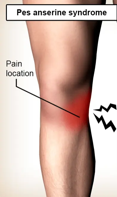
- Pain and tenderness in the back and outer side of the ankle.
- Swelling and inflammation around the subtalar joint.
- Stiffness in the foot and ankle.
- Difficulty walking on uneven surfaces or on inclines.
- Weakness of muscles around the joint
- instability in the affected foot
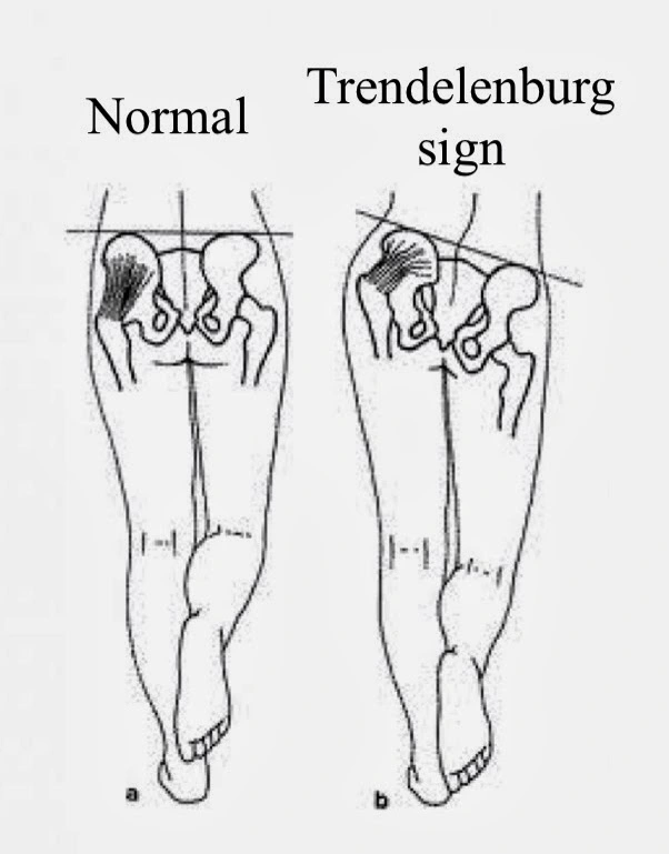
- Limping Gait
Differential Diagnosis:
When evaluating a patient with symptoms suggestive of subtalar osteoarthritis, it is important to consider other potential causes of similar symptoms. Here are some differential diagnoses to consider:
- Rheumatoid arthritis: Rheumatoid arthritis is an autoimmune disease that can affect multiple joints, including the subtalar joint. It is characterized by joint pain, swelling, and stiffness, typically affecting both sides of the body symmetrically. Rheumatoid arthritis can cause deformities and affect other joints beyond the subtalar joint.
- Gout: It is a form of arthritis caused by the buildup of uric acid crystals in the joints. It commonly affects the big toe joint, but it can also affect other joints, including the subtalar joint. Gout attacks are usually sudden and intense, causing severe pain, swelling, redness, and warmth in the affected joint.
- Ankle instability: Chronic ankle instability can result from repeated ankle sprains or ligamentous laxity. It can lead to recurrent episodes of pain, giving way, and a feeling of ankle instability. While subtalar joint involvement is less common, instability and abnormal foot mechanics can contribute to subtalar joint symptoms.
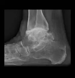
- Post-traumatic arthritis: Previous trauma, such as an ankle or foot fracture, can lead to post-traumatic arthritis. The subtalar joint may be affected, causing pain, stiffness, and difficulty with movement. X-rays or other imaging may reveal joint damage resulting from the injury.
- Tarsal tunnel syndrome: Tarsal tunnel syndrome occurs when the tibial nerve is compressed or irritated as it passes through the tarsal tunnel, located on the inner side of the ankle. This can cause pain, numbness, tingling, or a burning sensation in the foot and ankle, which may be mistaken for subtalar joint symptoms.
- Other arthritic conditions: Other types of arthritis, such as psoriatic arthritis or reactive arthritis, may involve the subtalar joint, along with other joints in the body. These conditions are often present with joint pain, stiffness, swelling, and other systemic symptoms.
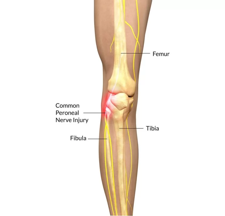
- Nerve impingement: Nerve impingement or compressions, such as from a herniated disc or peripheral neuropathy, can cause symptoms that radiate to the foot and ankle, mimicking subtalar joint pain.
Diagnosis:
Diagnosis of subtalar osteoarthritis typically involves a physical examination by a healthcare professional, including evaluating the range of motion and stability of the foot and ankle. Imaging tests such as X-rays or magnetic resonance imaging (MRI) may be used to assess the severity of joint damage and rule out other potential causes of symptoms.
Physical examination:
When a patient complains of subtalar discomfort, a thorough history and physical examination must be carried out. Uneven terrain makes it difficult to walk, which is a characteristic sign that is sometimes present but not always. Pain can be localized to the sinus tarsi area of the hindfoot’s posterolateral side or it can spread to the hindfoot’s posterior side along the posterolateral to the posteromedial axis. The ankle joint is thought to be the cause of discomfort when it occurs seldom in the front of the hindfoot. Subtalar joint motion might be misinterpreted for ankle joint motion, making it difficult to assess, however, contralateral evaluation can be helpful. a good hindfoot alignment
Years or even decades after the initial damage, clinical indications of pain and dysfunction may start to manifest. Patients typically complain of ankle and hindfoot discomfort that becomes worse with weight-bearing or exercise, as well as instability, stiffness, or edema that gets better with rest at first. Symptoms typically begin slowly and worsen slowly over time.
Imaging of subtalar osteoarthritis:
Weight-bearing radiography that uses both standard and specialty views, computed tomography that may be done using either a standard or weight-bearing approach, magnetic resonance imaging, and ultrasound assessment are all examples of imaging evaluation that must be customized to certain clinical settings. Radiographic imaging of the ankle usually kicks off imaging procedures. Most medical professionals take standing radiographs of the entire leg, including the foot. Some people could request Salzman or axial perspectives.
In addition to ruling out a coalition, a computed tomographic scan is helpful since it assesses the severity of the deformity. Single-photon emission CT has gained popularity in recent years because it makes it possible to locate the pain’s internal fixation, especially in posttraumatic situations when the pain’s origin is difficult to pinpoint using traditional imaging techniques. If necessary, magnetic resonance imaging can also be employed since plain films do not allow for the full characterization of non-ossified components such as articular cartilage, marrow tissue, and synovial fluid, making it possible to overlook any early signs of deterioration. The intricate forms of the bones and joints make it challenging to identify joint space narrowing or degeneration on standard standing radiographs without the use of special views.
Treatment of Subtalar Osteoarthritis:
Treatment options for subtalar osteoarthritis can vary depending on the severity of the condition and the individual’s symptoms.
The goal of nonoperative therapy should be to lessen the discomfort by restricting the mobility of the back foot. Traditional conservative therapies include weight reduction, painkillers, subtalar joint injections, shock-absorbent components like shoes or insoles, and specialized insoles designed to restrict hindfoot motion. Insoles with a central post can lessen the hindfoot’s tendency to evert, which may assist to alleviate subtalar discomfort. Over the past ten years, rocker-bottom footwear has gained popularity.
An effective therapy for ankle OA and other types of arthritis throughout the body is the intraarticular injection of glucocorticoids with or without anesthetic. When subtalar osteoarthritis is resistant to more conservative techniques, this therapeutic approach is frequently used. With post-traumatic ankle arthritis, the large variation in reported effectiveness is typically explained by the 30 to 80% success rate of needle placement when using just manual palpation as a guide. Utilizing ultrasonography to guide injections has reduced the frequency of difficulties caused by human mistakes and boosted the top limit of needle placement success rate from 32 to 97%.
Due to the shape of the foot and ankle, injections into the subtalar joint might be difficult. The posteromedial, posterolateral, and anterolateral methods are among the three that have been characterized and are often employed. The most typical technique is the posterolateral one. A long-axis method can be used to administer the injection when employing the posterolateral approach, which allows for continuous visualization of the whole needle shaft and tip.
Post-Operative Course:
Day 1
- Wrapped in a thick bandage and splint, with the foot elevated, iced, and taking painkillers
- Expect 12 to 24 hours of foot numbness followed by discomfort.
- Bloody drainage should be anticipated.
- Never remove the bandage.
Day 14
- A new dressing is placed, radiographs are obtained, and a boot is put on.
- No lifting for around 6 to 8 weeks.
6 weeks
- After removing the boot, x-rays are obtained.
- Continue your normal activities in the boot and gradually increase your weight bearing using your discomfort to guide you.
12 weeks
- Begin a workout regimen
- Most usually, orthotic arch supports are ordered.
Physiotherapy treatment of Subtalar Osteoarthritis:
- After an exercise, elevating the leg and applying an ice pack to the ankle and back of the foot will help relieve discomfort and swelling.
- You may be able to enjoy long walks without limping by having a little heel lift or cushion installed into the shoe to ease the strain on a tight ankle.
- An ankle brace, such as a strap around the ankle, or even securing the ankle to the backfoot might be helpful since it prevents the subtalar joint from moving around excessively.
- Purchase a new pair of comfy shoes and add extra padding. The ankles might not be put under stress as a result.
- Subtalar arthritis pain can be adequately managed with physical therapy or physiotherapy. Additionally, it strengthens the muscles around the ankle, aids in regaining lost range of motion, and also gives the foot stability.
- If the discomfort gets worse, using an assistive device like a walker or cane would be useful. It would be beneficial to lessen the tension and pain in the afflicted location by reducing the load on the joint.
General Factors of Postoperative Recovery:
- For at least six weeks, you won’t be able to walk on the leg. Follow-up x-rays and bone healing will establish a precise timeline.
- You’ll need to use crutches, a walker, a wheelchair, or a rollout sort of equipment to keep off your feet.
- After surgery, the leg will be wrapped in a stiff plaster cast for two weeks.
- At around two weeks, you’ll have your first follow-up appointment to have the sutures removed.
- At around two weeks, we’ll often put a detachable boot for you to wear.
- Two weeks after surgery on your left ankle, you should be able to operate an automated car. After three to four weeks, you can resume driving if the operation went well.
- Depending on your level of pain and the instructions provided to you, you may start putting some weight in the boot at around six weeks.
- For the ankle to restore its strength and range of motion, physical treatment is beneficial.
- Make an appointment with a physical therapist for one to two months.
- For around six months, there will be some little swelling in the foot, ankle, and leg.
- For almost a year following the procedure, your strength and mobility will continue to improve.
- After surgery, you should anticipate experiencing some discomfort and aching for four to six months.
- The rehabilitation process may benefit from orthotic arch supports.
Summary:
Subtalar osteoarthritis frequently results from repeated ankle sprains or trauma, which has a negative impact on the patient’s quality of life. Posttraumatic ankle osteoarthritis can occur in as many as 78% of those with persistent ankle instability. The connection between the talus and the calcaneus is referred to as the subtalar joint. It is a complex joint used in human movement and is crucial for propulsion and stress absorption. This joint is constructed to either give a flexible shock-absorbing structure or a hard propulsive structure to the foot.
The transverse tarsal joints become unlocked once the subtalar joint is everted, or in valgus, making the foot a flexible structure. The transverse tarsal joints lock when the subtalar joint inverts, creating a stiff lever arm that is helpful for mobility. The subtalar joint is separated into the talocalcaneonavicular joint and talocalcaneal joint by the sinus tarsi. This fact explains why, from the more traditional surgical methods that do not breach the sinus tarsi, only the posterior subtalar facet is visible.
FAQ:
What is subtalar osteoarthritis?
A kind of arthritis known as subtalar arthritis affects the subtalar joint, which is the joint in the backfoot that is located below the ankle joint. Uncomfortable hindfoot pain is a hallmark of the condition, which is made worse by standing and walking, particularly on uneven terrain.
What are osteoarthritic changes of the subtalar joint?
Arthritis in the subtalar joint’s cartilage is referred to as subtalar arthritis. A traumatic injury to the hindfoot, which is frequently observed following a fracture to the talus or calcaneus, is the most frequent cause of subtalar arthritis. Subtalar arthritis symptoms include discomfort and swelling in the backfoot.
Can ankle osteoarthritis be cured?
Although there is no known cure for arthritis, there are several therapy options that can lessen symptoms and decrease the disease’s progression. Many people with arthritis are able to manage their pain, stay active, and lead happy lives with the help of good medication.
Is subtalar arthritis permanent?
Gait and mobility impairments can result from subtalar joint disorders, especially when a person is walking on uneven terrain. Damage to the subtalar joint may become irreversible if untreated.
Can you fully recover from osteoarthritis?
Although osteoarthritis is a chronic ailment that cannot be treated, it doesn’t always become worse with time and in some cases, it even gets better. There are also several therapies available to lessen the symptoms. Simple solutions, such as routine exercise, can occasionally be used to treat mild symptoms.

4 Comments