
Brachiocephalic Vein
Introduction
The brachiocephalic vein, also known as the innominate vein, is a crucial component of the venous system in the human body. It plays a significant role in returning deoxygenated blood from the upper limbs, head, and neck to the heart for oxygenation.
This vein is notable for its unique anatomy and function, serving as an important junction before blood continues its journey towards the heart. Originating from the confluence of the subclavian and internal jugular veins on each side of the body, the brachiocephalic veins are located in the superior thoracic cavity. The right and left brachiocephalic veins usually come together to create the superior vena cava, a large vein that directly transports blood into the right atrium of the heart.
Apprehending the brachiocephalic vein’s layout and role is essential in comprehending the convoluted network that ensures efficient circulation throughout the body. Its anatomical position and connections make it a pivotal element in both medical and anatomical studies, highlighting its importance in cardiovascular health and clinical practice.
What is the anatomical location and pathway of the brachiocephalic vein?
The brachiocephalic vein, likewise known as the innominate vein, is a principal vein in the human body that is accountable for draining deoxygenated blood from the upper limbs, head, and neck. Here’s a detailed description of its anatomical location and pathway:
Origin and Formation:
- The brachiocephalic vein is formed by the union of the subclavian vein and the internal jugular vein on each side of the body.
- On the right side of the body, the right subclavian vein and the right internal jugular vein converge to form the right brachiocephalic vein.
- On the left side of the body, the left subclavian vein and the left internal jugular vein converge to form the left brachiocephalic vein.
Course and Pathway:
- Both brachiocephalic veins observe a short track in the superior mediastinum, the space within the thoracic cavity found between the superior thoracic aperture (thoracic inlet) and the level of the transverse thoracic plane (approximately at the level of the sternal angle).
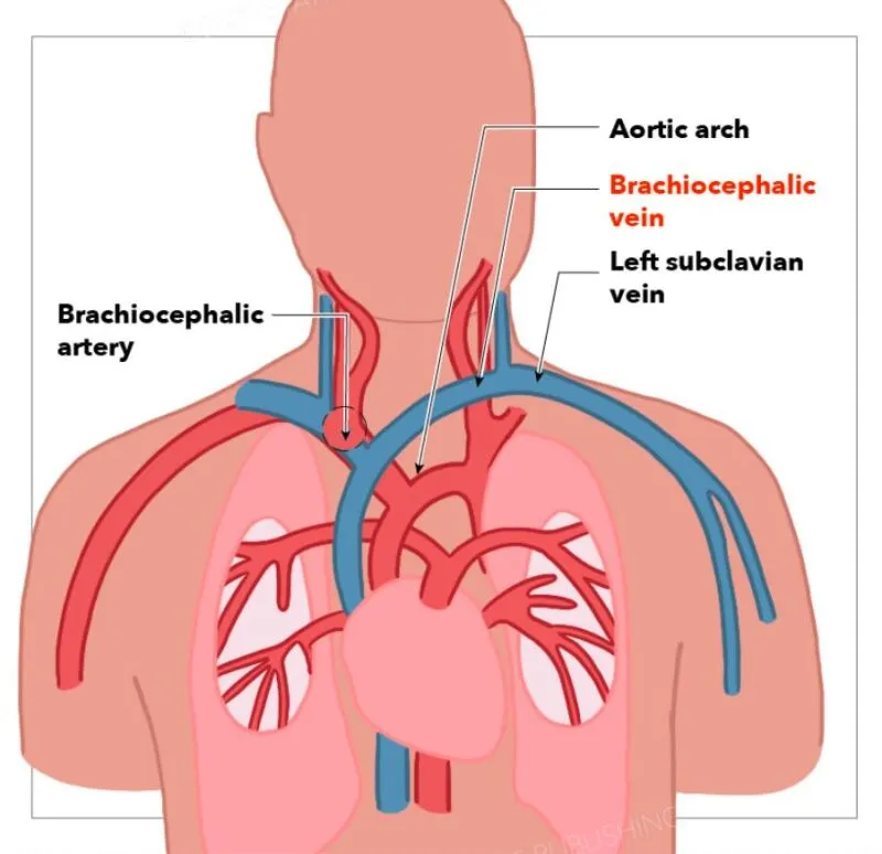
- The right and left brachiocephalic veins are situated anterior to the brachiocephalic arteries (which are branches of the aortic arch) and posterior to the sternum (breastbone).
- The veins then converge in the midline to form the superior vena cava.
Confluence and Termination:
- At the level of the first rib and just below the clavicle, the merging of the right and left brachiocephalic veins occurs, forming the superior vena cava (SVC).
- The superior vena cava is a large vein that extends upwards and ultimately carries deoxygenated blood from the upper body to the right atrium of the heart.
Anatomical Relationships:
- The brachiocephalic veins are closely affiliated with essential structures such as the sternum, trachea, esophagus, and main vessels including the aortic arch.
- They are situated in the superior mediastinum, which is the central compartment of the thoracic cavity housing critical structures of the heart and great vessels.
Understanding the anatomical location and pathway of the brachiocephalic vein is essential in grasping its role in the venous drainage of the upper body and its clinical relevance in various medical conditions affecting the thoracic region.
What is the size of brachiocephalic veins?
The brachiocephalic veins, also known as innominate veins, are relatively large veins in the human body. They are formed by the union of the subclavian vein (which drains the upper limb) and the internal jugular vein (which drains the head and neck) on each side of the body.
In terms of dimensions:
- Diameter: The diameter of the brachiocephalic veins can vary depending on the individual and physiological conditions. Generally, they are large enough to facilitate the combined flow of blood from the upper limb and head towards the heart.
- Length: The length of each brachiocephalic vein is relatively short, as they are formed by the union of the subclavian vein and internal jugular vein and quickly join to form the superior vena cava.
To provide a specific numerical measurement of their diameter, studies and medical references often do not provide exact sizes as they can vary widely among individuals. However, they are typically larger than most peripheral veins due to their role in conducting blood from major parts of the upper body to the heart.
Where does the brachiocephalic vein carry blood?
The deoxygenated blood from the upper limbs, head, and neck is brought towards the heart by the brachiocephalic veins. Here’s a detailed explanation of their path and function:
Right Brachiocephalic Vein (Innominate Vein):
- The right brachiocephalic vein is formed by the union of the right subclavian vein (which drains the right arm) and the right internal jugular vein (which drains the head and neck).
- It carries deoxygenated blood from the right arm, right side of the head, and right side of the neck.
Left Brachiocephalic Vein (Innominate Vein):
- The left brachiocephalic vein is assembled when the left subclavian vein (which organizes blood from the left arm) and the left internal jugular vein (which compiles blood from the head and neck) congregate.
- It carries deoxygenated blood from the left arm, left side of the head, and left side of the neck.
Superior Vena Cava (SVC):
- Above the heart, the right and left brachiocephalic veins unite to create the superior vena cava (SVC).
- The superior vena cava then carries the combined deoxygenated blood from both sides of the upper body to the right atrium of the heart.
In abridgment, the brachiocephalic veins play a vital role in carrying unoxygenated blood from the upper extremities, head, and neck to the heart, from where it will travel to the lungs which is traveled for oxygenation before being distributed to the rest of the body.
What role does the brachiocephalic vein play in circulating blood throughout the body?
By aiding the return of deoxygenated blood from the upper limbs, head, and neck back to the heart, the brachiocephalic veins are essential for blood circulation. Here’s how they contribute to the circulatory system:
Venous Drainage from Upper Limbs:
- The brachiocephalic veins receive deoxygenated blood from the upper limbs (via the subclavian veins) and from the head and neck regions (via the internal jugular veins).
- On the right side, the right brachiocephalic vein collects blood from the right subclavian vein (draining the right arm) and the right internal jugular vein (draining the right side of the head and neck).
- Blood from the left arm is collected by the left subclavian vein, and blood from the left side of the head and neck is collected by the left internal jugular vein. Both veins then carry the blood to the left brachiocephalic vein.
Confluence and Formation of Superior Vena Cava:
- Each brachiocephalic vein runs a short course in the superior mediastinum before merging with its counterpart in the midline.
- Above the heart, the right and left brachiocephalic veins come together to create the superior vena cava (SVC) in the heart.
- The superior vena cava is extends upward and sends oxygen-depleted blood directly to the right atrium of the heart.
Role in Systemic Circulation:
- The superior vena cava is one of the main veins that returns blood to the heart from the upper part of the body (above the diaphragm).
- Blood collected by the brachiocephalic veins contains waste products (deoxygenated blood) that need to be transported back to the heart for re-oxygenation and redistribution to the rest of the body via systemic circulation.
Clinical Importance:
- Conditions affecting the brachiocephalic veins, such as thrombosis (blood clot formation), stenosis (narrowing), or injury, can impair venous return from the upper body.
- Surgical procedures involving the brachiocephalic veins, such as central venous catheter placement or interventions to treat venous obstructions, highlight their critical role in clinical practice.
The brachiocephalic veins play a crucial role in draining blood from the upper extremities, head, and neck, helping to maintain efficient circulation by returning deoxygenated blood to the heart for oxygenation through the pulmonary circulation.
What are the primary tributaries that feed into the brachiocephalic vein?
The brachiocephalic veins receive blood from several major tributaries, which are the veins that drain blood from specific regions of the upper body. Here are the main tributaries that contribute to the brachiocephalic vein:
Subclavian Veins:
- The subclavian veins are major veins that drain the blood from the arms and shoulders.
- Specifically, the right subclavian vein drains the right arm and shoulder, while the left subclavian vein drains the left arm and shoulder.
- The subclavian veins and the internal jugular veins join to form the brachiocephalic veins.
Internal Jugular Veins:
- The internal jugular veins are enormous veins that drain blood from the brain, face, and neck provinces.
- The internal jugular vein gathers blood from the brain, head, and neck on both sides of the body.
- These veins also connect with the respective subclavian veins to create the brachiocephalic veins.
In overview, the leading tributaries contributing to the brachiocephalic veins are the subclavian veins and the internal jugular veins. These veins collect deoxygenated blood from the upper limbs (via the subclavian veins) and from the head and neck (via the internal jugular veins), respectively. The brachiocephalic veins are constructed by the convergence of these veins, and they then merge to assemble the superior vena cava, which assists the blood return to the heart for oxygenation.
What are common medical conditions or diseases that affect the brachiocephalic vein?
The brachiocephalic vein, like any major blood vessel in the body, can be affected by various medical conditions or diseases. Here are some common ones:
Thrombosis:
- The presence of a blood clot within the brachiocephalic vein is known as thrombosis. This can also obstruct blood flow and potentially lead to complications such as pulmonary embolism if the clot dislodges and travels to the lungs.
Stenosis:
- Stenosis is the narrowing of the brachiocephalic vein, which can restrict blood flow. It may result from conditions like atherosclerosis (buildup of plaque in the arteries) or external compression from adjacent structures.
Compression Syndrome:
- Thoracic outlet syndrome or other forms of compression syndromes can affect the brachiocephalic vein where it passes through tight spaces between muscles, bones, and ligaments in the thoracic outlet region. This compression can lead to venous congestion and symptoms like swelling, pain, or discomfort in the upper extremities.
Central Venous Catheter Complications:
- Placement of central venous catheters, particularly those inserted into the brachiocephalic vein, can lead to complications such as infection, thrombosis, or injury to the vein.
Trauma:
- Traumatic injury to the chest or neck region can cause damage to the brachiocephalic vein, leading to bleeding, thrombosis, or even rupture in severe cases.
Tumors or Masses:
- Tumors or abnormal masses in the chest or neck area can compress or invade the brachiocephalic vein, affecting blood flow and potentially leading to venous obstruction.
Inflammatory Conditions:
- Inflammatory diseases such as vasculitis (inflammation of blood vessels) can affect the brachiocephalic vein, causing inflammation, narrowing, or even blockage.
Congenital Anomalies:
- Rarely, congenital abnormalities of the brachiocephalic vein or its formation can lead to structural defects or functional abnormalities.
Management of these conditions typically involves medical therapy to treat underlying causes (such as anticoagulants for thrombosis), surgical interventions for severe cases (such as thrombectomy or venous bypass), or minimally invasive procedures (such as angioplasty and stenting for stenosis). Early diagnosis and appropriate treatment are crucial to prevent complications and ensure optimal venous function in affected individuals.
What are the treatment options for conditions affecting the brachiocephalic veins?
Treatment options for conditions affecting the brachiocephalic veins depend on the specific nature and severity of the condition. Here are some of the main treatment modalities used are as follows:
Anticoagulant Therapy:
- For conditions such as brachiocephalic vein thrombosis (blood clot), anticoagulant medications (blood thinners) are typically prescribed. These medications prevent the clot from growing larger and reduce the risk of complications like pulmonary embolism.
Thrombolytic Therapy:
- In cases where there is extensive thrombosis or acute occlusion of the brachiocephalic vein, thrombolytic therapy may be used. This implicates allocating medicines (thrombolytics) that dissolve the clot to restore blood flow.
Angioplasty and Stenting:
- Angioplasty is a minimally invasive approach utilized to feast stenosis (narrowing) of the brachiocephalic vein. During angioplasty, a balloon-tipped catheter is inserted into the vein and extended to broaden the narrowed area. In some circumstances, a stent (a small mesh tube) might also be positioned to maintain the vein open and enhance blood flow.
Surgical Interventions:
- In extreme cases or when other therapies are ineffective, surgical interventions might be essential. This can include thrombectomy (surgical removal of a clot), venous bypass surgery (redirecting blood flow around a blocked or narrowed vein using a graft), or reconstruction of the affected vein.
Management of Underlying Causes:
- Addressing underlying conditions that contribute to brachiocephalic vein disorders is essential. This might also affect managing disorders like thoracic outlet syndrome (compression of nerves or blood vessels in the thoracic outlet syndrome), feasting tumors or masses that compress the vein, or addressing structural abnormalities.
Compression Therapy:
- In cases where compression syndromes (e.g., thoracic outlet syndrome) are causing venous obstruction, conservative measures such as physical therapy, postural adjustments, or wearing compression garments may help alleviate symptoms and improve venous flow.
Monitoring and Supportive Care:
- Regular condition monitoring, imaging investigations to measure vein patency, and ongoing risk factor management (such as keeping a healthy weight, managing diabetes, and avoiding prolonged immobility) are essential for long-term care and recurrence prevention.
The choice of treatment leans on elements such as the underlying cause, the extent of the disorder, the patient’s prevailing health, and the presence of any complications. A multidisciplinary approach involving vascular specialists, interventional radiologists, and surgeons is often necessary to tailor treatment plans to individual patient needs and optimize outcomes.
How does the structure and function of the brachiocephalic vein vary from other significant veins in the body?
The structure and function of the brachiocephalic vein exhibit several distinctive features compared to other major veins in the body:
Anatomical Location and Formation:
- The brachiocephalic vein is unique in its location and formation. It forms by the convergence of the subclavian vein (which drains the arm) and the internal jugular vein (which drains the head and neck) on each side of the body.
- Unlike other veins that may have multiple tributaries or a more extensive network, the brachiocephalic vein is a relatively short segment in the superior mediastinum.
Unilateral Nature:
- The brachiocephalic vein is typically described in pairs: a right brachiocephalic vein and a left brachiocephalic vein. Individual pair unites to assemble a single superior vena cava, which then bears deoxygenated blood to the heart.
- In contrast, veins like the inferior vena cava have a more symmetrical and bilateral structure, collecting blood from both lower limbs and the abdominal organs.
Function in Venous Return:
- The primary function of the brachiocephalic vein is to facilitate the venous return of deoxygenated blood from the upper extremities, head, and neck to the heart.
- A larger role is played by other major veins, like the inferior vena cava, in gathering blood that has lost oxygen from the lower body and abdominal organs.
Relation to Surgical and Clinical Procedures:
- Due to its unique location and anatomy, the brachiocephalic vein is often a site for surgical interventions or clinical procedures. For example, it may be accessed for central venous catheterization or used as a conduit in certain vascular surgeries.
- In clinical settings, other large veins might not be used or accessed as frequently for comparable treatments.
Clinical Relevance and Variations:
- Variations in the anatomy of the brachiocephalic vein, such as anomalies or structural variations, can have implications for surgical planning and patient management.
- Understanding these variations is crucial for medical professionals to ensure safe and effective treatment of conditions affecting venous return from the upper body.
In conclusion, while the brachiocephalic vein performs similar functions to other major veins in facilitating blood circulation, its unique anatomy, formation, and clinical relevance distinguish it as an essential component of the venous system.
What evolutionary adaptations might have influenced the structure and function of the brachiocephalic vein over time?
The evolutionary adaptations that influenced the structure and function of the brachiocephalic vein (innominate vein) over time are likely tied to the changes in anatomy, physiology, and behavior in human ancestors. Here are some key evolutionary factors that may have influenced the development of the brachiocephalic vein:
Bipedalism:
- The evolution of bipedalism in humans led to significant changes in the venous system to accommodate the upright posture. This adaptation affected the structure and function of veins in the upper body, including the brachiocephalic veins.
- Veins like the brachiocephalic veins needed to efficiently return blood from the upper limbs and head against gravity, which influenced their size, location, and venous return mechanisms.
Increased Brain Size and Metabolic Demand:
- As human ancestors evolved larger brains and increased metabolic demands associated with cognitive functions, including enhanced social interactions and tool use, the circulatory system had to adapt to support these changes.
- Ensuring the efficient drainage of deoxygenated blood from the head into the brachiocephalic veins via the internal jugular veins became essential for supporting increased metabolic rates.
Thoracic Cavity and Heart Position:
- The growth and development of the brachiocephalic veins was influenced by changes in the thoracic cavity’s structure and the heart’s location in relation to other networks.
- The positioning of these veins in the superior mediastinum, anterior to the great vessels and posterior to the sternum, reflects adaptations to optimize venous return and circulation in bipedal humans.
Development of Vascular System Complexity:
- Over evolutionary time, there was likely an increase in the complexity and specialization of the vascular system to meet the physiological demands of larger, more complex organisms.
- The development of distinct venous pathways, including the formation of the brachiocephalic veins from the fusion of subclavian and internal jugular veins, represents an evolutionary adaptation to efficiently channel deoxygenated blood back to the heart.
Environmental and Behavioral Influences:
- Changes in environmental factors, such as temperature fluctuations and dietary adaptations, may have influenced cardiovascular adaptations, including venous structure and function.
- Behavioral factors, such as increased physical activity and social interactions, could have also influenced venous circulation patterns and adaptations in the upper body.
The changes in the brachiocephalic vein in humans demonstrate adjustments to walking on two legs, larger brain size, and energy requirements, alterations in chest structure, and the creation of a more intricate network of blood vessels. These adaptations have shaped the structure and function of the brachiocephalic vein to support efficient venous return and circulation in human ancestors and modern humans alike.
What happens if the brachiocephalic vein is blocked?
If the brachiocephalic vein is blocked, it can lead to several clinical consequences, depending on the location and severity of the blockage:
- Venous Insufficiency in Upper Limbs: Carrying blood that has lost oxygen from the head, neck, and upper limbs to the heart is one of the brachiocephalic vein’s essential functions. If blocked, it can lead to venous insufficiency in these areas.
- Swelling and Edema: A blockage in the brachiocephalic vein can cause swelling and edema (fluid buildup) in the arm on the affected side. This happens because blood cannot efficiently return to the heart, leading to increased pressure in the veins and fluid leakage into surrounding tissues.
- Collateral Circulation: In some cases, collateral veins may develop to bypass the blocked brachiocephalic vein, which can appear as visible veins on the chest or neck.
- Risk of Thrombosis: A blockage in the brachiocephalic vein can predispose to thrombosis (blood clot formation). This is because stagnant blood flow due to the blockage can lead to the formation of clots within the vein, potentially causing further complications such as deep vein thrombosis (DVT).
- Pulmonary Embolism: A pulmonary embolism, a potentially fatal disorder, can result from a clot that develops in the obstructed brachiocephalic vein and spreads to the lungs.
- Symptoms: Patients may experience symptoms such as pain, heaviness, warmth, or redness in the affected arm. There might also be observable swelling or prominent veins in the affected area of the body.
- Treatment: The underlying reason and severity of a blocked brachiocephalic vein determine how to treat it. It may include medications to dissolve clots (if present), compression therapy to reduce swelling, and in some cases, surgical interventions or catheter-based procedures to remove or bypass the blockage.
In summary, a blockage in the brachiocephalic vein can lead to venous insufficiency, swelling, risk of thrombosis, and potentially serious complications such as pulmonary embolism. Prompt evaluation and appropriate management are important to prevent further complications and restore normal venous circulation.
How might a blockage or thrombosis in the brachiocephalic vein impact blood flow in the upper extremities and head?
A blockage or thrombosis in the brachiocephalic vein can have significant implications for blood flow in the upper extremities and head due to its role in venous drainage from these regions. Here’s how such conditions might impact circulation:
Venous Congestion:
- The normal flow of deoxygenated blood from the upper extremities (arms) and head could also be obstructed by a blockage or thrombosis in the brachiocephalic vein.
- This obstruction leads to venous congestion, where blood accumulates upstream of the blockage, causing swelling (edema) and discomfort in the affected arm and potentially in the face or neck on the affected side.
Symptoms in Upper Extremities:
- Individuals may experience symptoms such as pain, heaviness, or a feeling of fullness in the affected arm.
- There may be visible swelling or engorgement of veins in the arm due to impaired drainage.
Symptoms in Head and Neck:
- Symptoms may also spread to the head and neck areas if the thrombosis affects the internal jugular vein.
- This can lead to swelling, pain, or discomfort in the face, neck, and head on the affected side.
- Furthermore, if there is elevated pressure in the veins that drain the head, patients may also encounter headaches or visual disturbances.
Complications:
- If the thrombus (blood clot) in the brachiocephalic vein or its branches dislodges, it can travel to the lungs (pulmonary embolism) or other parts of the body, causing serious complications.
- Chronic venous congestion can also lead to skin changes, ulceration, or even thromboembolic events if not managed promptly.
Functional Impairments:
- Reduced blood flow and oxygen delivery to the affected arm or head can impair normal function, leading to weakness or difficulty performing daily activities.
Management of a blockage or thrombosis in the brachiocephalic vein typically involves anticoagulation therapy to dissolve the clot and prevent further clotting, along with measures to relieve venous congestion and manage symptoms. In some cases, surgical intervention or minimally invasive procedures such as thrombectomy or venous stenting may be necessary to restore normal blood flow and prevent complications. Early diagnosis and treatment are crucial to minimize long-term effects and improve outcomes for affected individuals.
In what ways does the brachiocephalic vein adapt to its role in the circulatory system compared to veins in other animals?
The adaptation of the brachiocephalic vein in humans, compared to veins in other animals, primarily revolves around its structure, location, and function tailored to the unique physiological demands of bipedalism and the upright posture of humans. Here are several ways in which the brachiocephalic vein adapts specifically to its role in the human circulatory system:
Formation and Convergence:
The brachiocephalic vein is constructed when the internal jugular vein (which drains the top of the head and neck) and/or the subclavian vein (which drains the arm) congregate on either side of the body. The efficient drainage of deoxygenated blood from the upper extremities and head directly into the superior vena cava is facilitated by this special bilateral formation.
Superior Vena Cava Connection:
- Transporting deoxygenated blood directly to the right atrium of the heart, the human superior vena cava is a massive vein that emerges from the brachiocephalic veins. This direct connection is critical for efficient circulation and oxygenation of blood.
Anatomical Position:
- The brachiocephalic veins are situated in the superior mediastinum, anterior to the great vessels like the aortic arch, and posterior to the sternum. This location ensures a relatively straight and unobstructed pathway for venous return, optimizing blood flow from the upper body.
Surgical and Clinical Utility:
- Due to its accessibility and location, the brachiocephalic vein is often used in medical procedures such as central venous catheterization or as a conduit in certain cardiovascular surgeries. This adaptability reflects its importance in clinical practice and patient care.
Adaptations for Upright Posture:
- Humans are bipedal, which imposes unique challenges on the circulatory system compared to quadrupedal animals. The structure of the brachiocephalic vein, along with other veins in the upper body, is adapted to facilitate efficient venous return against gravity when standing upright.
Evolutionary Considerations:
- Over evolutionary time, the structure and function of the brachiocephalic vein have likely adapted to accommodate changes in human anatomy and physiology associated with bipedal locomotion and increased brain size.
In contrast, veins in other animals may vary in structure and function based on their specific anatomical and physiological adaptations. For example, in quadrupeds, veins may be structured to accommodate the different stresses and gravitational forces associated with four-legged locomotion. Understanding these adaptations provides insights into the specialized roles veins play in different species’ circulatory systems.
How does the brachiocephalic vein develop during embryonic and fetal stages?
The development of the brachiocephalic veins (innominate veins) during embryonic and fetal stages involves complex processes of venous morphogenesis and remodeling. Here’s an overview of how these veins develop:
Formation and Early Development:
- Initially, during the early stages of embryonic development (around the 5th to 6th week of gestation), venous plexuses begin to form throughout the embryo. These plexuses are networks of small veins that eventually coalesce and organize into larger vessels.
- In the area corresponding to the upper limbs and head, these plexuses give rise to the precursor veins that will eventually form the subclavian veins (draining the upper limbs) and the internal jugular veins (draining the head and neck) in the body.
Differentiation and Fusion:
- As development progresses, the venous plexuses differentiate into distinct venous segments, including the subclavian veins and internal jugular veins.
- On both sides of the body, the internal jugular vein and subclavian vein begin to unite or merge. The right brachiocephalic vein is formed by the meeting point of the right internal jugular vein and the subclavian vein. The left brachiocephalic vein is generated by the meeting point of the left internal jugular and/or the left subclavian vein.
Connection to Superior Vena Cava:
- As these vessels keep developing and increasing in size, the right and left brachiocephalic veins move toward the center and come together to create the superior vena cava.
- The main venous channel for collecting deoxygenated blood from the upper body and delivering it to the right atrium of the heart is formed by the superior vena cava.
Maturation and Functional Integration:
- Throughout fetal development and into early infancy, the brachiocephalic veins undergo further maturation and functional integration into the circulatory system.
- They adapt to the increasing metabolic demands of the growing fetus and later the newborn, ensuring efficient venous return and circulation.
Developmental Variations:
- Variations in the development of the brachiocephalic veins can occur, leading to anatomical anomalies such as duplicated or absent brachiocephalic veins. These variations may have clinical implications and require consideration in medical practice.
Understanding the embryonic and fetal development of the brachiocephalic veins is essential for comprehending their normal anatomy, potential variations, and clinical relevance in congenital conditions or developmental anomalies affecting the cardiovascular system. This developmental process underscores the intricate nature of venous morphogenesis and its role in establishing the mature circulatory system in humans.
Research and innovation of brachiocephalic vein
What current research is exploring the role of the brachiocephalic vein in cardiovascular health?
Current research exploring the role of the brachiocephalic vein (innominate vein) in cardiovascular health encompasses various aspects related to its anatomy, function, clinical implications, and therapeutic interventions. Here are some regions of the persistent analysis:
Venous Thrombosis and Stenosis:
- Studies are investigating the prevalence, risk factors, and outcomes associated with thrombosis (clot formation) and stenosis (narrowing) of the brachiocephalic vein.
- Research focuses on understanding the mechanisms leading to venous thrombosis in this vein and exploring novel diagnostic methods and treatments to manage these conditions effectively.
Anatomical Variations and Clinical Implications:
- Research continues to document anatomical variations in the brachiocephalic vein and their clinical significance.
- Studies aim to determine how variations such as duplicated or absent brachiocephalic veins impact venous drainage patterns and may contribute to venous disorders or complications during surgical procedures.
Surgical Interventions and Catheterization Techniques:
- Investigative efforts are directed toward optimizing surgical techniques involving the brachiocephalic vein, such as central venous catheterization and vascular access for medical interventions.
- Research explores the safety, efficacy, and complications associated with using the brachiocephalic vein for these procedures, particularly in critically ill patients.
Venous Flow Dynamics and Imaging Techniques:
- Advanced imaging modalities, including ultrasound, CT angiography, and MRI, are employed to study venous flow dynamics in the brachiocephalic vein.
- Research focuses on quantifying blood flow velocities, detecting abnormalities such as venous reflux or obstruction, and assessing the hemodynamic impact on cardiovascular function.
Genetic and Molecular Mechanisms:
- Molecular and genetic studies investigate the underlying mechanisms contributing to venous diseases affecting the brachiocephalic vein.
- The research aims to identify genetic markers associated with venous thrombosis susceptibility, vascular inflammation, or structural abnormalities, which may inform personalized medicine approaches in cardiovascular care.
Clinical Outcomes and Longitudinal Studies:
- Longitudinal studies and clinical trials evaluate the long-term outcomes of patients with brachiocephalic vein disorders, including prognosis, quality of life, and recurrence rates following therapeutic interventions.
- Research outcomes provide evidence-based recommendations for patient management and guide the development of clinical guidelines in cardiovascular health.
Overall, ongoing research into the role of the brachiocephalic vein in cardiovascular health spans from basic science investigations into venous physiology and anatomy to clinical studies exploring diagnostic and therapeutic advancements. These efforts aim to improve understanding, diagnosis, and treatment of conditions affecting this critical venous structure in humans.
How might advancements in medical technology impact the treatment of conditions related to the brachiocephalic vein?
Advancements in medical technology have the potential to significantly impact the treatment of conditions related to the brachiocephalic vein (innominate vein) by improving diagnostic accuracy, enhancing therapeutic interventions, and optimizing patient outcomes. Here’s how these advancements may influence treatment:
Advanced Imaging Modalities:
- High-resolution Ultrasound: Improvements in ultrasound technology allow for detailed visualization of venous anatomy, blood flow dynamics, and detection of thrombosis or stenosis in the brachiocephalic vein.
- Computed Tomography (CT) and Magnetic Resonance Imaging (MRI): These imaging techniques provide cross-sectional views of the brachiocephalic vein, facilitating accurate diagnosis of anatomical variations, venous obstructions, or structural abnormalities.
Minimally Invasive Procedures:
- Endovascular Techniques: Advances in endovascular interventions, such as angioplasty and stenting, enable minimally invasive treatment of brachiocephalic vein stenosis.
- Thrombectomy Devices: Innovative devices for mechanical thrombectomy can safely and effectively remove thrombi from the brachiocephalic vein, reducing the risk of complications associated with venous thrombosis.
Robotic Surgery and Navigation Systems:
- Robotic-assisted surgery and navigation systems enhance accuracy and control during complex surgical strategies involving the brachiocephalic vein.
- These technologies enable surgeons to perform delicate maneuvers with greater accuracy, potentially reducing operative risks and improving patient outcomes.
Advanced Catheterization Techniques:
- Catheter-based treatments, such as central venous catheter placement or retrieval, benefit from improved catheter design and navigation systems.
- Enhanced catheterization techniques ensure safer access to the brachiocephalic vein for therapeutic purposes, including the administration of medications or contrast agents.
Genetic and Molecular Therapies:
- Emerging therapies targeting genetic and molecular mechanisms underlying venous diseases may offer personalized treatment options for patients with inherited or acquired conditions affecting the brachiocephalic vein.
- These therapies aim to modulate vascular inflammation, promote vascular repair, or prevent recurrence of thrombotic events.
Telemedicine and Remote Monitoring:
- Telemedicine platforms enable remote consultation and monitoring of patients with brachiocephalic vein disorders, facilitating timely adjustments to treatment plans and improving access to specialized care.
- Remote monitoring technologies also enhance patient education and adherence to post-treatment protocols, reducing the likelihood of complications and hospital readmissions.
Advancements in medical technology continue to transform the landscape of cardiovascular care, offering new opportunities to enhance the diagnosis, treatment, and management of conditions related to the brachiocephalic vein. These innovations not only improve clinical outcomes but also promote patient safety, comfort, and quality of life through tailored and minimally invasive therapeutic approaches.
Creative and Hypothetical of the brachiocephalic vein
If the brachiocephalic vein had a unique ability or characteristic in a fictional story or alternate reality, what would it be?
In a fictional story or alternate reality, the brachiocephalic vein could possess a unique ability or characteristic that sets it apart from ordinary veins. Here’s an imaginative concept:
The Brachiocephalic Vein of Insight:
In this fictional world, the brachiocephalic vein is not just a conduit for blood but a vessel imbued with a mystical ability called the “Vein of Insight.” This unique characteristic allows it to serve as a conduit not only for deoxygenated blood but also for knowledge, wisdom, and intuitive understanding.
- Knowledge Transfer: The Vein of Insight can absorb and transmit knowledge and experiences from individuals it connects with. When someone touches or connects with the brachiocephalic vein, they gain insights and understanding related to the person’s memories, skills, or emotions.
- Intuitive Guidance: Those who are attuned to the Vein of Insight can receive intuitive guidance and foresight about future events or decisions. This ability aids in strategic planning, problem-solving, and navigating challenges by tapping into a collective consciousness.
- Healing and Balance: The Vein of Insight has healing properties, promoting physical and emotional balance in those it connects with. It can cleanse negative energies, restore vitality, and foster inner harmony.
- Guardian of Secrets: As a custodian of knowledge and wisdom, the Vein of Insight safeguards ancient secrets, mystical lore, and the history of civilizations. It functions as a repository for collective memories and cultural heritage.
- Symbol of Unity: In this alternate reality, the Vein of Insight symbolizes unity and interconnectedness among all beings. It highlights the interconnected nature of humanity and the universe, promoting empathy, understanding, and cooperation.
In the storyline, characters may seek out the Vein of Insight for its transformative abilities, facing challenges and adversaries who wish to exploit its power for selfish gain or to uncover hidden truths about the world. The brachiocephalic vein, thus transformed into the Vein of Insight, becomes a central element in the narrative, weaving together themes of knowledge, destiny, and the interconnectedness of all living beings.
How might a mythical creature or character be affected by a magical variation of the brachiocephalic vein?
A mythical creature or character affected by a magical variation of the brachiocephalic vein could experience unique abilities, transformations, or vulnerabilities tied to this mystical attribute. Here’s an imaginative exploration:
The Celestial Brachiocephalic Vein:
In this fantasy realm, certain mythical creatures or characters possess a variant of the brachiocephalic vein known as the Celestial Brachiocephalic Vein. This vein is infused with celestial energies, granting extraordinary powers and attributes to those who possess it.
- Flight and Levitation:
- Creatures or characters with the Celestial Brachiocephalic Vein can harness its magical energies to achieve flight or levitation. With grace and agility, they can navigate aerial landscapes while defying gravity and soaring through the air with ease..
- Enhanced Perception and Insight:
- The Celestial Vein augments sensory perception and grants profound insights into mystical and arcane phenomena. Those attuned to its energies can perceive hidden truths, foresee events, or discern the intentions of others with uncanny accuracy.
- Healing and Regeneration:
- The magical properties of the Celestial Brachiocephalic Vein facilitate rapid healing and regeneration. Wounds heal swiftly, and vitality is restored with the infusion of celestial energies, making its bearer resilient against physical and magical afflictions.
- Channeling Celestial Magic:
- The Celestial Vein serves as a conduit for celestial magic, enabling its bearer to wield powerful spells or abilities imbued with cosmic forces. This includes casting protective wards, summoning celestial beings, or channeling bursts of radiant energy in times of need.
- Guardian of Celestial Realms:
- Those entrusted with the Celestial Brachiocephalic Vein may bear the responsibility of safeguarding celestial realms or cosmic balance. They act as emissaries between mortal realms and celestial planes, ensuring harmony and protecting against malevolent forces.
- Bond with Celestial Creatures:
- Creatures possessing the Celestial Vein may form bonds with celestial beings such as angels, celestial dragons, or other ethereal entities. Their abilities are strengthened and their link to the heavenly realms is intensified by these bonds.
In the narrative, characters with the Celestial Brachiocephalic Vein navigate a world where mystical forces intertwine with mortal affairs. They may encounter challenges that test their mastery of celestial powers, confront adversaries seeking to harness or corrupt its magic or embark on quests to unlock the vein’s full potential. Ultimately, the Celestial Vein becomes a symbol of cosmic destiny, bridging mortal aspirations with celestial realms in a tapestry of magic and wonder.
Disclaimer: The information and education provided here are the only goals for which it is intended. Medical advice, diagnosis, or treatment should not be taken as true. Constantly pursue the guidance of a skilled healthcare specialist obeying any medical disorder or treatment. Employing any knowledge provided here is at your own risk. There is no guarantee of the accuracy, completeness, adequacy, or currency of the content. The brachiocephalic vein and related medical conditions should be discussed with a healthcare provider for personalized guidance and care. The importance of consulting healthcare professionals for medical advice is stressed in this disclaimer, and it aims to ensure that readers comprehend the constraints of informational content available online.
FAQs
What are the brachiocephalic veins?
The brachiocephalic veins, also known as innominate veins, are major veins in the human body that form from the convergence of the subclavian vein (draining the arm) and the internal jugular vein (draining the head and neck).
What number of brachiocephalic veins are there?
There are typically two brachiocephalic veins: the right brachiocephalic vein and the left brachiocephalic vein. Each pair connects to form the superior vena cava, which transports deoxygenated blood to the heart.
What is the function of the brachiocephalic veins?
The brachiocephalic veins are responsible for carrying deoxygenated blood from the upper extremities, head, and neck to the heart for oxygenation.
And to what does the brachiocephalic vein attach?
The brachiocephalic vein, also known as the innominate vein, connects to the superior vena cava. Specifically, it is formed by the union of the subclavian vein and the internal jugular vein on each side of the body.
How are brachiocephalic vein conditions diagnosed?
Diagnostic methods include imaging techniques such as ultrasound, CT angiography, and MRI to visualize the anatomy and blood flow in the brachiocephalic veins. The velocity of blood flow and any abnormalities can be assessed using Doppler ultrasound.
Are there variations in the anatomy of the brachiocephalic veins?
Yes, variations in the anatomy of the brachiocephalic veins, such as duplicated or absent veins, can occur. These variations may impact venous drainage patterns and may require consideration in clinical practice and surgical planning.
Can the brachiocephalic veins be used for medical procedures?
Yes, the brachiocephalic veins can be accessed for central venous catheterization, for example, in critically ill patients requiring intravenous medications or fluids. Surgical procedures may also utilize these veins as conduits in certain cardiovascular surgeries.
What research is currently exploring regarding the brachiocephalic veins?
Current research focuses on various aspects including the pathophysiology of venous thrombosis and stenosis, diagnostic advancements, therapeutic interventions, anatomical variations, and outcomes of treatments affecting the brachiocephalic veins.


One Comment