Axillary nerve
| | |

Axillary Nerve

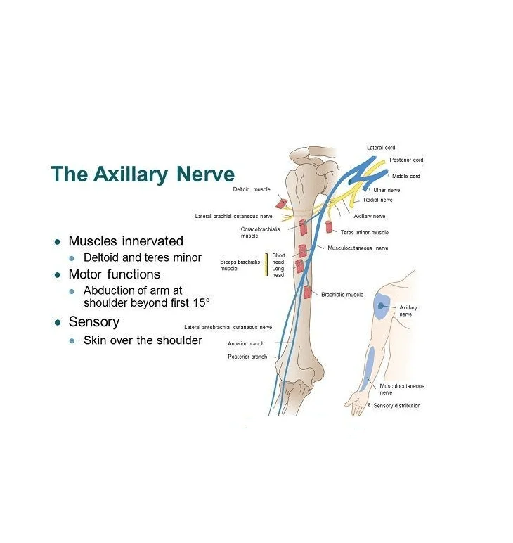
Axillary Nerve Anatomy The axillary nerve originates from the ventral rami of the C5 and C6 spinal nerves and extends as the smaller branch of the brachial plexus’s posterior cord. Its anterior supply the teres minor while its posterior branches supply the deltoid muscle. After action from the supraspinatus has already reached 15 degrees, the…

Food sources of Vitamin B 12
|

Vitamin B 12

What is a Vitamin B 12? Vitamin B12 is a water-soluble vitamin that is naturally present in some foods, added to others, and available as a dietary supplement and a prescription medication. Vitamin B12 exists in several forms and contains the mineral cobalt, so compounds with vitamin B12 activity are collectively called “cobalamins”. Methylcobalamin and…

Paraspinal Muscle

Paraspinal Muscle – Anatomy and Exercise

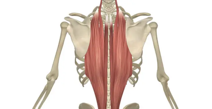
Anatomy: The paraspinal muscles, sometimes called the erector spinae, are three muscle groups that support your back. The paraspinal muscles are located on the left and right sides of your spine and are made up of three groups. These include: a) Iliocostalis b) Longissimus c) Spinalis A) ILIOCOSTALIS MUSCLE: The iliocostalis muscle is the most…

Femoral Nerve
|

Femoral Nerve: Anatomy, Course, Pathways, Function

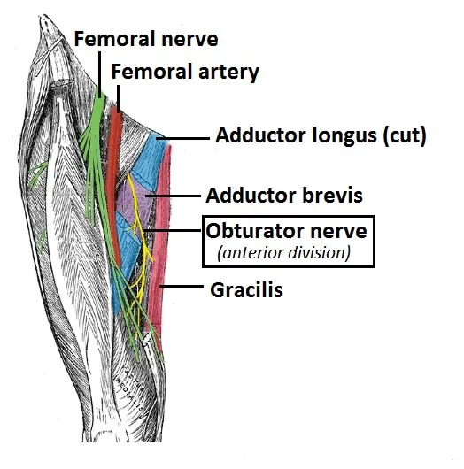
The femoral nerve is one of the major peripheral nerves of the lower limb. Descriptions: Nerve roots: L2-L4 Motor functions: Innervates the anterior thigh muscles that flex the hip joint (pectineus, iliacus, sartorius) and extend the knee (quadriceps femoris: rectus femoris, vastus lateralis, vastus medialis, and vastus intermedius). Sensory functions: Supplies cutaneous branches to the…

Abdominal Muscle
| | |

Abdominal Muscle – Anatomy and Exercise

Introduction The abdominal muscles form the anterior and lateral abdominal wall and consist of the external abdominal obliques, the internal abdominal obliques, the rectus abdominis, and the transversus abdominis. Acting together these muscles form a firm wall that protects the viscera and they help to maintain erect posture. In addition the contraction of these muscles…

Gluteus muscle
| | |

Gluteal Muscle Anatomy and Exercise

Anatomy of Gluteal Muscle Gluteus muscle, any of the large, fleshy muscles of the buttocks, stretching from the back portion of the pelvic girdle (hipbone) down to the greater trochanter, the bony protuberance at the top of the femur (thighbone). These include: 1) gluteus maximus2) gluteus medius3) gluteus minimus. 1) GLUTEUS MAXIMUS: This is a…

Median Nerve
| | |

Median Nerve: Anatomy, Origin, Course, Function, Clinical Importance

The Median Nerve is a nerve in humans and other animals in the upper limb. It is one of the five main nerves originating from the brachial plexus. Anatomy of Median Nerve: The median nerve originates from the lateral and medial cords of the brachial plexus and has contributions from ventral roots of C5-C7 (lateral…

calf muscle
| | |

Calf Muscle Anatomy: Anatomy and Exercise

Anatomy of the Calf muscle The calf muscle is located on the back of the lower leg and is actually made up of two muscles: The medial and lateral heads of the gastrocnemius muscle, which is the most superficial, are present. The gastrocnemius muscle belly is formed by the convergence of its two heads. The…

Hamstring Muscle
| | |

Hamstring Muscle: Anatomy And Exercise

Hamstring Muscle Anatomy The hamstrings are a group of muscles and their tendons at the rear of the upper leg. They include the biceps femoris, semitendinosus, and semimembranosus. The hamstrings flex the knee joint and extend the thigh to the backside of the body. They are used in walking, running, and many other physical activities….