Facial Muscle List
| | |

Facial Muscle List

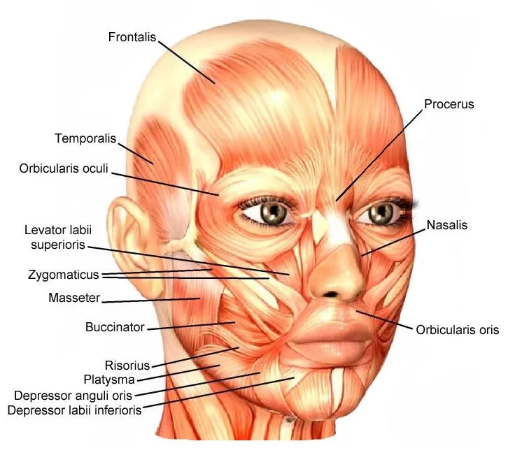
Facial Muscle Anatomy The facial muscles, also called craniofacial muscles, are a group of about 20 flat skeletal muscles, lying underneath the skin of the face and scalp. The muscles of facial expression (also known as the mimetic muscles) can generally be divided into three main functional categories: orbital, nasal, and oral. Contrary to the…

BRACHIAL PLEXUS Injury
| | | | | |

BRACHIAL PLEXUS INJURY

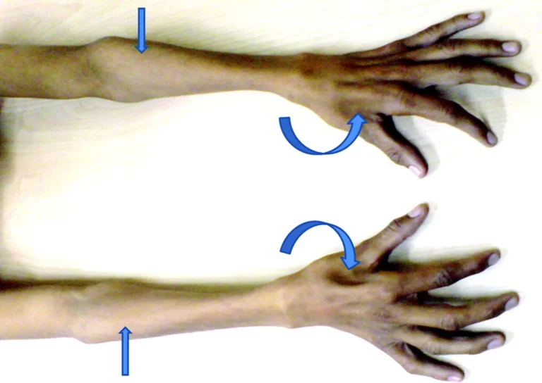
INTRODUCTION: A brachial plexus injury (BPI), also known as brachial plexus lesion, is an injury to the brachial plexus, the network of nerves that conducts signals from the spinal cord to the shoulder, arm and hand. These nerves originate in the fifth, sixth, seventh and eighth cervical (C5–C8), and first thoracic (T1) spinal nerves, and…

Facial Nerve
| | |

Facial Nerve: Anatomy, Physiology, Function and Clinical Importance

The facial nerve is the seventh cranial nerve, or simply CN VII. It emerges from the pons of the brainstem, controls the muscles of facial expression, and functions in the conveyance of taste sensations from the anterior two-thirds of the tongue. The nerves typically travel from the pons through the facial canal in the temporal…

Gluteal nerve
| |

Gluteal Nerve: Anatomy, Function, Importance

Gluteal nerve divides in superior & inferior gluteal nerve 1.Superior gluteal nerve 2.Inferior gluteal nerve 1.Superior gluteal nerve Anatomy of Superior Gluteal Nerve: The superior gluteal nerve (L4, L5, S1) passes through the sciatic notch above the piriformis muscle. It supplies the gluteus medius, minimus, and tensor fasciae latae muscles. The inferior gluteal nerve (L5,…

obturator nerve
| |

Obturator Nerve: Anatomy, Function, Importance

Obturator Nerve Anatomy The obturator nerve begins at the medial border of the psoas major muscle. It travels through the obturator foramen (an opening in the pelvic bone) before entering the thigh, where it branches into two parts, an anterior branch and a posterior branch. The nerve is part of a group of nerves called…

Axillary nerve
| | |

Axillary Nerve

Axillary Nerve Anatomy The axillary nerve originates from the ventral rami of the C5 and C6 spinal nerves and extends as the smaller branch of the brachial plexus’s posterior cord. Its anterior supply the teres minor while its posterior branches supply the deltoid muscle. After action from the supraspinatus has already reached 15 degrees, the…