Obesity Hypoventilation Syndrome (OHS)
What is Obesity Hypoventilation Syndrome?
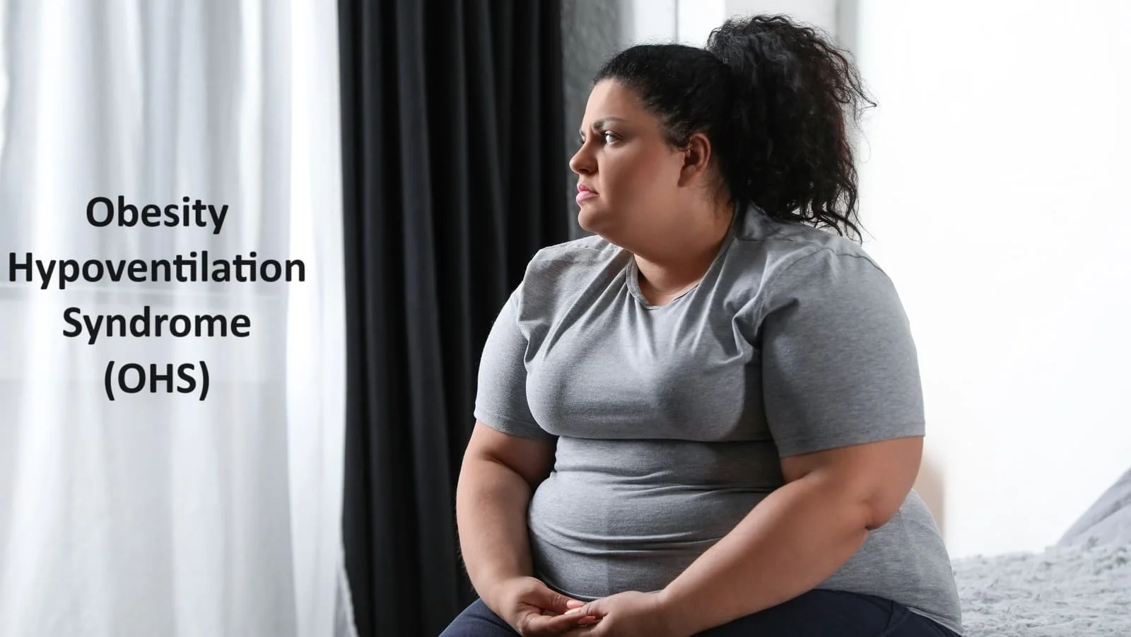
Obesity Hypoventilation Syndrome (OHS), also known as Pickwickian syndrome, is a medical condition characterized by a combination of obesity and hypoventilation (inadequate breathing). This disorder primarily affects individuals with obesity and involves abnormal respiratory function, resulting in elevated carbon dioxide (CO2) levels and low oxygen levels in the blood. OHS typically occurs in individuals with a body mass index (BMI) of 30 or higher and is more prevalent in middle-aged adults.
Due to the substantial mortality and morbidity risks associated with treatment delays, early diagnosis and treatment are crucial. Non-invasive positive airway pressure (PAP) therapy and weight loss are among the available treatment alternatives.
Long-term evidence about the efficacy of this therapy is few. In order to help identify and treat individuals with obesity-hypoventilation syndromes, this review summarizes present understandings of clinical presentation, diagnostic, and therapeutic procedures.
Introduction
Over the past three decades, the worldwide incidence of obesity has grown, posing a global health threat. According to a recent study, the prevalence of adult obesity in the US is 35.8% for adult women and 35.5% for adult males.
The combination of obesity (BMI > 40 kg/m2), hypoventilation during the day (PaCO2 > 45 mm Hg and PaO2 < 70 mm Hg at sea level), and sleep-disordered breathing (SDB) in the absence of another cause for hypoventilation (e.g., obstructive or restrictive lung disease, chest wall disorders such as kyphoscoliosis, neuromuscular disorders, and congenital central hypoventilation) is known as obesity hypoventilation syndrome (OHS).
Obstructive sleep apnea (OSA) accounts for about 90% of SDB in obese hypoventilation syndrome patients. The remaining 10% of patients exhibit sleep-related hypoventilation, which is defined by hypoxemia and is independent of the obstructive events of OSA, such as obstructive apneas or hypopneas.
Epidemiology
Although the actual frequency of OHS in the general population is unknown, estimates range from 0.3% to 0.15%. The prevalence of OHS in obese patients was shown to be substantially greater at 19 to 31 %. In a recent prospective and observational study, 9,480 patients at a tertiary care hospital participated in order to ascertain the prevalence of OHS in hospitalized patients. Individuals who had arterial blood gas measurements showing waking hypercapnia (PaCO2 > or equal to 45 mmHg) were included.
After analysis of 335 patients (3.4%) who met the inclusion criteria, it was shown that 35.5% of the patients had chronic hypoventilation, of which 27 patients, or 24.6%, had OHS. Other studies evaluating patients referred to sleep clinics with clinical symptoms of OSA and included a total of 1,326 patients discovered that OHS prevalence ranged from 11% to 20% overall. Studies conducted in the past that evaluated 1,927 patients who had a diagnosis of OSA determined that OHS was present in 9–14% of cases, with an overall prevalence of 11%.
As a result, it may be concluded that the prevalence of OHS varies and depends on the population under study, with hospitalized patients having the highest prevalence rate and population-based groups having the lowest. The hypercapnia requirement is more likely to be met by hospitalized patients, which has prompted some to propose that the definition of OHS should need to be reviewed.
Pathophysiology
Changes in alveolar ventilation in response to changes in metabolic rate and renal acid-base status keep the arterial pH in healthy persons within a specific range. Alveolar hypoventilation in OHS patients is encouraged centrally (by the “respiratory controller”) by a complicated interplay including aberrant respiratory mechanics, central respiratory drive, sleep-disordered breathing, and leptin resistance.
Alveolar hypoventilation during sleep causes nocturnal hypercapnia and hypoxia, which eventually causes the respiratory controller’s chemostat to reset, resulting in hypercapnia and hypoxemia throughout the day. Leptin resistance may exacerbate OHS’s hypercapnia. Leptin is essentially a respiratory stimulant that improves the function of the respiratory controller. Defects in upper airway neuromechanical regulation can result from leptin deficit or obesity-related leptin resistance. Nonetheless, PAP administered for a brief length of time can treat hypercapnia during the day.
Respiratory Mechanics
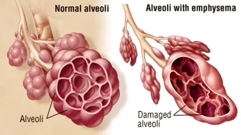
The respiratory system mechanics are significantly impaired in OHS patients compared to similarly obese eucapnic patients, whether or whether they have sleep-disordered breathing. The respiratory system is significantly burdened by central fat deposition in morbid obesity. This results in a dramatic decrease in lung volumes, increased airway resistance, and lung and chest wall compliance, all of which contribute to a higher work of breathing.
Compared to eucapnic obese people at similar BMI levels, OHS patients exhibit declines in vital capacity (VC), total lung capacity (TLC), and residual volume (RV). OHS patients had smaller lung volumes than OSA or eucapnic obesity patients because they have a higher degree of central obesity, as evidenced by larger neck circumferences and higher waist-to-hip ratios.
The observed increase in airway resistance and the considerable drop in lung and chest wall compliance are partially explained by tidal breathing at low resting lung volumes. Respiratory system compliance has been demonstrated to be roughly 20% lower in eucapnic obese persons and nearly 60% lower in patients with OHS when compared to participants of normal weight.
Breathing at low lung volumes can cause small airway closure during exhalation, which can limit expiratory flow and increase intrinsic positive and expiratory pressure (PEEPi). The work of breathing is further increased by such PEEPi, which in turn places an extra threshold strain on the inspiratory muscles before any inspiratory flow is generated.
When breathing spontaneously on room air, patients with OHS require substantially more work to breathe than either normal-weight persons or eucapnic obese patients with OSA. Supine position can increase upper airway inspiratory resistance to airflow and exacerbate respiratory system compliance and functional residual capacity (FRC). Compared to eucapnic OSA patients, who only exhibit resistance in the supine position, patients with OHS have increased upper airway resistance in both the sitting and recumbent positions.
Patients with OHS are far more likely to experience hypoxemia than people with eucapnic obesity. In morbidly obese people, low ERV exacerbates ventilation-perfusion mismatch, which is exacerbated in the supine position. Obese people adjust by breathing lower tidal volumes at greater respiratory rates, or “rapid shallow breathing,” which is energy inefficient, in order to maximize the oxygen cost of breathing. This breathing pattern exacerbates hypoxia and dead space ventilation. Due to their extended exposure to both daytime and nighttime hypoxia, people with OHS are more susceptible to pulmonary hypertension, congestive heart failure, and cor pulmonale.
Comparing OHS patients to eucapnic morbidly obese persons, respiratory muscle performance is compromised and deteriorates worse in the supine position. This observation has given rise to a number of theories. Potential reasons for diaphragmatic dysfunction include mechanical overstretching caused by belly obesity, persistent hypoxia, and hypercapnic condition, low levels of insulin growth factor (IGF-I), and decreased ventilatory drive.
Central Respiratory drive
At baseline, eucacapnic obese people had greater rates of CO2 generation and oxygen consumption. Increased respiratory drive to sustain alveolar ventilation and raise minute ventilation typically makes up for this.
However, those who have OHS are unable to adjust their respiratory drive to counteract the additional strain that comes with being overweight, which allows PCO2 to gradually rise. When compared to non-obese and eucapnic obese patients with or without OSA, central ventilatory response to hypoxia and hypercapnia is lessened in OHS patients.
The secondary cause of the reduced ventilatory response is the blunted neurological response to hypercapnia, which has been demonstrated to improve in the absence of a major weight shift within days or weeks of starting PAP therapy.
Sleep-disordered breathing
OHS is substantially linked to sleep-disordered breathing (90%) and OSA (most often), with sleep-related hypoventilation accounting for 10% of cases. Only a small percentage of individuals with severe OSA experience wake hypercapnia.
A meta-analysis of 15 studies revealed that, in comparison to eucapnic obese individuals with OSA, patients with hypercapnia had greater BMI and AHI, lower lung volumes consistent with restrictive lung physiology, and more substantial hypoxia on overnight pulse oximetry.
Leptin resistance and OHS
Adipose tissue produces the protein leptin. When compared to people without similar sleep disorders, obese people with greater blood leptin levels are found to have higher amounts of leptin. Leptin stimulates ventilation by acting on the central respiratory center. By preserving sufficient ventilation, elevated leptin levels in obese people may make up for the increased strain on the respiratory system. However, some people have less of this stimulatory action of leptin, which makes them more susceptible to hypoventilation and the ensuing hypercapnia.
Campo et al. discovered that elevated serum leptin levels are linked to decreased respiratory drive and hypercapnic response in obese people. Compared to eucapnic OSA patients with comparable leptin levels, hypercapnic OSA patients exhibit a considerably reduced hypercapnic ventilatory response. As a result, leptin resistance is assumed to play a role in the pathophysiology of OHS in humans in a manner akin to that reported in a leptin-deficient mouse model, which is characterized by abnormal respiratory muscle function, reduced lung volumes, compliance, and defects in upper airway neuromechanical control.
Symptoms of Obesity Hypoventilation Syndrome
Most Common symptoms of OHS are:
- Breathlessness
- Sluggishness or sleepiness
- Dizziness
- Fatigue, or extreme tiredness
- Headaches
- Depression
Your bed partner may observe the following signs while you’re asleep:
- Loud snoring.
- Choking or gasping.
- Pauses in your breathing.
Diagnosis
Your medical professional will do a physical examination and note your symptoms. Your body mass index will be determined by taking your height, weight, and measurements (BMI). Individuals that are obese have a BMI of 30 or higher.
Your physician can then suggest a few tests to aid in the diagnosis of OHS. These examinations could consist of:
- Arterial blood gas: To determine the amount of carbon dioxide and oxygen in your blood, your healthcare professional will draw blood from an artery.
- Pulse oximeter: To test the amount of oxygen in your blood, your healthcare professional will apply a sensor to your finger. Your blood’s level of carbon dioxide is not measured by this test. Additionally, it isn’t as exact as a blood sample in determining your blood’s oxygen content.
- Pulmonary function tests: These examine how well your lungs are working to rule out other potential reasons why you are having trouble breathing.
- Chest X-ray: This test can also rule out other potential reasons for dyspnea.
- Polysomnography, or sleep study: This test establishes whether you also have sleep apnea and, if so, the severity of your condition.
Treatment of Obesity Hypoventilation Syndrome (OHS)
The underlying pathophysiological causes of hypoventilation are the focus of OHS treatment. It can be broadly classified into three categories: medication, medically or surgically induced weight loss, and PAP therapy.
Positive airway pressure (PAP) therapy
PAP therapy significantly improves mortality, healthcare expenditures, and health-related quality of life. PAP therapy has been demonstrated to enhance blood gas levels during the day and sleep quality. Continuous positive airway pressure (CPAP) therapy or non-invasive positive pressure ventilation (NIPPV) with a bi-level PAP or other sophisticated PAP modalities are the two most common forms of PAP therapy.
Since CPAP therapy was originally advised to enhance nighttime gas exchange by preventing obstructive apneas and hypopneas and reducing the labor of breathing related to narrow airway closure and expiratory flow limitation, it is recommended that patients with OHS who have OSA. By decreasing the fluctuations in intrathoracic pressure required to sustain breathing, PAP treatment lowers the cost of breathing by improving FRC and negating the effects of PEEPi. Furthermore, PAP therapy increases oxygenation by widening the smaller airways, which reduces ventilation-perfusion mismatch.
Improvement in other physiological parameters, such as gas exchange during wakefulness and ventilatory response to hypoxia and hypercapnia, as well as improvement in symptoms and health-related quality of life, have been reported following the initiation of therapy over a few months in OHS patients. The majority of research showing CPAP therapy’s effectiveness in OHS patients has been small-scale and retrospective in design.
Non-PAP treatment modalities in OHS
NIPPV has been proven to be useful in patients with OHS by decreasing diurnal hypoventilation, quality of sleep, enhance gas exchange and symptoms associated with hypercapnia and mortality. Inadequate adherence and intolerance in certain patients restrict the benefits. It may not lessen the co-morbidities connected to obesity and has been demonstrated to have a minimal effect on weight loss. Options for non-PAP therapy include oxygen therapy, tracheostomy respiratory stimulants, weight loss, and exercise and rehabilitation.
It has been demonstrated that losing weight improves respiratory drive, respiratory muscle strength, lung function, and sleep-disordered breathing. Numerous researches have demonstrated the effects of weight loss by demonstrating notable enhancements in gas exchange and pulmonary function assessments. Such benefits are rarely realized with just medical and conservative treatments; substantial weight loss is required.
Medical Treatment
Medications may be regarded as NIPPV adjuncts or substitutes if they modify the ventilatory response to hypercapnia. Acetazolamide is a carbonic anhydrase inhibitor that can cause metabolic acidosis and lower serum bicarbonate, which can promote breathing. In 25 OHS patients recuperating in the intensive care unit (ICU) from acute hypercapnic respiratory failure, Raurich et al. examined the correlation between CO2 response, BMI, and plasma bicarbonate concentration, as well as the impact of acetazolamide on plasma bicarbonate and CO2 response. The CO2 response was lowest in patients with the greatest bicarbonate levels.
Acetazolamide was added, which increased the ventilatory response to hypercapnia and reduced the serum bicarbonate. It has been demonstrated that this ventilatory response is not high enough to treat OHS and is also varied. A recent Cochrane review of 30 randomized controlled trials involving 560 individuals with OSA found that medications like fluticasone, paroxetine, donepezil, and high-dose ondansetron plus fluoxetine improved subjective sleepiness during the day and baseline AHI. The short length and small sample size of the trials presented limitations. It was determined that there wasn’t enough data to suggest pharmaceutical treatment for OSA patients.
Exercise and Rehabilitation
For Obesity Hypoventilation Syndrome (OHS), exercise and rehabilitation are essential components of a comprehensive treatment plan. Because of their excess weight, OHS patients frequently experience respiratory difficulties.
However, with the right interventions, lung function, cardiovascular health, and general well-being can all be enhanced. The following factors should be taken into account for OHS patients’ rehabilitation and exercise:
Aerobic Exercise:

Walking, cycling, or swimming are examples of aerobic or cardiovascular exercises that can help with respiratory and cardiovascular health.
Building stamina and endurance can be achieved by gradual, supervised increases in aerobic activity. This is especially beneficial for people whose respiratory health is affected.
Respiratory Muscle Training:
Rehabilitation therapies for OHS can include specific exercises designed to strengthen respiratory muscles.
To improve respiratory function, a doctor may suggest respiratory muscle training, which includes exercises to increase the strength and endurance of the breathing muscles.
Weight Management:
A healthy weight can only be achieved and maintained by exercise, and weight loss is a critical part of treating OHS.
Regular physical exercise can help with sustainable weight loss when paired with dietary modifications, which lessens the strain on the respiratory system.
Pulmonary Rehabilitation:
Programs for pulmonary rehabilitation designed specifically for people with OHS may combine support, education, and physical activity.
Enhancement of exercise tolerance, improvement of lung function, and provision of tools for respiratory symptom management are the goals of these programs.
Strength Training:
Exercises involving resistance or strength training can enhance general muscular endurance and strength.
For people with OHS, it can be helpful to strengthen the muscles that sustain posture and are involved in breathing.
Monitoring and Individualization:
Exercise regimens for people with OHS should be closely watched and customized according to the patient’s needs, medical history, and degree of fitness.
The exercise program can be modified in accordance with results from routine evaluations of respiratory capacity and exercise tolerance.
Multi-disciplinary Approach:
A holistic approach to OHS management requires cooperation with healthcare providers such as dietitians, physiotherapists, and pulmonologists.
Exercise in conjunction with other therapy modalities guarantees a comprehensive and individualized approach for every person.
Before beginning any fitness program, people with OHS should speak with their healthcare provider. It is vital to have medical monitoring to guarantee safety, track advancement, and make any modifications. When paired with other therapy measures, a gradual and organized exercise program can make a big difference in the overall management and symptom improvement of people with Obesity Hypoventilation Syndrome.
Tracheostomy
By avoiding the upper airway, tracheostomy can lessen obstructive events linked to sleep disorders of breathing. Before Sullivan et al. effectively treated five patients with PAP therapy, the only treatment option for obstructive sleep apnea was tracheotomy. Since then, only a small percentage of patients getting home mechanical ventilation have had tracheostomy procedures performed.
The postoperative consequences of tracheostomy in critically ill morbidly obese patients with a BMI of 40 or higher have been previously reported by El Solh et al. They discovered that malpositioning due to an excess of adipose tissue around the neck and tracheostomy-related problems resulting from tube obstruction were both independently correlated with morbid obesity. Currently, tracheostomy is only used to manage OHS in cases where no other treatment works. There haven’t been any current studies assessing the effects of long-term home ventilation on tracheostomies.
Prevention
Maintaining a healthy weight will help lower your chance of having Open Heart Syndrome. If a CPAP or BiPAP was recommended by your physician, be careful to follow the instructions and continue the treatment.
Additionally, if you have an impending procedure or want to travel, discuss these with your clinician. You run a higher risk of experiencing severe problems in both of these scenarios.
Prognosis
With therapy, OHS symptoms may be lessened or eliminated completely. For some people, the symptoms can be alleviated by weight loss alone. Because treatment can enhance your quality of life, it is crucial. It also lessens the likelihood of developing new issues. By using a breathing apparatus early on, OHS mortality can be lowered by 10%.
If OHS is not treated, it can result in potentially fatal consequences such as heart and blood vessel problems. A deficiency of oxygen causes your heart to work harder. You run the chance of experiencing issues linked to sleep deprivation as well. Your risk of hospitalization rises and your quality of life declines without treatment.
For those who do not receive treatment, the prognosis is not good. Death from obesity-related hypoventilation syndrome usually results from untreated cases. Over an 18-month period, the death rate from OHS is 23% in individuals with underlying medical issues.
Complications
Obesity hypoventilation syndrome can lead to consequences from inadequate sleep if left untreated. These issues could include:
- Depression.
- Agitation and irritability.
- Increased risk of accidents.
- Issues with sex and intimacy.
OHS can potentially result in cardiac problems, such as:
- High blood pressure (hypertension).
- Right-sided heart failure.
- High blood pressure in your lungs (pulmonary hypertension).
When should I visit my Doctor?
You need to consult with a doctor if:
- You have additional OHS symptoms in addition to obesity.
- When you pause in breathing or snore loudly your bed partner notices.
- You have excessive daytime fatigue.
Conclusion
The obesity epidemic will inevitably lead to a rise in the prevalence of OHS. OHS is sometimes misdiagnosed and usually underdiagnosed. These people have a low quality of life connected to their health, a high mortality rate, longer hospital stays, and worsening co-morbidities associated with their disease if they go untreated and undetected. In order to significantly lower the mortality and morbidity of untreated OHS, it is imperative that early diagnosis and therapy be instituted.
Early diagnostic clues may be provided by identifying clinical traits and predicting factors during clinic visits and nocturnal polysomnograms. It is anticipated that a multimodal and multidisciplinary therapeutic strategy combining weight loss and rehabilitation programs with NIPPV will have a higher influence on the outcomes related to heart, metabolism, and HRQoL.
FAQ
How do you treat OHS syndrome?
Losing weight and managing your sleep-related breathing issues are two aspects of OHS treatment. Obstructive sleep apnea is one of the many symptoms and issues that can occasionally be resolved by weight loss alone. Losing weight is therefore the first step in treating your OHS.
Is obesity hypoventilation syndrome a restrictive lung disease?
One of the main respiratory effects of obesity is obesity hypoventilation syndrome. Testing for lung function usually reveals mild to moderate restrictive abnormalities.
What is the medical term OHS?
Some obese individuals experience impaired breathing due to obesity hypoventilation syndrome (OHS). It causes the blood’s levels of carbon dioxide to rise and oxygen to fall.
What is the difference between OSA and OHS?
Obesity and diurnal hypercapnia are hallmarks of obesity hypoventilation syndrome (OHS). The following are the characteristics of OHS that set it apart from obstructive sleep apnea (OSA): longer and more frequent episodes of hypoventilation during the night (perhaps with or without upper airway obstruction).
What medication is used for obesity hypoventilation syndrome?
Obesity-hypoventilation syndrome and central hypoventilation syndromes have been demonstrated to benefit from medroxyprogesterone’s enhancement of the central respiratory drive.
References
- Shetty, S., & Parthasarathy, S. (2015). Obesity Hypoventilation Syndrome. Current Pulmonology Reports, 4(1), 42. https://doi.org/10.1007/s13665-015-0108-6
- Professional, C. C. M. (n.d.). Obesity Hypoventilation Syndrome. Cleveland Clinic. https://my.clevelandclinic.org/health/diseases/24393-obesity-hypoventilation-syndrome