
Patella Alta
Definition
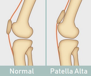
Patella Alta or high-riding patella mention to be an abnormally high patella in relation to the femur. The patella sits high on the femur where the groove is especially shallow. Here, the sides of the femoral groove provide only a tiny barrier to keep the high-riding patella in place.
This condition has been associated clinically with patellofemoral dysfunction or it is considered a predisposing factor for the development of patellofemoral pain (PFP). This makes the knee a little more stable and more prone to dislocation and anterior knee pain. The Patella Alta is also distinguished by the instability of the patella. In a few cases, the person is born with patella Alta, but it can also develop secondary to the knee injury such as a rupture of the patellar tendon.
What Is Patella Alta?
This is the condition where people are born with a kneecap (patella) positioned higher in the front of the knee than the average.
The patella, or the kneecap, is a little inverted (upside down) triangular bone that sits at the front of the knee. The kneecap is surrounded by the quadriceps tendon or rests in a dip on the front of the lower thigh bone, the femur, forming the patellofemoral joint.
This dip is called the patellofemoral groove, aka patella groove, trochlear groove, or intercondylar groove. As the knee moves, a patella slides up and down over this groove. Normal patella position with patella Alta aka high riding patella. The patellar tendon comes out from the bottom of the kneecap and connects the kneecap to the shin of the tibia. In the patella Alta, the kneecap sits higher than normal in the patella groove. Here, the groove is much shallower than further down, thus providing only a very minute barrier on each side of the kneecap.
As a result, the groove provides very small sideways stability for the kneecap. They are very frequently good athletes and seem to do well in the high jump and the triple jump, or basketball. The problem with patella Alta is that the knee cap is very mobile from side to side or it is predisposed to dislocation during sporting activities. Once the patella has dislocated several times it is known as recurrent dislocation and can be a major nuisance to the sporting individual causing them to give up all sporting activities. It has been the end of many basketball and football players’ careers, in whom the condition is common. In most people, the dislocation is incomplete, and then it is known as subluxation of the patella.
In a few cases, the patient may be able to dislocate the patella at will or they sometimes do it as a party trick. This is called habitual dislocation. As its name proposes that it can become a habit. It is not clever because it still creates little damage and will result in osteoarthritis in the longer term.
Both dislocation and/or subluxation are very painful and both result in damage to the hyaline cartilage under the patella and to the groove in which the patella runs (the trochlear groove of the femur) which after a time causes osteoarthritis of the patellofemoral joint with severe pain or weakness of the muscles.
Clinically Relevant Anatomy
The patella is the flat, inverted triangular bone, which is situated on the front of the knee joint. The patellofemoral joint is the part of the knee joint between the patella and the femoral condyles. The patellofemoral articulation fully depends on the function of the which are connected to the patella with a shared tendon.
The quadriceps femoris is separated into four different muscles with the same insertion on the patella: a rectus femoris (RF), the vastus lateralis (VL), and a vastus intermedius (VI) or the vastus medialis (VM). There is also a tendon that connects the bottom of the patella to the tibia, known as the patellar tendon. This tendon is very strong and allows the quadriceps muscle group to straighten the leg. These three bones are covered in articular cartilage which is an extremely hard, smooth substance designed to decrease friction forces. The patella lies in an indentation of the femur called the intercondylar groove. Patella dislocation is an often complication of patella Alta.
Why Is Patella Alta A Problem?
In general, when you bend and straighten the knee, the patella glides up and down in the middle of the patellofemoral groove. The groove is nice and deep, forming a good barrier at the sides to keep the patella extremely stable by limiting the sideways movement of the kneecap. But with patella Alta, because the kneecap is sitting higher than normal in the shallower part of the groove, there is less stability sideways.
The patella is therefore at risk of being pulled sideways over the low edge of the groove as the knee bends, and can partially or completely dislocate out of position. surround 30% of cases of recurrent patella dislocation are thought to be due to patella Alta. Any time the patella dislocates and subluxated (partially dislocates), the cartilage on the back of the kneecap is at risk of damage which can lead to patellofemoral pain, aka anterior knee pain, or chondromalacia. Having patella Alta also increases the risk of developing knee arthritis and inflammation in the infrapatellar bursa or a fat pad.
What Causes Patella Alta?
Though there is no exact etiology or cause for the existence of the patella Alta few of the factors can be claimed as reasons for the onset of this specific ailment. The frequent causes of patella Alta are:
- Congenital Defect: This defect is acquired during the development of the embryo. Under this condition, a person has had patella Alta conditions since its birth.
- Long Patellar Tendon: the individual with abnormally long patellar tendons, > 52mm, often suffers from patella Alta
- Knee Injuries: the Patella Alta may develop after a knee injury, typically in kneecap dislocation
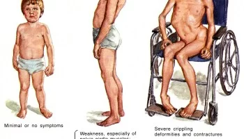
- Cerebral Palsy: Patella Alta may be a common abnormality with cerebral palsy, particularly in children who walk with bent knees.
- the Person’s Body Structure can be a cause of patella Alta. A person having a tall structure along with a thin built of the body is very much prone to this particular ailment.
- Patellofemoral Pain can cause patella alta. It is a type of syndrome which arises due to the contact of a knee cap with the thigh bone. Anyone found with this syndrome is at greater risk of this patella Alta disorder.
- Injuries of the Knee can cause patella Alta. This might be a primary cause for the patella Alta’s medical condition as various strenuous activities like sports can set the kneecap out. All of a sudden change in the direction during the activity could make it happen very common. A pulled-out kneecap would result in a dislocation of the patella which further can develop into high-ride kneecap conditions.
- Knee Twisting might also cause patella Alta. Any reason other than the sporty activity, under which often change or sudden change of direction is associated, such as jerks, could cause this patella Alta condition.
- Patella Alta can occur as the result of a sports injury, though, it is the large majority of the time it is a congenital or developmental condition that is unrelated to trauma. Its pathophysiology is not completely clear, but it is hypothesized that one of the causes of patella Alta is abnormally long patellar tendons (>52 mm).
Patella Alta Sign and Symptoms
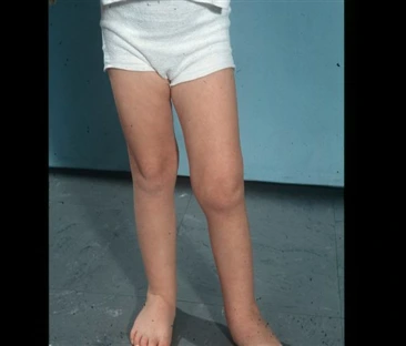
There are few signs and symptoms for patella alta, person suffering from the patella Alta condition has a dislocated kneecap which is, somehow, got oddly positioned at the higher side of the femur bone. This irregularity can create a few noticeable signs and symptoms, such as:
- Pain in the Patella is a great symptom of patella alta. The patient could complain of the pain in the patella portion, but it is a subject matter of proper diagnosis for patella alta condition as every patella pain could not be claimed as a high-ride condition.
- Chondromalacia Patella is additionally a symptom of patella alta. This condition, indicating patella alta, is identified by fissures and blisters around the patella cartilage that worsen because of the patella misalignments.
- Gait Instability might be a symptom of patella alta. People having unstable or irregular gait might have a condition of patella alta as it is primarily responsible for unsteady gait conditions.
- Dislocated Knee Cap is on other extensive symptom of patella alta. When the patella gets bent higher than its normal bending it pulls out the knee cap from its actual grooves and results in a condition of painful dislocation of the knee cap. Any of such conditions could be a reason for the patella alta ailment.
- Typical symptoms of patella Alta include:
- Instability: an individual with patella Alta often complain that their knee feels weak or unstable, particularly when walking or running
- Recurrent Kneecap Dislocation: knee dislocation may be a common problem for people with a high-riding of the patella. few people can push their kneecap in and out of position in the patellar groove at will causing it to dislocate and then relocate
- Anterior Knee Pain: the pain at the front of the knee, aka patellofemoral pain, is often with patella Alta, especially when walking up and down slopes, squatting, sitting for prolonged periods, or on stairs.
- Camel back Sign – generally, the patella points straight forward. And there is only one prominence famed and that is of tibia tuberosity. In patients within the high-riding patella, the patella points upward. In these patients, there is a prominence of the infrapatellar fat pad in extension along with tibial tuberosity, giving an impression of impact prominence such as camel’s back.
- Grasshopper Eyes – This is better appreciated in bilateral patella Alta. few patients may have externally rotated patellae consistent along with patella Alta and lateral tilt. There is often known as the grasshopper eyes sign.
Features and Clinical Presentation
- The Patella Alta could also be a positional fault defined most simply as a superior displacement of the patella within a trochlear groove of the femur. Patella Alta has been shown to be related to chondromalacia on the articular surface of the patella or pain. Patella Alta has also been involved in patellar osteoarthritis. The prevalence of patella Alta in people with patellar osteoarthritis was 6 times that of a person with normal patellar articular cartilage. As well, both Leung et al (1996) and Kannus (1992) noted that subjects with anterior knee pain demonstrated a significantly more superior patellar position within the affected knee relative to healthy, control knees.
- A common symptom of patellar injury and/or dislocation is acute pain after direct contact or a sudden change of direction. With sudden changes in direction, the femur medially rotates over the underside and stabilized the tibia. Under these conditions, athletes commonly feel the knee giving way, which may be a result of quadriceps inhibition from pain, a physiologic protective mechanism. with any knee flexion Rapid swelling, intense knee pain, and difficulty often occur. other dysfunctions with similar presentations or mechanisms of injury are meniscal and ligamentous injuries, particularly injuries of the anterior cruciate ligament.
Symptoms can also manifest as a steadily progressive sensation of anterior knee pain along with increased physical activity. Extreme physical activity increases JRFs across the knee. Such activities comprise inclined ambulation, squatting, prolonged sitting, or going up and down stairs. the Anterior knee pain is aggravated by activity is typical of chondral pathology. a Knee pain that improves during physical activity but returns after activity proposing tendinitis.
Associations Following conditions might also be associated with patella Alta and There are a lot of conditions that are called to be associated with patella Alta. For instance
- Patellofemoral instability
- Recurrent patellofemoral dislocation
- Neuromuscular diseases like poliomyelitis
- spastic cerebral palsy
- Osgood Schlatter disease
- Sinding Larsen Johanssen disease
- Patella tendon-lateral femoral condyle friction syndrome
- Chondromalacia patella
Effects of Patella Alta on Biomechanics
Instability and patellofemoral arthritis are two particular occurrences in patella Alta. Normally on knee flexion, the patella glides into a reciprocal groove within the front of the femur, called the trochlear groove. The groove keeps the patella stable within the middle of the knee [sidewise barrier] as it moves up and down. The patella during a straight knee sits above this groove, entering the trochlear groove at about 20 or 30 degrees of knee flexion. Till the time it enters into the groove, the patella has the potential to slip sideways especially laterally. If the patella is sitting too high then the patella will only take part within the trochlear groove later in the flexion arc (i.e. >20-30 degrees). This proposes that the patella is less stable for a greater percentage of the time. another thing is the greater windscreen wiper effect.
Differential Diagnosis
There are a lot of conditions that are known to be associated with patella Alta and these include:
- Patellofemoral instability
- Recurrent patellofemoral dislocation
- Neuromuscular diseases (poliomyelitis)
- Spastic cerebral palsy
- Osgood Schlatter disease
- Sinding Larsen Johanssen disease
- Patella tendon-lateral femoral condyle friction syndrome
- Chondromalacia patella
How Is Patella Alta Diagnosed?
To diagnose patella Alta, a doctor will start by examining your knee. They are going to look at the position of the kneecap in relation to the femur from various angles or with the knee in different positions.
Examination
The patient sits on the sting of the examination table with their feet on the bottom. knees are bent at 90 degrees, and thus the thigh is horizontally positioned. The vertical position of the patella height is best seen from the lateral. In Patella Alta, it is often seen that the partially tilted patella protrudes above the level of the thigh. This is more commonly remarkable In a unilateral Patella Alta. A patella Alta could also be noticed during inspection thanks to a so-called Camel hump patella, The knee has two striking bulges: one is the tibial tuberosity, and therefore the other one is the patella. The space distal to the patella or proximal to the fat body of Hoffa is distinguished by a notch.
Physical Examination:
The second diagnostic step may be a careful, complete, and essential physical examination. The aim of this examination is to reproduce the feature (pain/instability) and to locate the painful zone. The situation can indicate which structure is injured, it is truly helpful to compose the diagnosis or to plan the treatment.
Tests:
Fairbanks patellar apprehension test:
The test is positive when there is pain and muscle defensive contraction of lateral patellar dislocation with 2030 of knee flexion. The positive test indicates that lateral patellar instability is a crucial part of the patient’s problem. This might be so positive that the patient pulls the leg back when the therapist approaches the knee with his hand, preventing so any contact or the patient grabs the therapist’s arm. Greater than 100% sensitivity, 88.4% specificity, and overall accuracy of 94.1%.
Patellar glide test:
This test is used to evaluate the instability. A medial/lateral displacement of the patella greater than or adequate to 3 quadrants, with this test, is according to incompetent lateral/medial restraints. Lateral patellar instability is more common than medial instability.
Diagnostic Procedures
Patella Alta is usually defined with imaging-based measurements and rarely by description. Imaging comprises lateral radiographs, sagittal MRI, radiographic ratios measured on MRI, and patellar tendon length. cutoff values for patella Alta varied from > 1.2 to > 1.5 for ISI or from > 1.2 to > 1.3 for CDI. Both indices were seldom used on MRI; cutoff values were the same as those for conventional radiographs. On sagittal MRI, the patellar trochlear index was used most; cutoff values ranged from < 0.125 to 0.28. Eleven studies used patellar tendon length and situated it was increased (> 52 mm to > 56 mm).
Measurement of the patella alta
Patellar height measurement becomes important in the diagnosis of patella alta. Patella alta is a condition where the patella is high riding than its normal position. Measurement of patellar height can confirm or rule out the presence of patellar height anomalies. The following methods are used commonly:
- Insall-Salvati ratio
- Modified Insall-Salvati ratio
- Blackburne-Peel ratio
- Caton-Deschamps index (knee)
- Blumensaat method
Several methods are wont to determine the presence of patella Alta.
Insall-Salvati
It was the first to describe a method of establishing patella height on the basis of the ratio of the length of the patella tendon to the diagonal length of the patella on lateral radiographs. Various techniques, including those by Blackburne and Peel, Caton et al., and de Carvalho et al., have since been developed in an attempt to classify the patella position. A normal value of the patella is a ratio between 0.8 to 1.2. If the ratio is on top there is patella Alta.
Modified Insall-Salvati ratio
It is also applied on a lateral 30-degree of flexed knee radiograph but the measurements are slightly different. Distance from the inferior margin of the patellar articular surface (Instead of the lower pole of the patella in the original index) to the patellar tendon insertion and the Length of the patellar articular surface (Instead of the pole-to-pole distance).Modified Insall-Salvati ratio = A/B. The normal value is 1.25. greater than 2 which is considered diagnostic of patella alta.
The Blackburne-Peel index
This index can also be used to determine the presence of patella Alta. In this method, a lateral radiograph of the knee with 30 degrees of flexion is obtained. The lengths of three surfaces must be measured to work out if there is a patella Alta. The primary length is a vertical line between the top of the inferior aspect of the patella articular surface and the horizontal line of the tibial plateau. The second length is vertical and is that of the superior patella articular surface. Both vertical lines are measured on the posterior side of the patella articular surface. The third length may be a horizontal line on the tibial plateau. This technique is not easy on an MRI, because it requires measurement across different sections. A traditional value of the patella is a ratio between 0.5 and 1.0. When the ratio is higher than 1.0, there is a patella Alta.
The Caton-Deschamps index
It is the most commonly used radiographic technique for the evaluation of patellar height. This is measured on a lateral radiograph of the knee in 30 degrees of flexion. it is the ratio between the distance between The inferior most aspect of the patellar articular surface and the upper limit of the tibia, and The length of the patellar joint surface. Caton-Deschamps index can also be measured on a lateral radiograph or sagittal knee CT or MRI reconstructions with the knee flexed at an angle of 30º. This technique is easy to measure on an MRI. To determine the height of the patella. A normal value of the patella is a ratio between 0.6 to 1.3. When the ratio is higher than 1.3, there is a patella Alta.
Blumensaat method
This method is also used on the lateral view of an x-ray in 30 degrees of flexion. It uses the Blumensaat line or roof of the intercondylar notch as a reference line. The position of the lower pole of the patella (above or below the Blumensaat line) is reported. usually, a lower pole of the patella touches this line in 30 degrees of flexion. Moreover, the distance between the lower patellar pole and the Blumensat line in millimeters is noted.
How Do You Treat Patella Alta?
Patella alta can be treated in both ways either conservatively or surgically. Further details are explained below.
Nonoperative Treatment
Patellofemoral Instability: acute dislocation of the patella
Nonoperative treatment: It is a treatment that contains of immobilization followed by a period of structured rehabilitation. Immobilization is granted for the healing of the soft tissues, mainly the supporting structures on the medial side of the knee. Today brace treatment with early mobilization has become the norm, rather than the traditional immobilization of 3 to 6 weeks in a cylinder cast.
Most of the time these therapies start with the initial straight leg raises: Quadriceps setting exercises and/or three sets of 15 to 20 straight leg raises are done four or five times a day in the acute period. the Ice is applied for 20 minutes every two to three hours to decrease swelling. It is followed by a stationary bicycle for passive and active movement, and isotonic or isometric quadriceps strengthening. In the middle of three and eight weeks, the return to full activities was allowed when tenderness weakened or isotonic quadriceps strength was bilateral. In up to two to thirds of the knees the results were good to excellent, these results consist of a first-time acute dislocation, compared with only 50 % of those with recurrent dislocation. In universal, 73 % were satisfied with their knees, but 16 % were not and eventually decided to have surgical stabilization. Those with acute patellar dislocation can expect an extended rehabilitation period before returning to the sport, whether it is immobilized or not.
Patellofemoral Instability: Recurrent Dislocation of the Patella
Nonoperative treatment: Surgery is not necessarily required for the patient with patellofemoral malalignment or relaxation of the patella. Adequate results could be achieved with a conservative exercise treatment program. It is very specific in the rehabilitation program to strengthen the quadriceps muscle and vastus medialis obliquus (VMO). It is advised to follow the program which is the same as that followed after acute dislocation, but with more resistive exercises. This program also can be started early. In addition, a stabilization brace of the patella may help to stop chronic recurrent subluxation. evidence level: 3
Treatment for patella Alta aims to scale back knee pain and instability and restore full knee function. Non-operative treatment for a high-riding patella will comprise:
- Rest: from the aggravating activities to allow any inflammation to settle
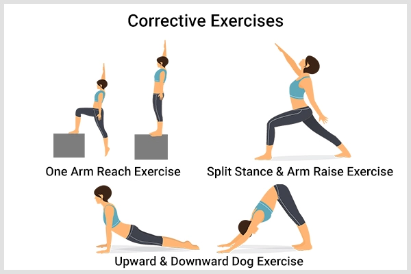
Strengthening Exercises:
strengthening the knee, kneecap, and buttock muscles can help to correct the patella position and thus reduce pain and improve knee stability with a high-riding patella. exercises are one of the good ways to cure knee pain and prevent it from coming back.
- Quad Clenches- it helps maintain and strengthen the quad’s muscles without moving the knee and helps enable full straightening of the knee. Lie flat down on your back or sit up. Leg and/or knee straight, Tighten the muscle on the front of the thigh by pushing your knee down. You should feel your thigh muscles clench. Hold for 3 secs. Repeat 10 to 20 x every 3 to 4 hours. If you are stumbled to get your knee to straighten fully, place a rolled-up towel underneath the ankle so that your leg is lifted slightly on the bed, Then do the exercise as described. Lifting the knee up slightly lets gravity assist the knee to straighten.
- Short Arcs- it helps to strengthen the quad’s muscles without requiring much knee movement and improve control. Lie flat on your back or sit up with your leg horizontally on a flat surface such as a bed. Place a rolled-up towel (approx 10 cm diameter) under the knee and/or Pull your toes towards you and clench your thigh muscles, then Slowly lift your foot up off the bed til your knee is straight (keep your knee resting on the towel). Hold for 3 to 5 secs and slowly lower repeat 10 to 20 times, 3 sets daily. You can challenge yourself further by Increasing the size of the towel under the knee, Adding a weight e.g. by wearing a shoe or using a light ankle weight, and progressing further by using a heavier weight.
- Straight Leg Raise(SLR) – Lie on your back with your hips straight and your legs laid out comfortably on the floor. Bend the knee of your unaffected leg at a 90-degree angle, planting the foot flatly on the floor. Stabilize the muscles on your straight leg by contracting your quadriceps (the group of muscles on the front of your thigh). Inhaling slowly then lift the straight leg six inches off the ground. Hold for three seconds. Exhaling slowly, lower the leg to the floor with control Relax and repeat 10 times more. When done correctly, you will feel the tension in your hip, thigh, and abdomen throughout the movement.
- The Clamshell – Lie on your one side with your feet and hips stacked, bend your knees to 90 degrees, and then your head resting on your right arm. Draw your knees in toward your body til your feet are in line with your butt. Place one hand on your other hip to ensure it does not tilt backward., This is your starting position. Keeping your abs engaged on your feet together, raise your one knee as far as you can without rotating your hip or lifting your other knee off the floor. Hold for 1 second, squeezing your glutes at the top of the move, before slowly lowering your one knee to the starting position. Continue for a total of 20 reps, then repeat on the other side.
- other exercise includes One Leg Standing, Hamstrings Clenches, The Bridge, Long Arcs, Knee Marching, Kick Backs, Heel Raises, and Buttock Kicks.
Knee strengthening exercises help to improve both the strength and control around the knee which is most important: For knee stability & control
- To ensure correct biomechanics in the feet, knees, hips, and back
- To reduce the risk of injury
- To allow full, pain-free movement
- To ensure full function
Physical Therapy:
Manual therapy could also help to improve the resting position of the kneecap. Manual gliding is performed to theoretically modify the resting height of the patella before knee extension, resulting in reduced pain in the knee. Correction of the positional fault of the patella by utilizing tape is a method to correct patellar alignment. Patients noted that they were having minimal difficulty with walking, Their average pain was reported to be a 12/10 on a VAS scale, and they noted decreased use of pain medication.
- Ice Packs: regularly applying ice packs could also help reduce pain and inflammation with symptomatic patella Alta – see the ice wraps section. Knee ice wraps are a good way to decrease inflammation, swelling, and pain. They could also help with recovery from an injury, or with longer-term knee conditions. They range from single-use disposable packs to specially designed ice bags, and inserts for knee braces with the higher-quality of cryotherapy. There are five different types of ice wraps covered here and you may compare models, read reviews, or find the best one for you:
- Ice Bags: Simply fill with ice or water and apply. Reusable material bag
- Compression or Ice Packs: Combine ice therapy with compression
- Reusable Ice or Heat Packs: Choose between cold or heat therapy
- Instant Ice Packs: Perfect for out and about. No prep time
- Cryocuff: on the Top of the range ice therapy. Particularly good after knee surgery
- Knee Brace: wearing a brace may also help to reduce the symptoms of a high-riding patella. Ideally, the brace should have a tubular section that sits above the kneecap to stop it from riding up. Knee braces provide different levels of support and fix the knee in a number of different ways. They also come in different materials or vary enormously in price. Bracing has been found to reduce pain and increase the patellofemoral contact area, in patients with patellofemoral dysfunction and anterior knee pain. although, during a study to assess the effect of a brace developed to prevent medial and lateral patellar subluxation, Shellock, et al (1994) demonstrated that the brace was not effective within the presence of patella Alta. Of the five knees that did not show any improvement in this study, four of them had patella, Alta. Because the brace utilized in this study only provided stabilization for medial and lateral patellar glide, it might not have been the most optimal brace design for individuals with patella Alta.
How do I tape my patella, Alta?
procedure
Start the tape in line with the middle way of the knee cap at the outer aspect of the knee. Using your thumb on top of the sports tape, kindly push the patella towards the inner aspect of the knee whilst simultaneously using your fingers to pull the skin at the inner side of the knee towards the patella. by the end of this taping technique at the inner aspect of the knee ensure you have created some wrinkling of the skin at the inner side of the knee. Repeat this process 1 to 3 times depending on the amount of support required. The goal of tapping is to create a mechanical realignment of the patella in the intertrochlear groove and reduce pain. Although patellar taping seems to reduce pain and improve the performance of individuals with PFPS, the exact mechanisms of these phenomena cannot be explained.
The Main Advantages of Patella Taping
Improvement in Disability, Effect on Quadriceps Function, Effect on Knee Joint during Gait, and Re-align the Patella. Principles of taping – Protection of the skin, Check the skin sensitivity of the person to be taped so that he is not allergic to the adhesive tape. Make sure there are no existing rashes or broken skin in the area to be taped. Hair removal in the area to be taped, It is better if the hair is removed 12 hours before the tape application to reduce skin irritation. Clean and prepare the skin. Tape may be used to: Stabilize or support an injury, Relieve pain by de-loading vulnerable or painful structures, and Facilitate normal movement, muscle action, or postural patterns. The padding of the sensitive areas within the adhesive tape.
Patellar Taping:
A taping could also help to correct the position of the patella. Taping is often used as an adjunct or temporary technique. The tape is frequently used by physiotherapists to – relieve your pain, improve joint stability, enhance athlete confidence, decrease injury recurrence, prevent injury, correct faulty biomechanics, inhibit muscle action, facilitate muscle action, reduce strain on injured or vulnerable tissues, enhance proprioception, compress in the presence of edema or lymphatic drainage.
Operative Treatment
To get back the actual position of the knee the process of surgery for the patella alta can be carried out. Osteotomy which is the advancement of tibial tuberosity is a surgical procedure that is usually applied to correct the deformity in the knee position. Whenever you undergo surgery for patella alta the movement of the knee is restricted and at least six weeks and this can be called a knee splint or sometimes knee braces. In a few cases, if the patient does not respond to any non-invasive cure then only surgical procedures for patella alta are carried out. Depending upon the severity of the patient’s condition there are numerous surgical options. Some of these are listed below:
Tibial Tuberosity Osteotomy: aka Tibial Tubercle Transfer
This is when the patellar tendon attachment is moved down, which in turn brings the patella down with it. The Q angle could also be corrected if necessary by moving the patellar tendon attachment inwards. Arthroscopy for patella alta is used to examine the knee joint as well as through small incisions, repairs are being made.
A tibial tuberosity osteotomy or TTT (Tibial Tubercle Transfer)
It is done by removing the tibial tuberosity from the front of the shin bone, moving it down, and reattaching it in its new position with a screw and wires. In order to align the position of the knee cap along with the femur such a method is implemented. To prevent recurrent dislocation as well as to make the more compact form of the knee cap this method is more used. the structures on the outer side of the knee are pulling the kneecap out of the position with a high riding patella then lateral release surgery may be carried out to loosen them. search out what surgery involves in the lateral release section. In lateral release surgery for patella alta compact tissue structures are loosened with the help of a surgical process. At present time you will find some of the controversial processes for lateral release.
Patellectomy:
This is where the patella is completely removed and is only indicated when other surgical methods have failed or where there is more severe patellofemoral arthritis. it is a process in which the knee cap is removed by carrying out a surgical operation. It can proffer permanent relief of pain as well.
Recovering From Surgery
Recovery Period or Healing Time for Patella Alta – Treatment mode defines the recovery period healing time in patella alta while a doctor’s assessment can also be used to get the approximate healing time in patella alta. You can seek your doctor’s advice for its recurrence as well as the recovery time for the patella alta.
Following surgery for patella Alta you will:
- Wear A Knee Brace: A hinged knee brace is generally worn for up to six weeks following surgery. The brace will limit the number of flexion at the knee to allow it to heal properly. A hinged knee brace is one of the simplest ways to reduce knee pain and instability. They offer much more protection and stability than the easiest pull up the sleeve and wrap surround knee braces on the market. Some hinged braces may be locked at different amounts of flexion and extension so you can control how much your knee bends and straightens. This could also is really useful in the early stages of recovery from knee injuries or surgery. Hinged knee braces might also be used for everyday activities or high-demand activities including sports and skiing.
- Use Crutches: For a primary couple of weeks, you will only take minimal weight through the operated leg so will need to use crutches. You will then gradually be able to increase the load through the leg over the next few weeks. Most of the individuals are off crutches for around six weeks. Getting up and down stairs need a combination of strength, mobility, knee flexibility, balance, and control
- Have Physical Therapy: Once you are allowed to start moving your knee, you will start physical therapy to assist or to regain full strength, stability, movement, and function in your knee. By around 3 months you may start gentle impact activities such as lunges and then progress on to gentle jogging. It generally takes approx six months to fully recover from patella Alta surgery and get back to high-impact activities and sports.
Medical Management
- Tibial tuberosity osteotomy is often performed in patients with patella Alta. With the surgery, they could also move the attachment of the patellar ligament downwards to the tibia. The patella is additionally attached to this ligament, thus the patella moves downwards. The quadriceps angle could also be increased with patella Alta, this will be corrected by shifting the bony attachment of the patellar ligament inwards.
- A couple of things that may happen after the surgery is done are infection, stiffness of the knee joint, nerve injury, and recurrent instability. The patient has to use crutches to walk after the surgery is completely done. This might also affect the movement of the knee, this is why physical therapy and exercise should be done after surgery in order to get rid of the pain and swelling. Muscle control would also increase with physical therapy. The recovery period must be around 3 to 6 months. evidence level: 4
Complications in Patella Alta
Instability and patellofemoral arthritis are two main complications in patella alta. A person having a condition of patella Alta are generally at a greater risk of having a further disorder of patellar instability. If the dislocation or bending of the patella is very higher in nature it got engaged around a flexion arc. The degree at angle is the bent is approximately 20 to 30 degrees. It implies that the patella is very less stable in long run.
yet, the patellar tendon which becomes longer has a higher windscreen wiper effect or you can say it performs higher action. It becomes quite unstable due to an increase in the size of the patellar tendon and thus it moves side by side. There is pressure exerted on the kneecap due to elevated riding patella which might give rise to a condition of knee pain, damage in articular cartilage as well as increased wear & tear. Ultimately, patellofemoral arthritis could also be faced in many situations as well.
Key Research
A study suggests that Patellar tendon tenodesis and tibial tubercle distalization result in the normalization of patellar tendon length, a stable patellofemoral joint, and good long-term knee function in patients with patella Alta. Perfect postoperative stability was established in 76.8% of patients
FAQ
Is patella alta a problem?
The problem with patella Alta is that the knee cap is very mobile from side to side or they are predisposed to dislocation during sporting activities.
Is patella alta a dislocation?
Individuals with patella Alta, a patella or kneecap that is situated higher up on the femur than normal are also at increased risk of dislocation, as the patella should travel a longer distance during flexion of the knee before engaging fully in the groove or track of the femur.
Is patella Alta hereditary?
Abstract. The presence of patella Alta has been linked to the recurrent dislocation of the patella and therefore the patellofemoral stress syndrome. It is not familiar whether patella Alta is an inherited or acquired trait.
Is patella Alta serious?
Around 30% of cases of recurrent patella dislocation are thought to flow from to patella Alta. Any time the patella dislocates or subluxated (partially dislocates), the cartilage on the back of the kneecap is at risk of damage which could also lead to patellofemoral pain, aka anterior knee pain, and chondromalacia.
What happens when you have patella Alta?
If the patella is sitting too high (patella Alta) then the patella will only engage within the trochlear groove later within the flexion arc (i.e. when the knee is bent, greater than the normal 20 to 30 degrees). This advised that the patella has the potential to be less stable for a greater percentage of time.
How does one know if they have patella alta?
The most important indication of patella alta is the dislocation of the kneecap. The patella may bend above the normal which can pull the knee out of its groove causing a dislocation. If you have got a misaligned patella it would create additional patellar issues because it causes the cartilage to wear down.
Can you play sports with the patella Alta?
They are very often good athletes and seem to do well in the high jump, t hop step and jump, and basketball. The problem within the patella Alta is that the knee cap is very mobile from side to side and is predisposed to dislocation during sporting activities.


7 Comments