
Vestibulocochlear Nerve (CN VIII)
Introduction
The vestibulocochlear nerve (auditory vestibular nerve), known as the eighth cranial nerve, transmits sound and equilibrium (balance) information from the inner ear to the brain.
The vestibulocochlear nerve is the eighth paired cranial nerve. It is comprised of two parts – vestibular fibres and cochlear fibres. Both have a purely sensory function.
Anatomy
The vestibular and cochlear portions of the vestibulocochlear nerve are functionally distinct, and so originate from different nuclei in the brain:
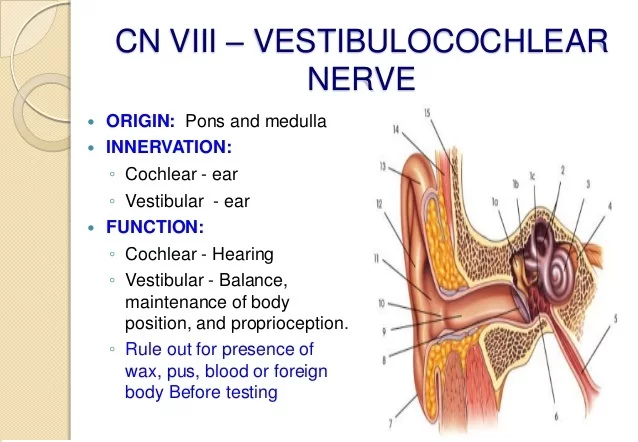
Vestibular component – arises from the vestibular nuclei complex in the pons and medulla.
Cochlear component – arises from the ventral and dorsal cochlear nuclei, situated in the inferior cerebellar peduncle.
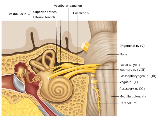
Both sets of fibres combine in the pons to form the vestibulocochlear nerve.
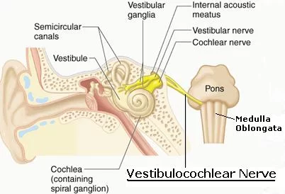
The nerve emerges from the brain at the cerebellopontine angle and exits the cranium via the internal acoustic meatus of the temporal bone.
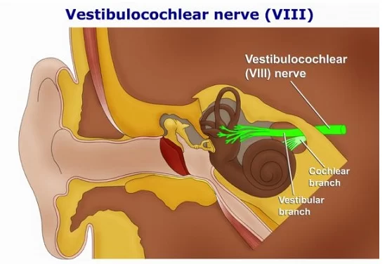
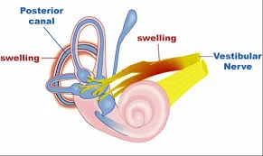
Within the distal aspect of the internal acoustic meatus, the vestibulocochlear nerve splits, forming the vestibular nerve and the cochlear nerve.
The vestibular nerve innervates the vestibular system of the inner ear, which is responsible for detecting balance.
The cochlear nerve travels to the cochlea of the inner ear, forming the spiral ganglia which serve the sense of hearing.
Structure and Functions of the Vestibulocochlear Nerve
Vestibular nuclei :
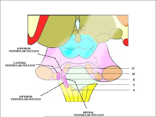
The vestibular nerve has four nuclei placed in the lateral corner of the rhomboid fossa and they are all special somatic afferent (SSA):
- The superior vestibular nucleus – Bechterew
- The lateral vestibular nucleus – Deiters
- The inferior vestibular nucleus – Roller
- The medial vestibular nucleus – Schwalbe
These nuclei belong to the vestibular component of the vestibulocochlear nerve and play a role in the function of balance, spatial orientation, and modification of muscle tone.
The vestibular nuclei are aligned within the rhomboid fossa in such a way that they form two vertical columns, one medial and one lateral.
In the medial column is the medial nucleus, whereas the inferior, lateral, and superior nuclei form the lateral column.
These nuclei contain the bodies of second-order neurons of the vestibular pathway.
First-order neurons are located within the vestibular ganglion at the fundus of the internal acoustic meatus.
The peripheral fibers of these neurons conduct signals from the receptors of the utricle, saccule, and semicircular ducts.
Each fiber splits into the superior and the inferior branch just before its ending.
The vestibular nuclei share multiple connections with the gray matter of the spinal cord, oculogyric nuclei (the nuclei of the CN III, IV, and VI), the reticular formation, and the cerebellum.
Signals from the vestibular ganglion and the cerebellum travel to the vestibular nuclei, and from there go to the motor nuclei of the spinal cord.
The lateral vestibulospinal tract goes to the ipsilateral lumbosacral segments of the spinal cord and is important for the maintenance of posture and walking.
The medial longitudinal fasciculus – through which it influences the movements of the neck and the upper limbs as a response to the stimuli that reach the vestibular nuclei.
Cochlear nuclei :
The cochlear nerve has two nuclei, and both are special somatic afferent (SSA):
- The posterior (dorsal) cochlear nucleus
- The anterior (ventral) cochlear nucleus
These nuclei are the most lateral of all the cranial nuclei. They are located medially to the inferior peduncle of the cerebellum at the lateral angle of the rhomboid fossa.
In these nuclei are the bodies of second-order neurons of the acoustic pathway. The first-order neurons of the acoustic pathway are located within the cochlear spiral ganglion in the inner ear.
The cochlear nerve enters the brainstem and terminates within its cochlear nuclei. Each cochlear fiber has two branches:
The anterior (ventral) branch conducts the impulses caused by the tones of the low frequency and terminates in the anterior cochlear nucleus
The posterior (dorsal) branch carries the information about the high-frequency tones and ends within the posterior cochlear nucleus
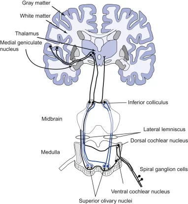
The neurons of the cochlear nuclei give rise to striatal fibers which create stripes of white matter.
These stripes communicate with the tertiary centers of the nervous system in which gathered information is integrated, interpreted, and consciously perceived.
The posterior nucleus sends two stripes: the posterior and the intermediate cochlear stripes, whereas the anterior nucleus sends the third, the anterior cochlear stripe.
All three stripes merge to form the lateral lemniscus.
Nearly half of the striatal fibers from the posterior and the intermediate cochlear stripes do not participate in the forming of the lateral lemniscus, nor do they cross to the contralateral side.
Instead, they synapse with the ipsilateral superior olivary nucleus and join the ipsilateral lateral lemniscus.
After the merging of the vestibular and cochlear root, the trunk of the vestibulocochlear nerve leaves the brain through the posterior cranial fossa traveling laterally to the abducens nerve and the facial nerve.
The vestibulocochlear nerve then extends anteriorly and laterally. Together with the facial and the intermediate nerve, it enters the internal acoustic meatus.
At the fundus of the internal acoustic meatus, the nerve splits into its two roots, vestibular and cochlear. Each root takes a different pathway and has different connections within the inner ear.
Special Sensory Functions of the Vestibulocochlear Nerve
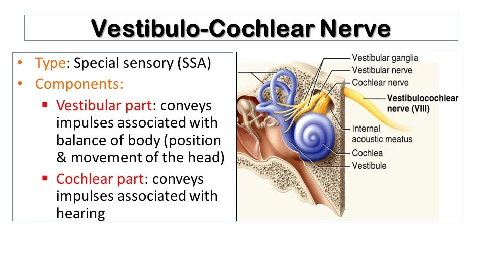
The vestibulocochlear nerve is unusual in that it primarily consists of bipolar neurons. It is responsible for the special senses of hearing (via the cochlear nerve), and balance (via the vestibular nerve).
Hearing :
- The cochlea detects the magnitude and frequency of sound waves. The inner hair cells of the organ of Corti activate ion channels in response to vibrations of the basilar membrane.
- Action potentials travel from the spiral ganglia, which house the cell bodies of neurons of the cochlear nerve.
- The magnitude of the sound determines how much the membrane vibrates and thereby how often action potentials are triggered.
- Louder sounds cause the basilar membrane to vibrate more, resulting in action potentials being transmitted from the spiral ganglia more often, and vice versa.
- The frequency of the sound is coded by the position of the activated inner hair cells along the basilar membrane.
Equilibrium (Balance)
- The vestibular apparatus senses changes in the position of the head in relation to gravity.
- The vestibular hair cells are located in the otolith organs (the utricule and saccule), where they detect linear movements of the head, as well as in the three semicircular canals, where they detect rotational movements of the head.
- The cell bodies of the vestibular nerve are located in the vestibular ganglion which is housed in the outer part of the internal acoustic meatus.
- Information about the position of the head is used to coordinate balance and the vestibular-ocular reflex.
- The vestibulo-ocular reflex (also called the oculocephalic reflex) allows images on the retina to be stabilized when the head is turning by moving the eyes in the opposite direction.
- It can be demonstrated by holding one finger still at a comfortable distance in front of you and twisting your head from side to side whilst staying focused on the finger.
Clinical Importance:
Examination :
Examinations that can be done include the Rinne test and the Weber test.
- Rinne’s test involves Rinne’s Right Test and Rinne’s Left Test since auditory acuity is equal in both ears. If Bone Conduction(BC) is more than Air Conduction(AC) i.e. BC>AC indicates Rinne Test is negative or abnormal.
- If AC>BC Rinne test is normal or positive. If BC>AC and Weber’s test lateralizes to the abnormal side then it is Conductive hearing loss.
- If AC>BC and Weber’s test lateralizes to the normal side then it concludes Sensorineural hearing loss.
Basilar Skull Fracture :
- A basilar skull fracture is a fracture of the skull base, usually resulting from major trauma.
- The vestibulocochlear nerve can be damaged within the internal acoustic meatus, producing symptoms of vestibular and cochlear nerve damage.
- Patients may also exhibit signs related to the other cranial nerves, bleeding from the ears and nose, and cerebrospinal fluid leaking from the ears (CSF otorrhoea) and nose (CSF rhinorrhoea).
Vestibular Neuritis:
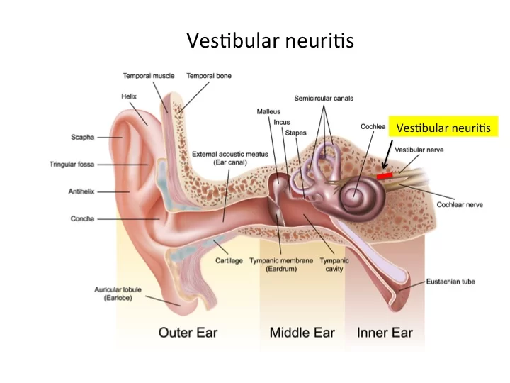
- Vestibular neuritis refers to inflammation of the vestibular branch of the vestibulocochlear nerve.
- The aetiology of this condition is not fully understood, but some cases are thought to be due to reactivation of the herpes simplex virus.
It presents with symptoms of vestibular nerve damage:
- Vertigo – a false sensation that oneself or the surroundings are spinning or moving.
- Nystagmus – a repetitive, involuntary to-and-fro oscillation of the eyes.
- Loss of equilibrium (especially in low light).
- Nausea and vomiting.
The condition is usually self-resolving. Treatment is symptomatic, usually in the form of anti-emetics or vestibular suppressants
Labyrinthitis :
- Labyrinthitis refers to inflammation of the membranous labyrinth, resulting in damage to the vestibular and cochlear branches of the vestibulocochlear nerve.
- The symptoms are similar to vestibular neuritis, but also include indicators of cochlear nerve damage:
- Sensorineural hearing loss
- Tinnitus – a false ringing or buzzing sound.

One Comment