
Fracture of Metatarsal Bone
Introduction
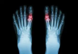
- One of the five metatarsal bones in each foot can shatter completely or partially, which is known as a metatarsal bone fracture. These long, thin bones are situated between the phalanges in the forefoot and the tarsal bones in the hindfoot, or between the toes and the ankle.
- Their slender body has a somewhat broader base, and at the end, they attach to the toe bones through a knob-like structure called the head. They begin at the middle of the foot and finish right before the toe webs. The closer ends of the metatarsal bones, which are located close to the center of the foot, form joints with other midfoot bones for the first through third metatarsals, however, these joints are not very mobile.
- On the other hand, the metatarsal and toe bones move a lot toward the other end of the bones close to your toes. The movement and location of each foot injury determine the appropriate course of treatment.
- The caput ossis metatarsi, corpus ossis metarisi, and foundation ossis metatarsi are the three components that make up each metatarsal bone. They create the ossa cuneiform on the proximal side and the os cuboideum on the medial and lateral sides. They articulate with the base of the proximal phalanges at the distal side.
- Another name for the tarsometatarsal joint is the Lisfranc line. The articular surfaces of the tarsometatarsal joints are comparatively flat, and their strong, short ligaments allow for slight translations and tilting movements. The three ossae cuneïforme are encircled by strong ligaments that constitute the basis of the os metatarsale. Between the medial side of M2 and the lateral side of C1, there is a robust dorsal ligament of lisfranc. The ligament system containing fibers between C2 and C3 is called lateral.
Anatomy of Bones
Osteology
- Shape and function similar to the metacarpals of the hand
- The first metatarsal has a plantar crista that articulates with sesamoids
- Widest and shortest
- Bears 30-50% of weight during gait
- The second metatarsal is the longest
- The most common location of stress fracture
Anatomy of Ligament
Ligaments
- The distal intermetatarsal ligaments in the second to fifth metatarsals preserve length and alignment with isolated fractures that result in the formation of interdigital (Morton’s) neuromas.
- Multiple metatarsal fractures cause the intermetatarsal ligaments to become less stable, which increases displacement.
Anatomy of Muscles
- muscular balance between extrinsic and intrinsic muscles
- extrinsic include
- Extensor digitorum longus (EDL)
- Flexor digitorum longus (FDL)
- intrinsics include
- Interossei
- Lumbricals
Blood supply
Dorsal and plantar metatarsal arteries
Biomechanics
Foot and Ankle Biomechanics.
Rate of occurrence
- Adults most frequently fracture their fifth metatarsal.
- The most frequent fracture in children under 4 years old is to the first metatarsal.
- Rarely do third-metatarsal fractures occur alone; 68% of them are linked to second or fourth-metatarsal fractures
- Population peak incidence occurs in the second and fifth decades of life.
- The metatarsals are the most common location of stress fractures in the human skeleton and are frequently injured in both acute and chronic contexts. The central and distal sections of the corpus ossalis metatarsalis II or III are the most often fractured metatarsal bones due to stress fractures. Less frequently occurring are stress fractures at the base of the first or second metatarsals, as well as relatively other metatarsal bones. Twenty percent of stress fractures in the lower extremities occur in runners, who are more likely to suffer metatarsal stress fractures than in any other sport. Stress fractures are most common in the second and third metatarsals because of the greater stresses these metatarsals endure during walking and running.
- There are two possible causes of metatarsal fractures: direct and indirect violence, and they exhibit a broad range of injuries from crush injuries with multiple fractures and significant soft tissue impairment to isolated, straightforward fractures of a single metatarsal. When a big object falls on an industrial worker’s foot, it frequently sustains direct injuries. When the forefoot is fixated and the leg and hindfoot are twisted, indirect damage takes place. The proportions appear as follows: Injuries to the spine: 48%
- Drop in altitude: 26%
- Injury from crushing: 12%
- Metatarsal fractures are more common in athletes, obese people, those with osteoporosis, people with rheumatoid arthritis, and people with diabetes.
- It also occurs in high-impact aerobic exercises, ballet, gymnastics, and running. Metatarsal stress fractures can be avoided by using shoe shock attenuation.
- Repetitive cyclic loading can result in chronic overloading, which increases the risk of a stress reaction and fracture, particularly in young athletes or military recruits.
- It has been demonstrated that age and the mechanism of injury affect the fracture pattern and degree of damage. This relationship can also be linked to age-related exercise levels and osseous growth.
Causes of Fracture of Metatarsal Bone
- The majority of fractures involving the corpus ossis metatarsals result from direct strikes or forces that twist. A stress fracture of the metatarsal corpus might result from a sudden increase in activity or from long-term overuse.
- Fifth metatarsal fractures most frequently result from falls from standing height or by twisting the ankle while the forefoot is immobilized. In this position, there is a longitudinal and torsional strain due to tension from the peroneus brevis tendon and a pulling force from the plantar aponeurosis lateral cord.
- Since the base of the fifth metatarsal is the terminus of the “supination fracture line,” an avulsion fracture of the metatarsal base (also known as a “tennis fracture”) may arise from inversion injuries to the foot.
- A fractured Jones bone is the most frequent fracture location, which happens when the patient’s weight is over the lateral aspect of the plantar flexed foot and is caused by vertical medial-lateral force at the fifth metatarsal base. Overuse, chronic stress, trauma, or a sharp change in direction with the heel off the ground are additional causes.
- Plantar flexion of the foot combined with ankle inversion is the usual cause of a tuberosity avulsion fracture. These fractures are frequently overlooked since the history frequently points to a lateral ankle sprain.
- Persistent overloading, particularly from jumping and pivoting movements in younger athletes, is frequently the cause of a diaphysial stress fracture.
- The majority of corpus ossis metatarsal fractures are caused by fatigue and are associated with long-term stress. It is the outcome of repetitive force, as demonstrated by soldiers, sportsmen, and ballet dancers. Walking does, after all, put additional strain and force on the second and third metatarsals. Consequently, second or third metatarsals frequently experience stress fractures and bone remodeling as a result of stress. It’s also aware of a high rate among recruits to the military compared to other metatarsal fractures, fractures affecting the first through fourth metatarsals are less prevalent. They should be given extra attention since they are frequently linked to injuries to the Lisfranc ligament complex. These vital ligaments preserve the foot’s arch and connect the metatarsals to the limbs. They also hold the metatarsal bases firmly in position.
- Crush injuries or direct strikes are the usual causes of proximal metatarsal fractures. They could also come from tripping over a foot that is flexed inward. The most frequent cause of a Lisfranc injury in athletes is axial stress applied to the plantar flexed foot.
- People who: abruptly raise their level of activity; engage in physically demanding activities; their feet, as marching (as in the military), dancing, leaping, or running.
- possess an illness of the neurological system that results in loss of sensation in the feet.
- osteoporosis (thin, weak bones)
- arthritis (inflamed joints).
- run over 30 kilometers a week.
- Metatarsal fractures account for 5–6% of all fractures treated in primary care. These are the most typical foot injuries. Approximately ten times as many occur as Lisfranc-dislocations. They are present in all racial groupings and equally among men and women.
- The fractures’ distribution:
- 5% of the first metatarsal
- Metatarsals 2nd: 12%
- 14% of third metatarsals
- 13% of fourth metatarsals
- Metatarsals five: 56%
- Fractures of several metatarsals: 15,6%
Different fractures
- There are three possible sites for a metatarsal fracture: the caput, corpus, or base of the ossis metatarsalis. In this way, we can distinguish between various fractures:
- fracture of the sub-capital.
- breakage of the metatarsalis corpus.
- breakage of the metatarsalis base.
- The proximal fifth metatarsal is fractured in three different ways. When categorizing proximal fifth metatarsal fractures, the junction of the basis ossis metatarsal IV and V is crucial.
- Fractures via avulsion: “Dancer’s fracture” is the term for an avulsion fracture on the fifth metatarsal bone.
- The fifth metatarsal bone is specifically susceptible to two types of fractures: the Jones fracture and the tuberosity avulsion (styloid) fracture. Tuberosity avulsion fractures are now recognized as “pseudo-Jones fractures.”
- The fracture of the unfused fifth metatarsal base apophysis is known as the diaphyseal stress fracture.
- Marcher’s fracture: Another name for this fracture is a metatarsal II and/or III fatigue fracture.
- The fractures from the first to the fourth metatarsals are located farther on.
- The base is the most typical location for fractures.
- the fifth metatarsal (Jones fracture), which is brought on by the forefoot inversion.
- Since the treatment for a Jones fracture differs greatly from that for fractures of the fifth metatarsal shaft, the fracture’s location needs to be thoroughly assessed.
- Walking puts additional strain on the second and third metatarsals, leading to a high frequency of stress fractures and bone remodeling from stress in these areas. This condition is also referred to as a “marcher’s fracture” because of its high prevalence among enlistees in the armed forces.
Typical indications of fractured metatarsals
- aching edema
- Pain palpable due to axial pressure
- Patients who suffer from metatarsal fractures often complain of pain during walking or difficulty bearing weight. It is sensitive to palpation and the forefoot is swollen. Only complex injury patterns, such as multiple toe dislocations and serial fractures, result in gross malformations.
Differential Diagnoses
Certain features vary depending on where the metatarsal fracture occurs:
- Broken bones ranging from the first to the fourth metatarsal: In youngsters, the first metatarsal growth center is situated proximally. It can be mistaken for a break.
- Proximal fifth metatarsal fractures: Inability to bear weight both immediately and for four steps while in the emergency room, as well as pain in the midfoot zone and one of the following: bone soreness at the base of the fifth metatarsal, bone tenderness at the navicular. These are the Ottowa Foot Rules, which establish whether a radiographic assessment of the foot is necessary.
- cracks in the shaft: These usually include discomfort, edema, ecchymosis, and trouble walking. At first, the agony only gets worse when you move. Severe swelling is common, particularly if the patient’s foot has not been raised. The fracture site is typically accompanied by point soreness. Pain is typically felt at the fracture site when an axial load is applied to the head of a fractured metatarsal. This procedure shouldn’t hurt patients who have soft tissue injuries. If the wound is not given time to heal, the pain will worsen, there will be additional swelling, and a fracture may even happen.
Diaphyseal stress fracture, Jones fracture, and tuberosity avulsion fracture:
- these three fractures result in lateral foot pain and make walking difficult. Acute fractures usually start suddenly and cause edema and ecchymosis. Pain from stress fractures typically increases gradually and gets worse with movement. Ensuring an accurate diagnosis of fifth metatarsal stress requires an understanding of the progressive emergence of symptoms.
cracks. Radiography may show growth plates or auxiliary bones that could resemble these fractures. Ossicles, which are tiny auxiliary bones, could hurt in the same area.
Early warning indications of stress fractures include soreness across the foot that subsides with rest and pain that is felt during activity. With time, the foot discomfort in one location will become more intense and persistent. You might feel some tenderness when you touch the fractured portion of the foot. It may also be enlarged. - Metatarsal fractures are diagnosed by x-rays, bone scans, and a physical examination of the foot. An assumption of a clinical diagnosis can be established if the patient presents with a typical history and relevant physical evidence.
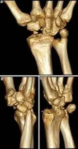
Typically, standard lateral, oblique, and anterior X-rays are enough to identify the fracture. When necessary, a CT scan or MRI is performed to rule out other injuries. There may also need to be a comparison with the opposite foot.
When one anticipates a stress fracture, a bone scan could be beneficial.
Patients with an acute metatarsal fracture (fracture corpus ossis metatarsals) typically complain of discomfort, ecchymosis, swelling, and trouble walking. - A broken metatarsal’s head hurts when an axial load is applied to it. It shouldn’t hurt for patients who have soft-only tissue damage. Radiographic findings: Two views oriented at a 90° angle to one another can be used to determine the fracture position. Oftentimes, oblique or modified lateral views are more beneficial. Early radiographs may not show fracture lines. In this instance, it is recommended to repeat the radiographs and the clinical assessment one to two weeks following the original injury.
- Proximal first through fourth metatarsal fractures: radiographic results Proximal fractures are frequently numerous, transverse, or oblique. In half of the cases with a Lisfranc ligament injury, a conventional foot series may be normal.
- Weight-bearing anteroposterior and lateral radiographs are required in this situation. In stage III injuries, the lateral image shows a lost arch height, while the anteroposterior view shows a widening of the space between the first and second metatarsal heads (stage II or III). When a stage I injury is suspected clinically and radiography is normal, a radionuclide bone scan can be used to diagnose the condition with remarkable accuracy.
- The unfused fifth metatarsal base apophysis fracture is a type of fracture that usually affects children aged 9 to 14. The lucent line associated with an unfused apophysis is invariably oriented longitudinally, in contrast to the fractures that occur in this region. In circumstances where there is uncertainty, contralateral foot comparison radiographs can be useful.
- Acute proximal fifth diaphysis fracture:
We can distinguish between a tuberosity avulsion fracture and a lateral ankle sprain by using the Ottawa ankle rule. If the foot seems normal and there is point discomfort over the fifth metatarsal, it may be an ankle series or sprain.
Results of radiography for the Jones fracture: Acute fracture of the proximal diaphysis-corpus ossis metatarsi quinti junction. The fracture line is acute and runs into or in the direction of the point where MT 4 and MT 5 articulate. A Jones fracture can be either acute (sudden) or stress (small, hairline break that happens over time). - Styloid (tuberosity) fracture: Clinical observations The fifth metatarsal’s long axis is perpendicular to the radiolucency that is observed. The fracture never extends into the joint between the fourth and fifth metatarsals (=different from Jones fractures), and it may be intra- or extraarticular (cuboid-metatarsal articulation). This type of fracture generally happens close to the base of the fourth and fifth metatarsal joints. It affects the styloid process tip where the peroneus brevis and plantar aponeurosis attach. With its wide lateral insertion, the peroneus brevis tendon may exacerbate subsequent dislocation.
- Stress fractures: It’s possible that patients who have a normal history and relevant results don’t require the testing. Findings on plain radiographs are rarely seen until two to six weeks after the onset of symptoms. The diagnosis can be verified with an MRI or bone scan.
Metatarsal diaphysis stress fracture, Torg type II. An older stress fracture will show a wider fracture line and partial or whole obliteration of the medullary canal, while an early stress fracture will show cortical thickening.
Diagnostic Procedures
Certain features vary depending on where the metatarsal fracture occurs:
- Broken bones ranging from the first to the fourth metatarsal: In youngsters, the first metatarsal growth center is situated proximally. It can be mistaken for a break.
- Proximal fifth metatarsal fractures: Inability to bear weight both immediately and for four steps while in the emergency room, as well as pain in the midfoot zone and one of the following: bone soreness at the base of the fifth metatarsal, bone tenderness at the navicular. These are the Ottowa Foot Rules, which establish whether a radiographic assessment of the foot is necessary.
- Cracks in the shaft: These usually include discomfort, edema, ecchymosis, and trouble walking. At first, the agony only gets worse when you move. Severe swelling is common, particularly if the patient’s foot has not been raised. The fracture site is typically accompanied by point soreness. Pain is typically felt at the fracture site when an axial load is applied to the head of a fractured metatarsal.
- This procedure shouldn’t hurt patients who have soft tissue injuries. If the wound is not given time to heal, the pain will worsen, there will be additional swelling, and a fracture may even happen.
- Diaphyseal stress fracture, Jones fracture, and tuberosity avulsion fracture: these three fractures result in lateral foot pain and make walking difficult. Acute fractures usually start suddenly and cause edema and ecchymosis
- Pain from stress fractures typically increases gradually and gets worse with movement. Ensuring an accurate diagnosis of fifth metatarsal stress requires an understanding of the progressive emergence of symptoms.
- Radiography may show growth plates or auxiliary bones that could resemble these fractures. Ossicles, which are tiny auxiliary bones, could hurt in the same area.
- Early warning indications of stress fractures include soreness across the foot that subsides with rest and pain that is felt during activity. With time, the foot discomfort in one location will become more intense and persistent. You might feel some tenderness when you touch the fractured portion of the foot. It may also be enlarged.
Examination
Every patient suspected of having a metatarsal fracture should have a neurovascular examination.
Bony discomfort is typically caused by the fracture. It is important to thoroughly examine the skin for any signs of an open fracture, such as wounds.
- skin tenting over a fracture that is misplaced
Skin that is devitalized and may necrotize; common fracture findings (e.g., edema); uncommon findings (e.g., deformity).
Particular to each metatarsal fracture
Acute fractures of the metatarsals
Pain is typically experienced at the fracture site when an axial load is applied to the head of a fractured metatarsal. To accomplish this, align the toe with the matching metatarsal and press the toe inward toward it. The fracture site may be revealed by sequential palpation.
- It is beneficial to evaluate discomfort while assessing the toes’ dorsal and plantar flexion. from distinguishing between a tendon injury and a fracture. Direct palpation should cause more discomfort if a fracture is present than assessing tendon function by resisting dorsiflexion and plantar flexion.
- Breaks in the first through fourth proximal metatarsals
similar to acute fractures of the metatarsals. Whenever a patient complains of pain near the Lisfranc ligament, it’s important to keep a high degree of suspicion to identify Lisfranc’s injury. - Acute Jones fractures of the proximal fifth diaphysis: Edema and ecchymosis; pain on the outside of the foot anterior to the ankle
Acute tuberosity (styloid) fracture: - There is edema and ecchymosis at the point of maximum tenderness, along with tenderness near the base of the metatarsal.
fracture of the metatarsal shaft due to stress
Point tenderness over the fracture; discomfort in the event of axial loading on the metatarsal head; presence of tenderness over the fracture site; possible presence of edema and ecchymosis.
Treatment of Fracture of Metatarsal Bone
Immediate Treatment
- If a crush or twisting injury caused your metatarsal fracture, you probably won’t be able to bear the discomfort long enough to wait to see a doctor. You might choose to visit your healthcare physician or an emergency department. Taking X-rays is often how the diagnosis is made.
- The emergency department doctor will usually put you on crutches, put you in a splint (half cast), give you pain medication, and tell you to see an orthopedic surgeon or your primary care physician for follow-up care if the bone did not poke through the skin. For the first two to three days, you should also try to keep your foot as elevated as possible and use ice to assist in reducing any swelling.
Surgical Treatment
- General Care
Surgery is not usually necessary for treating metatarsal fractures. You may use a walking boot, a cast, or even a shoe with a firm sole. The bones that are broken will determine how much pressure you can apply to your foot. Your Surgeon will help this decision for you. - Your pain will subside over 8–12 weeks as your broken bone(s) mend. With time, you might be able to exert more pressure on your foot.
It will be recommended that you cease the activity that resulted in the stress fracture of your metatarsal. You’ll likely be told to avoid using your foot for four to six weeks, or perhaps longer until the discomfort goes away.
- Surgery is beneficial for certain metatarsal fractures. These include fractures that have broken through the skin and any fractures that are too wide apart to heal or function correctly in the future. This is particularly valid for first metatarsal fractures.
- The bones can frequently be straightened and kept in place with temporary pins if surgery is required. In roughly six to ten weeks, these pins might be taken out of the office. In certain cases, realigning your bones may require cutting the top of your foot, after which the bone will be fixed using screws and metal plates.
Complications
- Metatarsalgia, a consequence resulting from mal-union or residual deformity, is the most frequently observed side effect following non-operative therapy of metatarsal fractures. Non-union, delayed union, and posttraumatic abnormalities such as digitus quintus varus, and arthrosis are among the other prevalent consequences.
- Nonunion, implant failure, hardware irritation, and sural nerve injury are among the less frequent problems.
Medical Supervision
- An urgent referral is required in cases of vascular compromise, open fractures, compartment syndrome, concomitant neurologic deficits, devitalizing skin, or skin that is in danger of devitalizing.
- An inappropriate location for a shaft fracture, a displaced styloid fracture of metatarsal 5, and a displaced fracture of a single metatarsal when the fracture is close to the metatarsal head should all result in an immediate referral.
- Additionally, if the first metatarsal fracture is not non-displaced, there will be discomfort over the Lisfranc ligament, preventing damage from being ruled out, there are several shattered metatarsals, and the outcome of the subsequent therapy is not adequate.
- When the bones are not likely to keep in alignment or have a disturbed alignment, surgery is necessary. After surgery, screws, plates, or other similar hardware will hold the bones in place. Surgery is necessary to repair the bone and clean the wound when a piece of bone pierces the skin.
Physical Therapy Treatment
- The location and kind of the fracture determine how long the fracture needs to be treated.
Protection, rest, ice, compression, and elevation are crucial during the initial days until the swelling stabilizes.
For the first exercises in rehabilitation, the afflicted limb should bear little to no weight while the bones continue to calcify and mend appropriately. - To reduce discomfort and inflammation and to encourage an increased range of motion in the smaller metatarsal and tarsal joints, therapists will use manual therapy to the ankle and plantar aspect of the foot. It is necessary to use ice to minimize inflammation and swelling.
- The physical therapist may apply electrotherapy, hydrotherapy, soft tissue massage, joint mobilizations, and, in the future, strength, flexibility, and balance exercises.
Active mobilizations such as plantar and dorsal flexion, as well as inversion and eversion of the foot and ankle, should be encouraged in the patient (and should be performed at home!). - Exercises for mid-therapy: these vary depending on the location and degree of fracture. Strengthening and stretching exercises can help promote ankle flexion and extension. an increase in weightlifting activities.
Developing proprioception and strength, potential exercises: - Position a towel on the floor, place a weight on it, and have the patient place their foot on the towel from their metatarsal joints. Request that the patient merely reflect to bring the weight closer.
- Place the patient’s foot on an Airex pad and instruct him to dig his toes into it.
The patient tiptoes around. The patient can perform this with very little weight bearing while seated in a chair. The patient’s ability to bear weight is increased by allowing them to do this exercise standing. - Completing therapy and release: The patient must be able to walk for extended periods without assistance and maintain full use of the affected foot.
- Training tools for the foot include the Airex pad, Sissel pillows, balancing boards (e.g., Wobble board, Bosu ball), and air exhalation pads.
Prognosis
- If given enough rest and immobilization, metatarsal fractures that are not displaced or only slightly displaced have a fair prognosis. Because they are situated in cancellous bone, metatarsal base fractures have a strong chance of healing. First metatarsal fractures have a fair chance of healing since there is a sufficient quantity of cancellous bone.
- Because of their natural stability and the quantity of vascularity from the surrounding musculature, central metatarsal fractures heal quickly. Because of the numerous displacement forces and limited vascular supply, the Jones fracture has a poor prognosis for healing. Avulsion fractures of the fifth metatarsal tuberosity have a better propensity to heal than Jones’s fracture.
Recovery
- Your foot must be strong enough to endure a lot of strain because it bears the whole weight of your body when you walk or run. metatarsal fractures frequently heal fully, allowing patients to return to their pre-injury activities devoid of issues. In certain cases, they may not heal and need surgery, bracing, or activity adjustment.
- Even when a patient’s metatarsal fracture heals fully, some experience pain in their feet.
Summary
The long bones in your foot that join your ankle to your toes are called metatarsals. They also aid with equilibrium when walking and standing. An acute (sudden) fracture, or break, in one of the bones, might result from an abrupt hit, a violent twist to your foot, or from overuse.
FAQ
Does a fractured metatarsal allow you to walk?
It will hurt to walk on a fractured foot. With a fifth metatarsal fracture, you might be able to walk carefully, but immobilization is necessary for the fracture to mend properly.
What is the average healing time for a metatarsal fracture?
The majority of fractures heal perfectly after six weeks. Your symptoms, which might include pain or discomfort, stiffness, decreased strength, and swelling, might take up to six months to fully go away. If you smoke or have diabetes, your bones might repair more slowly.
Reference
- Metatarsal Fracture. (n.d.). Orthopaedic Trauma Association (OTA). https://ota.org/for-patients/find-info-body-part/3723#/+/0/score,date_na_dt/desc/
- Weatherford, B. (n.d.). Metatarsal Fractures – Foot & Ankle – Orthobullets. https://www.orthobullets.com/foot-and-ankle/7032/metatarsal-fractures
- Metatarsal Fractures. (n.d.). Physiopedia. https://www.physio-pedia.com/Metatarsal_Fractures


2 Comments