
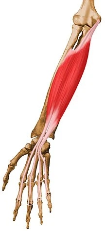
Flexor digitorum superficialis muscle
What is the Flexor digitorum superficialis muscle?
The largest muscle in the forearm’s anterior compartment is the flexor digitorum superficialis. Along with the pronator teres, flexor carpi radialis, flexor carpi ulnaris, and palmaris longus, it is a member of the forearm’s superficial flexors. This muscle is alternatively categorized as a distinct middle/intermediate layer of the anterior forearm, located between the superficial and deep groups, according to some sources.
The flexor digitorum superficialis is divided into two heads according to its origin sites: a radial and humeroulnar head. Its huge solid midsection runs distally toward the wrist, where it parts into four ligaments and connects to the center phalanges of the hand’s second through fifth digits. These tendons can be easily palpated on the distal part of the forearm due to their superficial location.
The flexion of the digits 2 to 5 at the metacarpophalangeal and proximal interphalangeal joints is this muscle’s primary action.
Origin of Flexor digitorum superficialis muscle
There are two heads to the flexor digitorum superficialis muscle:
The medial epicondyle of the humerus and the medial border of the coronoid process of the ulna, the common origin of wrist flexors, form the humeroulnar head.
The anterior oblique line of the radius’s shaft is where the radial head begins.
Insertion
The flexor digitorum superficialis divides into two planes of superficial and deep muscle fibers as it runs down the forearm:
The middle and ring fingers receive tendons from the superficial plane, which further divides.
Four of the nine ligaments in the carpal passage are the four flexor digitorum superficialis ligaments, which pass deeply to the cross-over carpal tendon. The part of the superficial plane that connects with the ring finger tendon is joined by a muscular slip on the deep plane. The superficial plane then divides to supply tendons for the index and little fingers. The flexor digitorum superficialis slips tendinously into the palm and eventually inserts onto the middle phalangeal bases of digits 2 through 5, on the volar surface of the hand, after passing posteriorly around each side of the tendons of the flexor digitorum profundus
Relations
The forearm’s flexor digitorum superficialis connects the flexor digitorum profundus and flexor pollicis longus muscles superficially and the pronator teres, palmaris longus, flexor carpi radialis, and flexor carpi ulnaris muscles deeply. The median nerve and ulnar artery travel through the muscular arch formed by the two heads of the flexor digitorum superficialis.
Innervation
Muscular branches of the median nerve that originate from the roots C8 and T1 of the medial and lateral cords of the brachial plexus innervate the flexor digitorum superficialis. The muscles’ skin is supplied by the roots C6-8 and T1.
Blood supply
The ulnar artery and its anterior recurrent branch provide the flexor digitorum superficialis with the primary arterial blood supply. The anterior and lateral surfaces of the muscle are supplied by radial artery branches, in addition to ulnar artery branches; and the median artery also receives branches from its posterior surface.
Function of Flexor digitorum superficialis muscle
The flexion of the digits 2 to 5 at the proximal interphalangeal and metacarpophalangeal joints is the primary function of the flexor digitorum superficialis. The flexor digitorum superficialis, in contrast to the flexor digitorum profundus, has distinct muscle slips for each of the four digits. As a result, each digit can be individually flexed at its proximal interphalangeal joints. Additionally, the flexor digitorum superficialis facilitates wrist flexion.
Clinical significance
Carpal tunnel syndrome: Numbness, paresthesia, and pain in the thumb, index finger, middle finger, and medial side of the ring finger are common symptoms of carpal tunnel syndrome. The median nerve, along with the tendons of the flexor digitorum superficialis, flexor digitorum profundus, and flexor pollicis longus, is compressed as it travels through the carpal tunnel. A combination of factors that reduce the space around the median nerve, such as injury-related swelling, inflammation, or a neoplasm, is the most common cause of carpal tunnel syndrome. Once in a blue moon, carpal passage disorder can be brought about by a deviant muscle midsection emerging from the ligament of flexor digitorum superficialis that packs the middle nerve.
Swan Neck Deformity: It is the hyperextension of the proximal interphalangeal (PIP) joint with flexion stance of the Plunge joint that most usually happens auxiliary to rheumatoid joint pain, cerebral paralysis, connective tissue problems like Ehlers-Danlos condition, or injury.
Syndrome of the anterior interosseous nerve (AIN): The motor nerve that runs deep between the flexor pollicis longus and flexor digitorum profundus is called the anterior interosseous nerve. The median nerve at the level of the anterior interosseous nerve (AIN) branch that causes AIN syndrome was also found to be compressed by a tight, fibrous arch of the flexor digitorum superficialis.
Variation of the flexor digitorum superficialis that does not have a single tendon for the little finger: Knowledge of this variation may be useful during hand surgery as well as in the preoperative diagnosis of problems with the tendons that run through the carpal tunnel and the muscles on the forearm’s front side. Because this muscle is responsible for the skill actions of the fingers, it also plays an important role in the postoperative rehabilitation phase.
Assessment
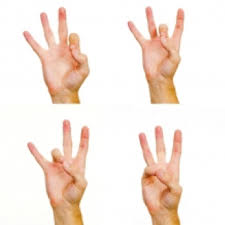
The patient is asked to flex the proximal interphalangeal (PIP) joint of one of the digits from the second to the fifth to test flexor digitorum superficialis, while the other three digits are held in extension to activate the flexor digitorum profundus.
Evaluation of the clinical variables of the flexor digitorum superficialis muscle from the first to the fifth finger:
In practice, two methods are common and accurate:
The test modified by Baker: The examiner asks the subject to flex the proximal interphalangeal (PIP) joint of the little finger by itself while the examiner holds the other fingers extended. After that, the little and ring fingers were allowed to flex together to see how the proximal interphalangeal (PIP) joint flexion had improved. On the off chance that an individual can’t flex the proximal interphalangeal (PIP) joint of a little finger while different fingers are controlled in expansion however ready to flex the proximal interphalangeal (PIP) joint of the fifth digit while the fourth digit is delivered, then, at that point, it shows the shortfall of the fifth ligament of Flexor digitorum shallow.
Another test requires the participants to closely align their palms and corresponding fingers with one while relying on the forces exerted by their own hands. If there is a connection between the little finger and the adjacent fingers, the influence of the adjacent fingers can be apparent while the little finger is instructed to flex.
Flexor digitorum superficialis exercise
Finger opposition
Finger dexterity is tested in this exercise. To make a squeeze grasp, on the other hand, contact your thumb with each finger and hold for as long as two seconds all at once.
Inch grip
The finger flexors are the focus of this grip-strengthening exercise. Clutch two lightweight plates by squeezing them together.
Bulb grip
The finger flexors are the focus of this grip-strengthening exercise. Grip the base of a kettlebell with your hands and fingers to hold onto it.
Fat grip
The finger flexors are the focus of this grip-strengthening exercise. Grip a weight plate with your hands and fingers to hold onto it.
Hook grip
The finger flexors are the focus of this grip-strengthening exercise. By letting the handle of a weight, such as a kettlebell or a dumbbell, hang off your fingers, you can hold onto the weight.
Bulb grip curls
This is a modified biceps curl that trains grip strength with a kettlebell. Feeding your hand through the loop and holding onto the base of the weight is the best way to grip a kettlebell. Curl the weight by flexing the elbow to its full range of motion, keeping it positioned directly below your shoulder.
Standing finger curl
This is a reinforcing exercise for the finger flexors. Begin by holding a portable weight with one arm while standing up. Pull the weight up by slowly extending your fingers so that you can only hold it with your fingers. Afterthen, curl your fingers into a fist.
Banded finger flexion
The finger flexors can be strengthened with this isometric exercise. Pull down with your non-target hand on an exercise band while looping it around the target finger with the palm facing up. Per repetition, hold for up to 7 seconds and rest for 5 seconds. As necessary, repeat.
Banded bulb grip curls
This is a more advanced version of the bulb grip curl that trains grip strength with a kettlebell. Feeding your hand through the loop and holding onto the base of the weight is the best way to grip a kettlebell. Step on a band that has been looped around the handle of the kettlebell for additional resistance. Curl the weight by flexing the elbow to its full range of motion, keeping it positioned directly below your shoulder.
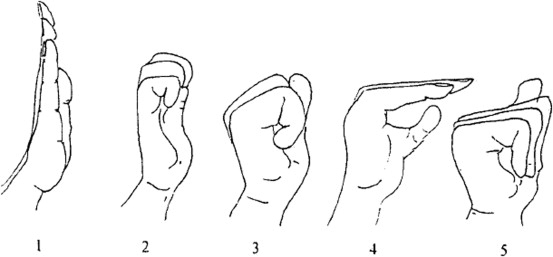
Carpal tunnel tendon glide
This is a movement exercise for the wrist tendon. Arm raise to 90 degrees of shoulder flexion and wrist extension to 90 degrees of extension Crease your hand so the fingers are straight, however, are twisted at the main arrangement of knuckles. The second set of knuckles should remain flexed while the first set of knuckles should be extended. Last but not least, fully extend the final set of knuckles on each finger. Then carry on as necessary.
FAQ
How is the superficialis flexor digitorum different from the profundus flexor digitorum?
The flexor digitorum superficialis, in contrast to the flexor digitorum profundus, has distinct muscle slips for each of the four digits. As a result, each digit can be individually flexed at its proximal interphalangeal joints.
Where is the FDS ligament found?
Tendons of the flexor digitorum superficialis (FDS) run down the forearm and through the carpal tunnel. These tendons, like the flexor digitorum profundus tendons, move along the fingers and the hand in sheaths.
Where is the flexor digitorum superficialis palpated?
If the palmaris longus tendon is present, place your fingers on it. As you help the patient relax the pinch, press your fingers medially into the ulnar valley. The patient should now be directed to alternately flex and relax their fingers. In the ulnar valley, palpate the tendons.
What is the most typical injury to the flexor tendon?
Lacerations (cuts) are the most common cause of flexor tendon injuries. The flexor tendons can be damaged if the forearm, hand, or wrist are lacerated. When a flexor tendon injury occurs, the fingers, thumb, or wrist may be unable to bend.
What is no man’s region close by?
It’s the area of the hand where the flexor digitorum superficialis and flexor digitorum profundus change their positions while passing through a synovial sheath with little tolerance, about from the midpoint of the middle phalanx to the distal palmar area.
3 Comments