
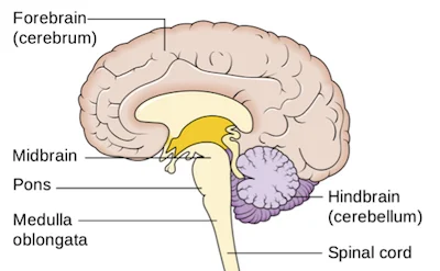
Midbrain (Mesencephalon)
The mesencephalon or midbrain is the greatest rostral (front) segment of the brainstem that joins the cerebellum and pons with the forebrain. For most of its portion, the midbrain sits in the posterior cranial fossa, crossing the hiatus of the tentorium cerebelli.
The mesencephalon or midbrain is the smallest segment of the brainstem. However, it comprises numerous important structures that make it necessary for the proper functioning of the body:
It comprises the transmit the nuclei comprises in the processing of auditory and visual knowledge
It domiciles the nuclei of three cranial nerves: the oculomotor nerve (CN III), trochlear nerve (CN IV), and one of the nuclei of the trigeminal nerve (CN V) through which it controls or regulates the movements of the eye and sensation of the face
It gives the passageway for the pathways that run between the cerebral cortex and spinal cord
Gross anatomy
The midbrain is present between the thalamus (rostrally) and pons (caudally). The lateral sides of the midbrain are enclosed and covered by the hippocampal gyri of the brain.
Anterior (ventral) surface
The anterior surface of the midbrain is marked by the two trunks called the cerebral peduncles. The peduncles are comprised of numerous pathways that run between the cerebral cortex and the spinal cord.
As the peduncles intersect caudally towards the pons, they bounce a fossa on the anterior surface of the midbrain, called the interpeduncular fossa. The floor of this fossa is made up of the posterior pierce substance, which is a layer or coating of gray matter pierced by the branches of the posterior cerebral artery that supply the midbrain or mesencephalon. The components of the interpeduncular fossa are the following:
The terminal (end) bifurcation or divergence of the basilar artery into the posterior cerebral arteries
The emerging or appearing roots of the oculomotor nerve (CN III)
The posterior communicating arteries
The midbrain is nearly or closely related to the optic tract. The tract horizontally crosses or intersects the crura cerebri and winds around them to reach or extend the posterior surface of the midbrain.
The posterior surface or region of the midbrain is called the tectum(roof) of the midbrain. The tectum is characterized by four tubercles on its surface which present inferior to the pineal gland. The upper pair or a couple of tubercles are the right and left superior colliculi, while the lower pair or couple are the right and left inferior colliculi. Because of this organization or alignment, the tectum is also called the quadrigeminal plate, while the colliculi are collectively called the corpora quadrigemina. The colliculi are split from each other or each one by the cruciform sulcus. Rostrally(front), the sulcus extends or continues from the depression of the pineal gland, while its caudal end is continued(enlarged) by the frenulum of the superior medullary velum. The frenulum separates or splits the inferior colliculi one from the other. The trochlear nerve (Cranial nerve IV) can be seen coming out from either side of the frenulum.
Internal anatomy
The midbrain comprises two major parts: cerebral peduncles and tectum. The cerebral peduncles comprise the crura cerebri and tegmentum. They are separated or split from each other by a darkened band called the substantia nigra. The dorsal portion of the tegmentum is crossed by the cerebral aqueduct, which connects the third and fourth ventricles of the brain. The tectum presents dorsal to the tegmentum and cerebral aqueduct, and it comprises the nuclei of the superior and inferior colliculi.
Cerebral peduncles
On the cross-section of the midbrain, we can see that the cerebral peduncles comprise the ventral and dorsal areas. The ventral area of each crus is called the crus cerebri, and comprises the white matter from the cortex. The dorsal area of the crura is continuous with each other and is called the tegmentum of the midbrain. The tegmentum comprises certain neural pathways, reticular formation, and cranial nerve nuclei. The tegmentum is separated or split from the crura cerebri by the substantia nigra.
Crus cerebri
The crus cerebri is the greatest ventral area of each cerebral peduncle. It is collected from three descending pathways that have particular arrangements. These pathways are also overall called the longitudinal pontine fibers:
Corticospinal tract
Corticospinal and corticonuclear pathways run toward the spinal cord and cranial nerve nuclei, respectively. cooperatively these pathways occupy or attend the central two-thirds of each crus cerebri.
Corticopontine pathways are split into the frontopontine and temporopontine tracts based on from which part of the cerebral cortex they rise or originate. The frontopontine tract occupies or attends the medial sixth of the ipsilateral crus, while the temporopontine tract composes or frame the lateral sixth of the ipsilateral crus.
Tegmentum (Pretectal area)
The mesencephalic tegmentum also called the pretectum, is the central part of the midbrain. It comprises the reticular and cranial nerve nuclei, as well as different neural pathways. These structures span different levels of the midbrain.
Midbrain reticular formation
The reticular formation is a structure of phylogenetically old nuclei that controls basic and vital autonomic functions. The reticular formation is spread from the whole brainstem. The mesencephalic portion function n of the reticular formation is present within two clusters that are obtained anterolateral to the periaqueductal gray, respectively. Each cluster is split into three columns with particular functions:
Gigantocellular reticular nuclei (medial column) – include in motor correlation
Raphe nuclei (median column) – include pain and mood control.
Parvocellular reticular nuclei (lateral column) – include in the control of breathing
Periaqueductal gray matter
The periaqueductal gray (PAG) is a mass of neuronal bodies that surrounds the cerebral aqueduct. The major role of periaqueductal gray is to regulate pain by releasing or letting out endogenous opioids, similar to enkephalin, serotonin, and dynorphin.
Periaqueductal gray matter
The Periaqueductal gray matter (PAG) connects to the somatosensory regions of the cerebral cortex, as well as with the lower spinal cord centers. In the wide sense of speaking, Periaqueductal gray matter (PAG) is the place where the difference between the expected and real recognize pain is made. This function is very important for basic survival, as it permits a person to learn about and ignore behaviors that are potentially life-threatening and dangerous.
Cranial nerve nuclei
The nuclei of the oculomotor (CN III), trochlear (CN IV), and trigeminal nerves (CN V) are situated near the periaqueductal gray matter (PAG).
Nucleus of the oculomotor nerve
The nucleus of the oculomotor nerve is a widespread somatic efferent nucleus. It is situated ventral to the periaqueductal gray. This is a motor nucleus that gives the fibers for the supply of all extraocular muscles except for the superior oblique and lateral rectus muscles.
The accessory oculomotor nucleus (Edinger-Westphal) is a widespread visceral efferent nucleus, present just dorsal to the nucleus of the oculomotor nerve. It is a parasympathetic nucleus that supplies the ciliary and sphincter pupillae muscles and permits the miosis of the pupil. These nuclei play an important role or function in pupillary light reflex and arrangement of the eye.
The nucleus of the trochlear nerve is a general somatic efferent nucleus. It is situated in the most ventral portion of the periaqueductal gray (PAG), and its role is to supply the superior oblique muscle.
The mesencephalic nucleus of the trigeminal nerve is a general somatic afferent nucleus. It expands via the lateral portion of the periaqueductal gray, from the pons up to the region of the superior colliculus of the midbrain. The role of this nucleus is to collect proprioceptive information from the muscles of the face.
Red nucleus
The red nucleus (nucleus ruber) is an ovoid mass situated dorsomedial to substantia nigra at the region of the superior colliculus. It stands for this name because of the reddish color that this nucleus has on fresh or natural specimens, due to its high iron content.
- The red nucleus consists of two parts:
- The caudal (magnocellular) portion
- The rostral (parvocellular) portion
The main inputs to the red nucleus derive from the primary somatomotor and somatosensory regions of the cerebral cortex, as well as from the cerebellum. These inputs are carried via the corticofugal and cerebellorubral tracts, appropriately.
The major achievement of the red nucleus is the rubrospinal tract. It gets originates from the magnocellular portion of the nucleus, decussates, and terminates or ends within the cervical portion of the spinal cord. The major role of this tract is to adjust or maintain the movements of the upper limbs in order to maintain or restore the balance of the body. For example, the back-and-forth movements of the arm that we help while we walk are controlled by the rubrospinal tract.
The parvocellular portion of the nucleus collects the fibers from the dentate nucleus of the cerebellum. It then predicts to the inferior olivary nucleus by running via the central tegmental tract that passes through the ventral midbrain. The neurons of the inferior olivary nucleus then predict back to the cerebellum, forming the cerebello-rubro-olivary circuit. The role of this circuit is to coordinate with the cerebellar system of motor control. It is known that the damage or injury of this circuit results in tremors (shaking) and motor discoordination.
The midbrain gives the passage for numerous ascending pathways. specifically, these are the medial lemniscus, lateral lemniscus, superior cerebellar peduncles, medial longitudinal fasciculus, trigeminal lemniscus, and spinal lemniscus.
The superior cerebellar peduncles (brachium conjunctivum) are a couple of white matter bundles that connect the cerebellum with the midbrain. They arise from the cerebellum and enter the midbrain at the region of the inferior colliculi. The fibers are situated centrally in the ventral portion of the mesencephalic tegmentum. They decussate at the inferior colliculi region, forming a horseshoe-shaped commissure of the Wernekinckes area. After decussating, the fibers split into ascending and descending bundles.
Ascending pathways
The ascending bundle comprises the cerebellothalamic tract and cerebellorubral tracts. The former continues its pathway via the brainstem to expand the ventrolateral nucleus of the thalamus, while the closing synapses with the contralateral red nucleus. A small quantity of the ascending fibers synapses with the periaqueductal gray and reticular formation.
The descending bundle comprises the fibers that synapse with the reticular development of the pons and medulla, as well as with the olivary nuclei.
The midbrain comprises; one fasciculus, five sensory pathways, and four lemnisci. particularly, they are the medial longitudinal fasciculus (MLF) and the medial, trigeminal, lateral lemnisci and spinal, The medial longitudinal fasciculus is situated just dorsal to the intersection of the superior cerebellar peduncle. This pathway connects the Edinger-Westphal, vestibular, reticular, oculomotor, trochlear, abducens, and spinal accessory nuclei. through these connections, it permits the conjugate eye movements, as well as the related movements of the head and neck.
The four lemnisci pass via the lateral margin of the tegmentum. From ventral to dorsal, they are divided in the following order:
Medial lemniscus – this is a sensory pathway that is pursued on the dorsal column of the spinal cord, developing the dorsal column-medial lemniscus pathway with it. This sensory system is in charge of conveying information about vibration, slight skin stretch, touch, proprioception, temperature, alteration in texture, discrimination, and pressure to the thalamus.
Trigeminal lemniscus (trigeminothalamic tract) – it conducts the tactile, pain, and temperature sensations from the head to the thalamus.
Spinal lemniscus (spinothalamic tract) – it passes or conveys pain, temperature, non-discriminative touch, and pressure information to the thalamus.
Lateral lemniscus – it conveys the auditory information from the cochlear nucleus to the contralateral (opposite)inferior colliculus.
Substantia nigra
The substantia nigra is a semilunar lamina of heavily pigmented neurons situated between the crus cerebri and the mesencephalic tegmentum. The differentiable dark color derives from the pigment neuromelanin. The substantia nigra range all the levels of the midbrain, from the pons to the subthalamus. Functionally, the substantia nigra is a segment of the extrapyramidal motor system, via which it contributes to the control or regulation of movements by connecting or joining with the basal ganglia.
The substantia nigra comprise two segments that share different connections with other portions of the nervous system:
Dorsal pars compacta are comprised of neurons that construct the neurotransmitter dopamine. It connects with the striatum through the nigrostriatal pathway and facilitates its function. The loss of the dopaminergic neurons of the pars compacta plays a central role or function in the development or formation of Parkinson’s disease and other parkinsonian syndromes.
Ventral pars reticulata, are comprised of the neurons that mainly manufacture the inhibitory neurotransmitter GABA. This portion of the substantia nigra collects the information from the striatum, after which it further predicts the thalamus.
Tectum
The tectum, or the quadrigeminal plate, comprises two couple of relay nuclei, all together called the corpora quadrigemina.
The superior colliculi are included in the processing of visual information. They collect afferents mainly from the retina while sending their production to the lateral geniculate bodies. The role of the superior colliculi is to facilitate the ocular reflexes that mainly have preservative functions, for example, covering the eyes upon a sudden onset of brightness.
The inferior colliculi are connected with the cochlear nuclei and are included in the processing of auditory information. They predict the medial geniculate bodies and are included in reflexive movements induce by auditory stimuli. An example of such movement is turning the head toward the origin of a sudden and unexpected sound (e.g. explosion).
Blood supply
Functions of the midbrain
The midbrain gives the passage or entrance for the main descending pathways of the cerebral cortex. specifically, the corticospinal and corticobulbar tracts that permit us the voluntary movement of the head and body.
It functions as a conduit or channel for the main ascending pathways from the spinal cord which conduct the sensory stimuli from the body and head to the brain. specifically, these are the medial, lateral, spinal, and trigeminal lemnisci.
It residence the mesencephalic part of the reticular formation which, together with other portions of the brainstem, controls pain, mood, and breathing.
encourage survival tendency by facilitating the recognition of definitely dangerous behaviors.
residence the substantia nigra, it comprises a portion of the extrapyramidal motor system and thus it includes the control of voluntary movements.
It permits the movements of the eye by the residence of the nuclei of the oculomotor and trochlear nerves. besides, it facilitates the correlated functioning of these nuclei.
associate with the visual and auditory nuclei, it permits ocular and auditory reflexive movements.
The tectum of the midbrain collects its blood supply from the superior cerebellar artery.
The central portion of the tegmentum is supplied or contributed by the posterior cerebral artery and paramedian branches of the basilar artery, while the lateral portion of the midbrain is nourished or maintained by the posterior cerebral artery solely.
Clinical relations
Infarction of the midbrain
Any injury to the pons and midbrain has the prospective to disrupt critical or censorious body functions. When the infarction or lesion affects the tegmentum, the clinical signs will rely on or depend on the affected cranial nerves. A patient could present with the loss of dorsal column procedure on the contralateral side due to medial lemniscus damage or injury.
Parinaud’s syndrome
injury or damage to the tectum of the midbrain may present as Parinaud’s syndrome. In this scenario or framework, the patient will be unable or inadequate to gaze upwards or downwards, as the corpora quadrigemina will be affected.
Weber’s syndrome
An aneurysm in the posterior cerebral artery at the region of the circle of Willis can result in Weber’s syndrome. The patient will have weakness in the tongue, palate, and lower face of the opposite side (contralateral side )due to damage or injury of the corticobulbar fibers. Since the corticospinal tract may also be compromised or composed, the patient may also have hemiparesis or half-side weakness of the muscle of the contralateral trunk (opposite trunk) and extremities. Paralysis of the opposite (contralateral)) CN III, ptosis, complete or finalized internal ophthalmoplegia (paralysis of the intrinsic eye muscles) on the same side, and depression and abduction of the eyes can also be observed or noticed.
Argyll Roberston pupil (“prostitute’s pupils”)
Clinical findings that normally take place in patients with late-stage neurosyphilis, however, can take place in diabetic neuropathy, stroke, multiple sclerosis, or alcohol intoxication. It’s described by bilateral miotic (small) and irregular pupils which construct briskly when converged or merged but do not react to bright light, therefore, displaying light-near dissociation or separation. The exact cause behind this is still unknown but is thought it’s because of the bilateral damage or injury of the pretectal nuclei of the midbrain.
FAQ
What is the midbrain important for?
The midbrain is related to vision, hearing, motor control, sleep/wake, temperature arousal, and alertness acting as a sort of transmitting station for auditory and visual information.
What happens if the midbrain is damaged?
Damage or injury to the midbrain can result in a broad variety of movement disorders, difficulty with hearing and vision, and difficulties with memory. Because the midbrain residence the hypothalamus, it also plays a main role in automatic body functions.
How many types of the midbrain are there?
There are three main regions of the midbrain – the colliculi, the tegmentum, and the cerebral peduncles
What is the other name of the midbrain?
The midbrain is also called the mesencephalon. It connects the forebrain to the pons. It makes the brain stem along with the hindbrain.
What is the blood supply of the midbrain?
The blood supply to the midbrain is through branches of the basilar artery, which bisect at the level of the third cranial nerves into the right and left posterior cerebral artery (PCA).
Is the midbrain responsible for sleep?
The basal forebrain, near the anterior and bottom of the brain, also encourages sleep and wakefulness, while the portion of the midbrain acts as an arousal system.
What are the diseases of the midbrain?
specific syndromes related to midbrain pathology include the Weber, Benedikt, Nothnagel, Claude, and Parinaud syndromes. The oculomotor and trochlear cranial nerves also reside in this
At what age does the midbrain develop?
At three to four weeks after conception or fertilization, the neural groove closes into a tube, and three definite areas a forebrain, midbrain, and hindbrain begin to take form.
Does the midbrain control balance?
And while the balance system occupies several portions of the brain, the main portion of the brain that controls balance is the cerebellum.
What is the size of the midbrain?
The midbrain is the small, constricted portion, which connects the pons and cerebellum with the thalamus and cerebral hemispheres. It is the smallest of the brainstem, almost 2 cm in length, and most of it is situated in the posterior cranial fossa [3, 5].
What cells are in the midbrain?
The adult midbrain comprises two main anatomically defined populations of dopaminergic neurons, situated in the ventral tegmental area (VTA) and in the substantia nigra pars compacta (SNc)
Is the midbrain the largest?
The midbrain is the smallest area of the brain and is situated most centrally within the cranial cavity. Limbic System – the limbic system is frequently referred to as the “emotional brain”, or ‘childish brain’. It is obtained buried within the cerebrum and comprises the thalamus, hypothalamus, amygdala, and hippocampus.
Why is it called the midbrain?
The midbrain or mesencephalon is the forward-most area of the brainstem and is related to vision, hearing, motor control, sleep and wakefulness, alertness, and temperature regulation.
What is the smallest part of the brain?
The brain stem is the smallest and is situated under the cerebellum, expanding downward and back toward the neck. The cerebral cortex is the outside area of the cerebrum, also called the “gray matter”. It gives rise to the most complex intellectual thoughts and controls or manages body movement.
What is the midbrain in psychology?
the relatively small area of the upper brainstem that connects or joins the forebrain and hindbrain. It comprises the tectum (and related to inferior and superior colliculi), tegmentum, and substantia nigra. the other name of the midbrain is mesencephalon.
What nerves come from the midbrain?
(III) The oculomotor nerve and trochlear nerve (IV) converge from the midbrain, the trigeminal nerve (V), abducens nerve (VI), facial nerve (VII) and vestibulocochlear nerve (VIII) from the pons, and the glossopharyngeal (IX), vagus (X), accessory (XI) and hypoglossal (XII) converge from the medulla.
Why is the midbrain reduced in humans?
The midbrain is damaged by some developmental disorders, involving cobblestone lissencephaly (type II lissencephaly), in which neurons fail to move between the 12th and the 24th week of gestation, resulting in a lack or deficiency of formation or development of grooves and folds in the brain surface.
Is thalamus a midbrain?
The thalamus is a paired or match gray matter structure of the diencephalon situated near the center of the brain. It is above the midbrain or mesencephalon, allowing or permitting nerve fiber connections to the cerebral cortex in all directions — each thalamus connects to the other through the inter thalamic adhesion
Is the midbrain responsible for speech?
the brain has many portions but speech is primarily controlled by the largest area of the brain, the cerebrum. The cerebrum can be split into two parts, called hemispheres, which are connected by a band of nerve fibers called the corpus callosum. the speech is typically governed by the left side of the cerebrum.
How is the midbrain activated?
MidBrain (Interbrain) has to be aroused by stimulating a hormonal discharge. In the human body, it is the pituitary gland that controls the hormone secretions and this function has to be aroused. For this, it is compulsory to activate the neighboring Pineal Gland.
What is a midbrain stroke?
Definition. A brainstem stroke occurs when the blood supply to the base of the brain is blocked or stopped. This can affect many functions in the body, similar as breathing, heart rate, and blood pressure.


2 Comments