Vastus Medialis Muscle Pain
The Vastus Medialis Trigger Points and Referred Pain?
Two trigger points can appear in the vastus medialis muscle.
The lower vastus medialis pain with trigger point is the more typical of the two and is located just above and to the inside of the knee joint. It directs to an aching pain to the front of the knee that may extend up into the lower medial thigh area. After a few weeks, the pain from this trigger point can diminish but is returned by surprising weakness in the muscle that leads to the knee unexpectedly buckling while a person is walking.
The upper vastus medialis trigger point is found a few inches above the lower trigger point, on the inside part of the lower thigh. It directs pain down the inside of the thigh to just above the knee.
What Causes Vastus Medialis Muscle pain?
The following circumstances or activities may activate or reactivate these quadricep muscle pain:
- The quadriceps muscle group as an entirety is typically overloaded by “catching oneself” after stumbling or unexpectedly stepping into a hole. In these situations, the muscles undergo a strong eccentric (lengthening) contraction as they try to decelerate the body’s full importance during the fall.
- A new exercise program that contains squats or deep knee flex may also overload these muscles.
- Unaddressed trigger point activity in the hamstrings can lead to chronic tightness in these muscles. As the biomechanical double to the hamstrings, the quadriceps will continually develop trigger point activity in reaction to the chronic muscle tension in the hamstrings.
- A patient that has tension in their soleus muscle will usually compensate for this when trying to squat down and lift something by overloading their quadricep muscles. The tight soleus controls them from flexing their ankle (dorsiflexing) correctly while squatting down.
- Skiing, and skiing accidents, are particularly hard on the quadricep muscles in general.
- Trigger point activity in the vastus medialis is seen more continually in those people who too pronate their foot (toes outward) and in those people with Morton’s Foot Structure (see Connected Disorders).
- Common sporting movements such as jogging, running, soccer, football, and basketball can also overload this muscle.
- Car accidents where the person’s knee hits the steering wheel can activate the vastus medialis trigger points.
Symptoms of Vastus medialis muscle pain
Three common symptoms of vastus medialis injury are:
- Pain with the inner thigh and at the front and inside of the knee
- Continued pain in the knee joint
- Buckling of the knee (or when the knee allocates out)
Other symptoms include:
- Cramping or tightness in the thigh
- Tenderness when touching the site
- Swelling
- Bruising
Foot pronation on the involved side (which is when toes on the affected leg will point externally)
Pain is usually defined as an ache and can hurt both during movement and at rest. A hardly damaged muscle will produce intense pain. However, after several weeks, the pain may reduce only to be superseded by knee weakness during movement.
This muscle will show any of the following symptoms or clinical findings:
- Patients complain of toothache-like pain that they experience deep in the knee joint, particularly when trying to sleep.
- In time, they may also experience a buckling knee disorder that is indicated by sudden weakness in the knee while walking that leads to them falling.
- The toes on the foot of the manufactured leg will continually be pointed to the outside (pronation).
Vastus Medialis Muscle:
Introduction
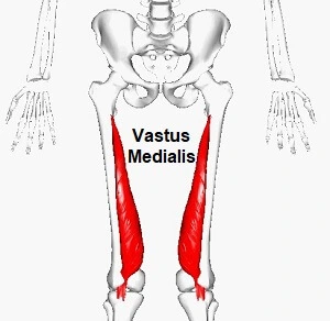
The Vastus medialis muscle is a considerable quadriceps muscle situated on the inner side of the thigh, also known as the teardrop muscle. It is the considerable medial of the 3 muscles of the quadriceps (thigh) group. It is placed in front of the rectus femoris & behind the vastus lateralis.
The muscle is liable for the straightening of the knee joint. It is a part of the extensor mechanism of the knee.
The muscle is more completely working when the knee is at a nearly 30 angle, particularly when the leg is partly extended. Therefore, quadriceps exercises that include a last 30 degrees of angle motion exercise are the best to strengthen this muscle.
Physiotherapists concentrate more on the strengthening of the Vastus medialis as approximated to other Quadriceps groups of muscle mainly due to its work on the end range of knee extension (Last 30 Degree Knee Extension).
Origin
It arises from the the lower part of the intertrochanteric line and along with the spiral line to the medial lip of the linea Aspera
the medial intermuscular septum & the aponeurosis of the adductor Magnus.
Insertion
It inserts into the medial side of the quadriceps tendon, merging with rectus femoris, vastus lateralis, & vastus intermedius muscles, surrounding the patella, then by the patellar ligament into the tibial tuberosity.
Nerve Supply
Femoral Nerve (A component from the posterior part) – Nerve Roots are from L2, L3 & L4.
Blood supply
Vastus medialis is provided by three muscular branches of the femoral artery. It also gets some minor service from the deep femoral & down genicular arteries.
Structure
The vastus medialis is a muscle in the anterior compartment of the thigh, & is one of the 4 muscles that create the quadriceps muscle. The other muscles are the vastus lateralis, vastus intermedius & rectus femoris. It is the most medial of the vastus group of muscles. The vastus medialis originates medially along the whole length of the femur, & connects with the other muscles of the quadriceps in the quadriceps tendon.
The vastus medialis muscle arises from a continuous line of attachment on the femur, which starts on the front & middle side (anteromedially) on the intertrochanteric line of the femur. It continues down & back (posteroinferiorly) along the pectineal line & then plunges along the inner (medial) lip of the linea Aspera & onto the medial supracondylar line of the femur. The fibers connect onto the patella’s inner (medial) part of the quadriceps tendon & the inner (medial) border.
The obliquus genus muscle is the numerous distal portion of the vastus medialis muscle. Its particular activity plays a vital role in keeping the patella part & limiting damage to the knee. With no clear illustration, it is simply the most distal type of fiber of the vastus medialis.
Function
The vastus medialis is one of four muscles in the anterior compartment of the thigh. It is applied in knee extension, along with the other muscles that make up the quadriceps. The vastus medialis muscle also donates to the proper tracking of the patella.
A conflict of the vastus medialis muscle into two groups of fibers has been hypothesized, a long & rather inline group of fibers with the quadriceps ligament, the vastus medialis longus, and a shorter & more obliquely oriented set of fibers, the vastus medialis obliquus. There is however inadequate proof to confirm or refute this hypothesis conclusively.
Examination
Palpation
It can be palpated along its whole length. Distally, the quadriceps tendon can be palpated connecting to the proximal edge (base) of the patella.
Related Knee Pain Disorders
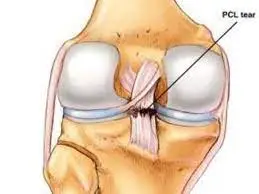
Knee Joint Injury: Strains or tears of the ligaments and menisci are quite typical in professional and recreational sports. As with any joint, the knee joint designs are sensitive to abnormal stress from chronic tension in the muscles that drag the joint (i.e. the quadriceps, hamstring, and gastrocnemius muscles). Many of these damages could be avoided with some regular (maintenance) trigger point treatment to the vastus lateralis, vastus medialis, and hamstring muscles. This is particularly true for the athletes at both ends of the activity spectrum (the poorly conditioned and highly prepared athletes). Many of these damages occur from physical trauma to the leg and knee and are thus inescapable in the traditional sense.
Knee pain is considered to be primarily connected to specific quadriceps muscle weakness or fatigue, particularly in the vastus medialis obliquus (VMO). It is called fatigue & can be led by many various mechanisms, ranging from the collection of metabolites within muscle fibers to the generation of inadequate motor control in the motor cortex. Characteristics of the vastus medialis, including its angle of insertion, correlate with the reality of knee joint pain (patellofemoral pain syndrome). However, this syndrome is difficult & definitive.
Foundering & fatiguing of the vastus medialis obliquus (VMO) causes mal-tracking of the patella & following damage to surrounding structures creating advanced force on the knees, often resulting in damages such as patellofemoral pain syndrome, and anterior cruciate ligament rupture, chondromalacia, and tendinitis. Via the use of an analysis of muscle activity of the VMO via the use of electromyography, and appropriate rehabilitative plans electromyography, experimenters can evaluate & record the electrical movement produced by the skeletal muscle of the vastus medialis obliques to examine the biomechanics & detect any possible abnormalities, weakness, or fatigue.
With & goals can be specified to not only correct the already specified abnormality but even prevent such damages if tested sooner. Containing injuries is important as well as teaching proper training techniques to ensure no valgus collapse forces are causing unexpected stress on other structures of the knee, causing asymmetry, & predisposing that person for injury.
Deficiency of the vastus medials is associated with patellar maltracking & patellofemoral pain. A system of treatment tries to restore the balance between vastus medialis & lateralis, which requires strengthening of the oblique fibers of the medial, as well as an examination of the degree of movement supination & pronation of the foot.
VMO strengthening has become a less popular method for the treatment of anterior knee pain as the proof supporting isolated activities has been criticized for its poor quality. Afterward, students doubt the presence of VMO & have found that any quadricep activity will similarly activate the vast muscles. Strengthening further up the kinetic chain has been proposed as a more practical approach, Khayambashi et al. confirmed that hip strengthening was more useful for improving patellofemoral pain than knee strengthening.
Patellofemoral Dysfunction: This disease is indicated by abnormal tracking on the knee cap (patella) during motions of the knee joint. The contraction of the vastus lateralis and vastus medialis muscles must be conformed properly for the patella to track generally. Trigger points in either of these muscles can compromise this coordination, causing the knee cap to be replaced laterally.
Phantom Limb Pain: Because the pain from trigger points is generally experienced in another region of the body, it is often liable for phantom limb pain in people that have had a limb amputated. These people may be told that their pain is “not real” and is manufactured in their mind, but in reality, their pain is as real as anyone else’s pain and has its origin in trigger points in the remaining portion of the limb or trunk. Directed pain doesn’t require the body part targeted by the pain to exist, as it’s just a neurological reflex created to tell the brain to modify one’s physical activity so the muscle holding the trigger point can rest and recover.
Nerve Entrapment: Pain on the medial part of the knee may be generated by entrapment of the saphenous nerve.
Morton’s Foot Structure: A deviation of the boney structure of the foot where the bottom of the second toe (next to the big toe) is farther along than the base of the big toe. Generally, thick callus forms on the foot where the second toe connects to it. This disease causes side-to-side rocking or instability of the foot and knee and may overload the vastus medialis muscle as it attempts to compensate for the knee instability.
Growing Pains: Unexplained knee and thigh pain in children is often attributed to “growing pains” when trigger point movement in the quadriceps (and other muscles) is the actual cause. Children are continually pushing their bodies to new limits and can easily overfill the leg muscles while playing.
Treatment of Vastus Medialis Muscle Pain
Treatment of Rectus Femoris will rely on the severity of the condition. When treating acute rectus femoris the principles of RICE (rest, ice, compression, and elevation) should be started.
Rest: avoid the activities that make the pain (jumping, running, going up or down stairs, kneeling, and squatting.)
Ice: use ice on the tendon or area of inflammation. It is one of the quickest ways to reduce swelling, pain, and inflammation. Apply it right away and then at breaks for about 20 minutes at a time. Do not apply ice instantly to the skin.
Compression: such as an ace bandage to help take the stress off the impaired muscle may be helpful. When using ice, use light compression. This is particularly helpful if swelling is present.
Elevation: promote the area to help decrease swelling.
Crutches for ambulation or moving around may be required in the case of ruptures or important damage to the rectus, hamstrings, or adductors.
Mild
In mild cases rest, ice and drug may be enough to relieve the pain. Once the pain is relieved, physical therapy is suggested to develop a series of stretching and strengthening exercises to prevent the reoccurrence of the injury. Return to the movement should be gradual to prevent a flare-up of symptoms.
Moderate to Severe
If the problem continues, consulting with your healthcare provider should be the next step. Your physiotherapist will perform a comprehensive evaluation to decide what tendon(s) is involved, the severity of the condition, and the best course of treatment.
Medical treatment:
Avoiding the movements that produce pain or stress in the engaged tendon is the first line of treatment.
- RICE: Rest, Ice, and Compression Elevation should be used to relieve the stress on the bursa.
- NSAIDs (Nonsteroidal anti-inflammatory drugs) to relieve pain and inflammation.
- Injection of steroids may be required to decrease inflammation of the involved tendon
- Immobilization, strapping or bracing may be helpful to rest the tendon and promote healing.
Physiotherapy Treatment
Physiotherapists are specialists, educated and trained to assist interventions.
Your physiotherapist will perform a thorough evaluation to evaluate and select the following:
Tendon: a sequence of tests will be conducted to select which tendon is affected.
Strength: opposed testing is performed to resolve if there is associated weakness or strength inequalities
Flexibility: tight muscles can donate to poor mechanics and weakness creating inequalities and making the hip more unsuspecting to tendinitis.
Technique: Often it is the way we perform movements (run, jump, cycle, or row) that may generate a problem. Examine and observe the activities you participate in, that may have initiated the problem to enhance technique.
Training: examine your training program and any impulsive changes that may have precipitated or led to the present condition.
Alignment or footwear: a physiotherapist will evaluate your leg lengths, foot mechanics, and alignment to see if there are any inequalities. Checking for proper footwear is a crucial part of balancing the stresses applied to your legs and body.
Physical therapy for rectus femoris/quadriceps tendinitis must remain traditional at the onset to avoid aggravating the condition. Emphasis will be on rest, decreasing inflammation, and increasing blood circulation for healing. Once the initial inflammation has been decreased, a program of stretching and strengthening will be initiated to correct flexibility to the muscles involved and enhance strength to reduce stress on the tendons and the hip. Taping or strapping to rest and relieve the stress placed on the tendon and promote healing may be required. Your therapist is taught these clear taping techniques.
Common Physiotherapy interventions in the treatment of rectus femoris pain include:
Manual Therapeutic Technique (MTT): hands-on care including soft tissue massage, stretching, and joint mobilization by a physiotherapist to enhance alignment, mobility, and range of movement of the knee and hip. The use of mobilization procedures also allows for modulating pain.
Therapeutic Exercises (TE) include stretching and strengthening exercises to regain range of movement and strengthen muscles of the knee and lower extremity to support, stabilize and reduce the stresses placed on the bursa & tendons of the hip joint.
Neuromuscular Reeducation (NMR) to restore peace, retrain the lower extremity and enhance movement techniques and mechanics (for example, running, kneeling, squatting, and jumping) of the active lower extremity to reduce stress on the bursa and tendons in daily movements. Taping, strapping, or bracing may be useful to rest the tendon and stimulate healing.
Modalities include the use of ultrasound, electrical stimulation, ice, cold laser, and others to decrease pain and inflammation of the tough tendon and bursa.
A home program that contains strengthening, stretching, and stabilization activities and instructions to help the person complete daily tasks and move to the next functional level.
Vastus Medialis muscle Stretching exercise:
After the follow of electrotherapy for 2-3 days for release to muscle pain by the physiotherapist then the therapist is indicated to stretch for release to muscle tightness.
This stretching is used when your pain is released & when you feel comfortable.
This Stretching exercise allows you to release muscle pain & tightness
- The Lying Quad Stretch
- The Simple Quad Stretch
- The Kneeling Quad Stretch
- Vastus Medialis Stretch
The Lying Quad Stretch:
You are lying in a face-down position & supporting the head on the left hand.
Alternatively, you can lie on the side to complete this stretch.
After some seconds, pull the right foot toward the butt & flex the left knee joint to stabilize yourself.
Then Grasp onto your ankle joint & keep this stretching position for 30 seconds.
Repeat it 3 times at 1 time & do the 3 times each day.
The Simple Quad Stretch:
You are standing on your left leg & with 1 knee joint touching the other.
You can maintain a chair & the wall to support the continued if needed.
Hold the right foot & use your right hand & pull it towards your butt.
Must Be sure to press your chest up & hips joints forward.
Try not to stress about pushing the foot too close to your backside.
But your focus must be on feeling the stretch in your quad muscle & making your hip joint forward to get a good hip flexor muscle stretch.
Maintain this position for 30 seconds.
Repeat it 3 times at 1 time & do the 3 times per day.
The Kneeling Quad Stretch:
This stretching exercise begins with a high lunge position, with the left foot forward.
Then Carefully plunge your right knee joint to the ground & take a moment the balance.
When you are ready, reach back with your right arm & hold your ankle joint or toes.
Maintain this stretching position for 30 seconds but must keep the body steady.
Then Slowly come back into the lunge position & opposite from the left foot to the right foot.
Repeat this Stretching 3 times at 1 time & do the 3 times per day.
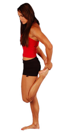
Vastus Medialis Stretch:
Stretching Exercise Of Quadriceps In Standing Position
You are standing holding a chair & the wall for balance
Bring your heel up behind you, hold your ankle joint & pull your heel towards your buttock, till you are feeling a stretch, without leaning forwards
It is done the stretch all four of the quads muscles but to stretch the vastus medialis muscle by moving the foot across the body towards the other buttock.
You can Improve the stretch by pushing your hip joint forwards in this exercise.
Repeat this Stretching 3 times at 1 time & do the 3 times per day.
Vastus medialis muscle Strengthening Exercises:
After the follow of electrotherapy & massage for 2 -3 days release muscle pain the physiotherapist then the therapist suggested to you strengthen exercises release to muscle weakness.
This strengthening exercise is always suggested when you feel to remove pain & when you feel comfortable.
This all-strengthening exercise helps you with muscle weakness & pain:
- Lying Pigeon Progression
- The Frog Pose
- Floor extension
- Lateral heel drop
- Step downs
- Leg extension
- Single leg raises
- Terminal knee extensions (TKEs)
- Vastus Medialis activation Exercise
- Ball Clench Extensions
- Twisted Leg Raise
- Ball Bridges
- Ball Wall Squats
- Isometric Contraction of the vastus medialis muscle
- Seated Isometric vastus medialis muscle & Adduction
- Externally Rotated ½ Squats
- Wall/Ball Squat
- Split Squats/Static Lunges
- Step-Ups
Lying Pigeon Progression:
In the Lying Pigeon Progression first position, a mat on the floor & you are lying face down.
Then must be Secure to place an opposition band around the affected foot, with the extra band in a reachable area.
Grab the band with the left hand & must keep the right leg extended or flex the left knee joint.
Must keep your toes indicated toward the ceiling.
Then use the opposition band to pull forward till you feel the stretch.
Maintain this exercise position for 10 seconds.
Repeat this strengthening 10 times in 1 time & do the 3 times per day.
The Frog Pose:
The Frog Pose exercise is start by lying on your stomach means in a prone position & supporting the torso up on your elbow joints.
Flex both of your knee joints &, and reach back to grab onto your feet.
You sense the stretching at this point.
Then Change the fingers to point the same way as your toes, then carefully lift your elbow joint to point to the roof.
Push the chest up as high as achievable.
This exercise is Controlled completely when you feel any discomfort in the hip or knee joint.
Maintain this exercise position for 10 seconds.
Repeat this strengthening 10 times in 1 time & do the 3 times per day.
Floor extension:
You are sitting down on the ground with a tall posture.
Your shoulder joint should be dragged down the back with your chest proud.
Then Flex your left knee joint toward your chest with your left foot flat on the ground.
Raise your leg in front of you with your foot pointing slightly out to the side then.
Maintain under the left knee joint with both hands interlocked & must keep your muscle bent for the duration of this exercise.
Do the exhale without failing the posture & leaning out from the wall, and lift the right leg in the air as high as possible.
Maintain this position for 10 seconds.
Then inhale & slowly lower down to your starting position.
Repeat this strengthening 10 times in 1 time & do the 3 times per day.
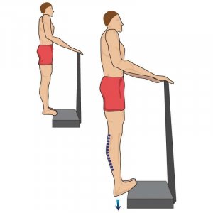
Heel drop:
You are standing tall with your left leg straight but not locked & your foot is resting on a little action.
Do the right knee joint slightly flexed & your left foot should be flat on the ground.
Your right knee joint must be running over the toes.
Then Squeeze your core muscle for equilibrium.
exhale & push up off the right leg till both legs are completely straightened.
Try to keep your hip joint status as you step up.
Inhale then tighten your left vastus medialis muscle & slowly return to your starting place.
Maintain this exercise place for 10 seconds.
Repeat this strengthening 10 times in 1 time & do the 3 times per day.
Step downs:
You are standing with your right foot on the stage & your left foot off to the side.
Do the inhale & Flex the vastus medialis muscle.
Then flex your right knee joint till your left foot is flat on the ground.
Must be Also, try to keep your hip joint level at all times.
Do the exhale & commit your core muscle.
Then force off your foot & return to your starting position.
Maintain this exercise position for 10 seconds.
Repeat this strengthening 10 times in 1 time & do the 3 times per day.
Leg extension:
Sitting on a chair & scoot yourself to the front of the seat.
Then wrap an antagonism band around your ankle joint & feed the band beneath the chair, which you reach back & hold with your hand.
Do the exhale & in one movement then slowly extend your leg to full extension out in front of you.
Then inhale & contract your muscle & slowly lower the leg back down to 30 degrees.
Maintain this exercise position for 10 seconds.
Repeat this strengthening 10 times in 1 time & do the 3 times per day.
Single leg raises:
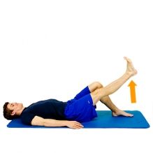
You are lying on the back with your knee joint flexed & foot flat on the mat.
Fully extend your right leg out in front of you must be putting an ankle joint weight on your thigh.
Squeeze your core muscle & contract the vastus medialis muscle & raise the right leg about 2 inches off the mat.
Must Support elevated the leg duration of this exercise.
Make sure you are not arching your back.
You do not place any space between the back & the mat.
Maintain this exercise position for 10 seconds.
Repeat this strengthening 10 times in 1 time & do the 3 times per day.
Terminal knee extensions (TKEs):
You Tie an antagonism band around a sturdy commentator & slide the other end up to slightly above the back of your right knee joint, meeting the commentator.
Step back till the band is tight.
Then Straighten your left leg & must keep your right knee joint slightly flexed.
Do the exhale & push your right knee joint back to match your left knee joint & magnify the contraction in your vastus medialis muscle.
Maintain this exercise position for 10 seconds.
Repeat this strengthening 10 times in 1 time & do the 3 times per day.
Vastus Medialis activation Exercise:
You are sitting erect in a chair, with your knee joint flexed.
Place the ball between your knee joint & your feet flat on the ground.
Then Place your thumbs on the soft, squashy area on the inner side of the knee joint and just beyond your kneecap–patella & press down firmly.
And then Clench your glutes & gently squeeze the ball.
Make sure the move comes from your knee joint rather than the inner thigh.
If you do not feel stretch must be, try clenching your buttocks, clenching your knee joint & squashing the backs of your thighs down into the chair
Maintain this exercise position for 10 seconds.
Repeat this strengthening 10 times in 1 time & do the 3 times per day.
Ball Clench Extensions:
You are lying on your back with a rolled-up towel under your knee joint & place the ball between your knee joint.
Then Clench your buttocks & slowly squeeze the ball & lift one heel off the floor till the knee joint is straight.
Must be Keep clenching the ball & keep it for 10 seconds then gradually return to starting position.
Repeat this strengthening 10 times in 1 time & do the 3 times per day.
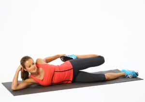
Twisted Leg Raise:
You are lying on your back with one leg extended out straight & the other knee joint bent.
It brings the pressure off the lower back as you work the straight leg.
Turn your foot outwards about 20 into external rotation & raise the foot till your thighs are parallel.
Maintain this exercise position for 10 seconds.
Repeat this strengthening 10 times in 1 time & do the 3 times per day.
Must be Keep the leg bent outwards in this exercise which is helpful to you start the vastus medialis muscle.
Ball Bridges:
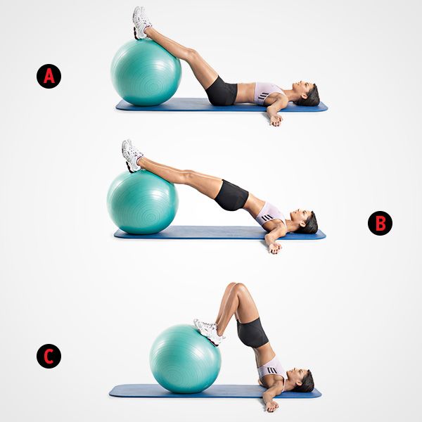
You are lying on your back with your knee joint bent, feet are hip space apart.
Put the ball between your knee joints.
Then Clench your glute muscles & slowly squash the ball.
Lift your buttocks as high as possible without arching your back.
Maintain this exercise position for 10 seconds.
Repeat this strengthening 10 times in 1 time & do the 3 times per day.
Ball Wall Squats:
Standing with your back against a wall & squashy ball between your knees joints.
Away from the wall & toes are suggesting forwards.
Clench your glutes muscle & gently squash the ball to start the vastus medialis muscle then slowly slide down the wall, & flexing your knee joint.
Maintain this exercise position for 10 seconds.
Repeat this strengthening 10 times in 1 time & do the 3 times per day.
Isometric Contraction of the vastus medialis muscle :
You are Sitting position on your bed & ground with the legs out straight & place a towel beneath your knee joint.
Flex your quads muscle with the hip joint and leg slightly externally rotated.
Maintain this contraction for 10 seconds & place your fingers on your VMO to ensure your vastus medialis muscle is triggering & shooting.
Repeat this strengthening 10 times in 1 time & do the 3 times per day.
Seated Isometric Vastus Medialis Muscle & Adduction:
You are sitting on a chair & platform where your feet suspend freely.
Put a ball between your thighs & squeeze the ball together triggering your vastus medialis muscle.
Maintain this muscle contraction for 10 seconds.
Repeat this strengthening 10 times in 1 time & do the 3 times per day.
Externally Rotated ½ Squats:
You are standing with your legs shoulder-width apart with the knee joint & feet externally turned.
Squat halfway down & come up nice & gradually which is focusing on activating the vastus medialis muscle to get you back up to a standing position.
Repeat this strengthening 10 times in 1 time & do the 3 times per day.
Wall/Ball Squats:
You are placing a swiss ball on your back against the wall.
Then slowly squat down into a near-seated position so that your thighs are similar to the floor.
Gradually come back up & avoid locking your knee joint.
Repeat this strengthening 10 times in 1 time & do the 3 times per day.
Split Squats/Static Lunges:
Split Squats/Static Lunges exercise is to start with your feet shoulder-width separated & take one large step forward.
You can put your hands on your hip joint.
To make this exercise harder must maintain dumbbells by your side.
With an erect posture, lunge down & up without your knee joint at the front moving in front of your great toe.
But Focus on placing most of the weight via your front heel & don’t let your knee joint buckle in.
Repeat this strengthening 10 times in 1 time & do the 3 times per day.
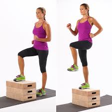
Step-Ups:
Standing in front of a bench & chair.
Step up onto a platform & move from the gluteal muscle, not from your toe.
Provide your knee joint is not buckling inwards.
It is moved/pushed out.
Then slowly step down making sure your knee joint is tough.
Repeat this strengthening 10 times in 1 time & do the 3 times per day.
Prognosis
In general, patients react well to conservative treatment of hip tendinitis. It is essential that once the pain and inflammation are decreased, and motion and strength are restored, the patient gradually returns to full activities. Instruction in daily exercises or sports performance helps decrease a reoccurrence of tendinitis. In most cases, a full recovery from the activity will take from 2-6 weeks counting on the rigor of the tendinitis.
As a preventative measure individuals should:
- Warm Up: Warm up before physical activity is crucial in preparing the muscles and tendons for the work instructed of them. A 5-10 minute warm-up elevates the body temperature, improves the circulation to the muscles and tendons, and raises the heart rate. Elevating your body temperature and increasing circulation will also make the muscles and tendons more flexible for stretching.
- Stretching: stretching regularly in complement to before and after activity will decrease the chances of developing tendinitis. Stretching will also enhance and maintain the elasticity and flexibility of muscles and tendons. Maintain stretches for 20 seconds and do not bounce. Remember, as tendons get older they lose flexibility. It is part of the aging procedure.
- Strength: performing a regular strength schedule will keep muscles strong sufficiently to absorb the stresses placed on them. Just driving or playing a sport does not ready your muscles for the result forces involved in these activities. Remember, as people age, they unaffectedly get weaker.
- Training: avoid sudden gains in your training program. Slowly progress your training program to avoid injury.
- Foot Wear: in weight-bearing or impact activities proper footwear is important to reduce and disseminate forces properly.
FAQ
Why would my vastus medialis impair?
The VMO is also generally overloaded with repeated use in the following circumstances: suddenly increasing your volume of running or cycling a new (or impulsive increase) in an exercise program concerning repetitive squats, lunges, leg extensions or wall sits.
How long does vastus medialis take to recover?
A person with grade 1 damage will likely heal within 1–2 weeks if they rest the muscle as much as possible. It can take longer to heal from a grade 2 or 3 strain, in some cases over 1 month.
How long does a VMO strain take to recover?
Rehabilitation phase: 72 hours to 6 weeks
After the initial inflammatory reaction occurs, it’s time for your tissues to start reconstructing. Full healing can take up to six weeks or more.
Can tight VMO lead to knee pain?
Many people suffer from anterior knee pain and many times, that pain is generated by a tight VMO (teardrop) muscle.
What occurs when VMO is weak?
When the VMO is weak, the other quadriceps muscles pull the kneecap to the outside part of the groove. This can lead to rubbing and extra wear and tear of the joint consistencies in the outside or lateral patellofemoral joint region.
Does walking backward strengthen VMO?
For example, one of the primary causes of a runner’s knee is a defect in the vastus medialis oblique (VMO), so adding some backward walking or running into your recovery program may be a useful way to improve strength and stability in this muscle.